Kombucha tea, a trendy fermented beverage, inspired researchers to develop a new way to generate tough, functional materials using a mixture of bacteria and yeast similar to the kombucha mother used to ferment tea.
With Army funding, using this mixture, also called a SCOBY, or symbiotic culture of bacteria and yeast, engineers at MIT [Massachusetts Institute of Technology] and Imperial College London produced cellulose embedded with enzymes that can perform a variety of functions, such as sensing environmental pollutants and self-healing materials.
The team also showed that they could incorporate yeast directly into the cellulose, creating living materials that could be used to purify water for Soldiers in the field or make smart packaging materials that can detect damage.
“This work provides insights into how synthetic biology approaches can harness the design of biotic-abiotic interfaces with biological organization over multiple length scales,” said Dr. Dawanne Poree, program manager, Army Research Office, an element of the U.S. Army Combat Capabilities Development Command, now known as DEVCOM, Army Research Laboratory. “This is important to the Army as this can lead to new materials with potential applications in microbial fuel cells, sense and respond systems, and self-reporting and self-repairing materials.”
The research, published in Nature Materials was funded by ARO [Army Research Office] and the Army’s Institute for Soldier Nanotechnologies [ISN] at the Massachusetts Institute of Technology. The U.S. Army established the ISN in 2002 as an interdisciplinary research center devoted to dramatically improving the protection, survivability, and mission capabilities of the Soldier and Soldier-supporting platforms and systems.
“We foresee a future where diverse materials could be grown at home or in local production facilities, using biology rather than resource-intensive centralized manufacturing,” said Timothy Lu, an MIT associate professor of electrical engineering and computer science and of biological engineering.
Researchers produced cellulose embedded with enzymes, creating living materials that could be used to purify water for Soldiers in the field or make smart packaging materials that can detect damage. These fermentation factories, which usually contain one species of bacteria and one or more yeast species, produce ethanol, cellulose, and acetic acid that gives kombucha tea its distinctive flavor.
Most of the wild yeast strains used for fermentation are difficult to genetically modify, so the researchers replaced them with a strain of laboratory yeast called Saccharomyces cerevisiae. They combined the yeast with a type of bacteria called Komagataeibacter rhaeticus that their collaborators at Imperial College London had previously isolated from a kombucha mother. This species can produce large quantities of cellulose.
Because the researchers used a laboratory strain of yeast, they could engineer the cells to do any of the things that lab yeast can do, such as producing enzymes that glow in the dark, or sensing pollutants or pathogens in the environment. The yeast can also be programmed so that they can break down pollutants/pathogens after detecting them, which is highly relevant to Army for chem/bio defense applications.
“Our community believes that living materials could provide the most effective sensing of chem/bio warfare agents, especially those of unknown genetics and chemistry,” said Dr. Jim Burgess ISN program manager for ARO.
The bacteria in the culture produced large-scale quantities of tough cellulose that served as a scaffold. The researchers designed their system so that they can control whether the yeast themselves, or just the enzymes that they produce, are incorporated into the cellulose structure. It takes only a few days to grow the material, and if left long enough, it can thicken to occupy a space as large as a bathtub.
“We think this is a good system that is very cheap and very easy to make in very large quantities,” said MIT graduate student and the paper’s lead author, Tzu-Chieh Tang. To demonstrate the potential of their microbe culture, which they call Syn-SCOBY, the researchers created a material incorporating yeast that senses estradiol, which is sometimes found as an environmental pollutant. In another version, they used a strain of yeast that produces a glowing protein called luciferase when exposed to blue light. These yeasts could be swapped out for other strains that detect other pollutants, metals, or pathogens.
The researchers are now looking into using the Syn-SCOBY system for biomedical or food applications. For example, engineering the yeast cells to produce antimicrobials or proteins that could benefit human health.
Earlier this week, RaftsTheGame (@TheRaftsGame) popped up on my twitter feed, which was excellent timing since it’s getting close to Christmas in a year (2020) when I imagine a lot of people may be home and inclined to play games.
The people (rafts4biotech) who produced Rafts The Game (also called Rafts!) are involved in a research project funded by the European Union’s Horizon 2020 programme,
RAFTS! Create the bacterium of your dreams
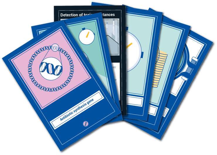
Have you ever wondered what it would be like to be a genetic engineer? Now’s your chance to find out! Rafts! is a card game in which your aim is to design a bacterium while trying to overcome the challenges of research work.
If you are a researcher, look no further – Rafts! enables you to finally share your academic struggles with those friends who don’t have a clue of what you do!
THE GAME
In Rafts! you race to become the first scientist to create a bacterium that can do incredible things: cleaning an oil spill, detecting toxic compounds, producing blood for donations… Sounds like science fiction? More like a regular day at the lab!
But don’t get carried away – nobody said conducting research was easy! Hard work alone isn’t enough if you don’t have the right genetic instructions as well as a combination of money, time as well as food for your bacterium. You’ll have to collect all of these resources to finish the masterpiece that is your bacterium.
In this laboratory people play dirty, so don’t forget to keep an eye on your colleagues – they are all trying to achieve their objectives, and sometimes you will compete for the same resources. Don’t hesitate to strike back!
THE CARDS
There are three types of cards in Rafts!: action cards help you gather the resource cards that you will need to achieve the goal in your objective card. Bring your mouse on top of a card to know what it can do!
…
GET YOURS
Ready to become the biotech wizard you’ve always wanted to be? You’re just a click away from building the bacterium of a lifetime!
Download Rafts! for free and print it yourself – or let your local print shop do it for you:
For anyone curious about the source for the game, here’s a bit about rafts4biotech, from the homepage,
Engineering bacterial lipid rafts to optimise industrial processes
Context
Bacteria are used in the biotechnology industry to produce a wide range of valuable compounds. However, the performance of these microorganisms in the demanding industrial conditions is limited by the toxicity of some compounds and the complex metabolic interactions that occur within the bacterial cells.
Challenge
Generating new synthetic microorganisms that will solve productivity hurdles and yield a great variety of economy-value compounds. These modified strains will be used as standardised microbial chassis platforms to fit industry needs.
Solution
The R4B solution relies on confining the production of compounds to specific areas of the microbe’s membrane called lipid rafts. This recently-discovered regions present an ideal setting that will avoid interferences with bacterial metabolism and viability.
…
Given that at least one of the COVID-19 vaccines (Pfizer-BioNTech?) is wrapped in lipid nanobodies and, now, with this mention of lipids, it seemed like a good idea (for me) to learn about lipids. Here’s what I found in the definition for lipid in The free Dictionary,
a group of substances comprising fatty, greasy, oily, and waxy compounds that are insoluble in water and soluble in nonpolar solvents, such as hexane, ether, and chloroform.
If you’re hoping for a Rumpelstiltskin reference (there is more about the fairy tale at the end of this posting) and despite the press release’s headline, you won’t find it in this August 10, 2020 news item on Nanowerk,
When nanocellulose is combined with various types of metal nanoparticles, materials are formed with many new and exciting properties. They may be antibacterial, change colour under pressure, or convert light to heat.
“To put it simply, we make gold from nanocellulose”, says Daniel Aili, associate professor in the Division of Biophysics and Bioengineering at the Department of Physics, Chemistry and Biology at Linköping University.
The research group, led by Daniel Aili, has used a biosynthetic nanocellulose produced by bacteria and originally developed for wound care. The scientists have subsequently decorated the cellulose with metal nanoparticles, principally silver and gold. The particles, no larger than a few billionths of a metre, are first tailored to give them the properties desired, and then combined with the nanocellulose.
“Nanocellulose consists of thin threads of cellulose, with a diameter approximately one thousandth of the diameter of a human hair. The threads act as a three-dimensional scaffold for the metal particles. When the particles attach themselves to the cellulose, a material that consists of a network of particles and cellulose forms”, Daniel Aili explains.
The researchers can determine with high precision how many particles will attach, and their identities. They can also mix particles of different metals and with different shapes – spherical, elliptical and triangular.
In the first part of a scientific article published in Advanced Functional Materials, the group describes the process and explains why it works as it does. The second part focusses on several areas of application.
One exciting phenomenon is the way in which the properties of the material change when pressure is applied. Optical phenomena arise when the particles approach each other and interact, and the material changes colour. As the pressure increases, the material eventually appears to be gold.
“We saw that the material changed colour when we picked it up in tweezers, and at first we couldn’t understand why”, says Daniel Aili.
The scientists have named the phenomenon “the mechanoplasmonic effect”, and it has turned out to be very useful. A closely related application is in sensors, since it is possible to read the sensor with the naked eye. An example: If a protein sticks to the material, it no longer changes colour when placed under pressure. If the protein is a marker for a particular disease, the failure to change colour can be used in diagnosis. If the material changes colour, the marker protein is not present.
Another interesting phenomenon is displayed by a variant of the material that absorbs light from a much broader spectrum visible light and generates heat. This property can be used for both energy-based applications and in medicine.
“Our method makes it possible to manufacture composites of nanocellulose and metal nanoparticles that are soft and biocompatible materials for optical, catalytic, electrical and biomedical applications. Since the material is self-assembling, we can produce complex materials with completely new well-defined properties,” Daniel Aili concludes.
Here’s a link to and a citation for the paper,
Self‐Assembly of Mechanoplasmonic Bacterial Cellulose–Metal Nanoparticle Composites by Olof Eskilson, Stefan B. Lindström, Borja Sepulveda, Mohammad M. Shahjamali, Pau Güell‐Grau, Petter Sivlér, Mårten Skog, Christopher Aronsson, Emma M. Björk, Niklas Nyberg, Hazem Khalaf, Torbjörn Bengtsson, Jeemol James, Marica B. Ericson, Erik Martinsson, Robert Selegård, Daniel Aili. Advanced Functional Materials DOI: https://doi.org/10.1002/adfm.202004766 First published: 09 August 2020
This paper is open access.
As for Rumpelstiltskin, there’s this abut the story’s origins and its cross-cultural occurrence, from its Wikipedia entry,
“Rumpelstiltskin” (/ˌrʌmpəlˈstɪltskɪn/ RUMP-əl-STILT-skin[1]) is a fairy tale popularly associated with Germany (where it is known as Rumpelstilzchen). The tale was one collected by the Brothers Grimm in the 1812 edition of Children’s and Household Tales. According to researchers at Durham University and the NOVA University Lisbon, the story originated around 4,000 years ago.[2][3] However, many biases led some to take the results of this study with caution.[4]
…
The same story pattern appears in numerous other cultures: Tom Tit Tot in England (from English Fairy Tales, 1890, by Joseph Jacobs); The Lazy Beauty and her Aunts in Ireland (from The Fireside Stories of Ireland, 1870 by Patrick Kennedy); Whuppity Stoorie in Scotland (from Robert Chambers’s Popular Rhymes of Scotland, 1826); Gilitrutt in Iceland; جعيدان (Joaidane “He who talks too much”) in Arabic; Хламушка (Khlamushka “Junker”) in Russia; Rumplcimprcampr, Rampelník or Martin Zvonek in the Czech Republic; Martinko Klingáč in Slovakia; “Cvilidreta” in Croatia; Ruidoquedito (“Little noise”) in South America; Pancimanci in Hungary (from A Csodafurulya, 1955, by Emil Kolozsvári Grandpierre, based on the 19th century folktale collection by László Arany); Daiku to Oniroku (大工と鬼六 “A carpenter and the ogre”) in Japan and Myrmidon in France.
An earlier literary variant in French was penned by Mme. L’Héritier, titled Ricdin-Ricdon.[5] A version of it exists in the compilation Le Cabinet des Fées, Vol. XII. pp. 125-131.
The Cornish tale of Duffy and the Devil plays out an essentially similar plot featuring a “devil” named Terry-top.
All these tales are Aarne–Thompson type 500, “The Name of the Helper”.[6]
…
Should you be curious about the story as told by the Brothers Grimm, here’s the beginning to get you started (from the grimmstories.com Rumpelstiltskin webpage),
There was once a miller who was poor, but he had one beautiful daughter. It happened one day that he came to speak with the king, and, to give himself consequence, he told him that he had a daughter who could spin gold out of straw. The king said to the miller: “That is an art that pleases me well; if thy daughter is as clever as you say, bring her to my castle to-morrow, that I may put her to the proof.”
When the girl was brought to him, he led her into a room that was quite full of straw, and gave her a wheel and spindle, and said: “Now set to work, and if by the early morning thou hast not spun this straw to gold thou shalt die.” And he shut the door himself, and left her there alone. And so the poor miller’s daughter was left there sitting, and could not think what to do for her life: she had no notion how to set to work to spin gold from straw, and her distress grew so great that she began to weep. Then all at once the door opened, and in came a little man, who said: “Good evening, miller’s daughter; why are you crying?”
…
Enjoy! BTW, should you care to, you can find three other postings here tagged with ‘Rumpelstiltskin’. I think turning dross into gold is a popular theme in applied science.
Gold stars for everyone who recognized the loose paraphrasing of the title, Love in the Time of Cholera, for Gabrial Garcia Marquez’s 1985 novel.
I wrote my headline and first paragraph yesterday and found this in my email box this morning, from a March 25, 2020 University of British Columbia news release, which compares times, diseases, and scares of the past with today’s COVID-19 (Perhaps politicians and others could read this piece and stop using the word ‘unprecedented’ when discussing COVID-19?),
How globalization stoked fear of disease during the Romantic era
In the late 18th and early 19th centuries, the word “communication” had several meanings. People used it to talk about both media and the spread of disease, as we do today, but also to describe transport—via carriages, canals and shipping.
Miranda Burgess, an associate professor in UBC’s English department, is working on a book called Romantic Transport that covers these forms of communication in the Romantic era and invites some interesting comparisons to what the world is going through today.
We spoke with her about the project.
What is your book about?
It’s about global infrastructure at the dawn of globalization—in particular the extension of ocean navigation through man-made inland waterways like canals and ship’s canals. These canals of the late 18th and early 19th century were like today’s airline routes, in that they brought together places that were formerly understood as far apart, and shrunk time because they made it faster to get from one place to another.
This book is about that history, about the fears that ordinary people felt in response to these modernizations, and about the way early 19th-century poets and novelists expressed and responded to those fears.
What connections did those writers make between transportation and disease?
In the 1810s, they don’t have germ theory yet, so there’s all kinds of speculation about how disease happens. Works of tropical medicine, which is rising as a discipline, liken the human body to the surface of the earth. They talk about nerves as canals that convey information from the surface to the depths, and the idea that somehow disease spreads along those pathways.
When the canals were being built, some writers opposed them on the grounds that they could bring “strangers” through the heart of the city, and that standing water would become a breeding ground for disease. Now we worry about people bringing disease on airplanes. It’s very similar to that.
What was the COVID-19 of that time?
Probably epidemic cholera [emphasis mine], from about the 1820s onward. The Quarterly Review, a journal that novelist Walter Scott was involved in editing, ran long articles that sought to trace the map of cholera along rivers from South Asia, to Southeast Asia, across Europe and finally to Britain. And in the way that its spread is described, many of the same fears that people are evincing now about COVID-19 were visible then, like the fear of clothes. Is it in your clothes? Do we have to burn our clothes? People were concerned.
What other comparisons can be drawn between those times and what is going on now?
Now we worry about the internet and “fake news.” In the 19th century, they worried about what William Wordsworth called “the rapid communication of intelligence,” which was the daily newspaper. Not everybody had access to newspapers, but each newspaper was read by multiple families and newspapers were available in taverns and coffee shops. So if you were male and literate, you had access to a newspaper, and quite a lot of women did, too.
Paper was made out of rags—discarded underwear. Because of the French Revolution and Napoleonic Wars that followed, France blockaded Britain’s coast and there was a desperate shortage of rags to make paper, which had formerly come from Europe. And so Britain started to import rags from the Caribbean that had been worn by enslaved people.
Papers of the time are full of descriptions of the high cost of rags, how they’re getting their rags from prisons, from prisoners’ underwear, and fear about the kinds of sweat and germs that would have been harboured in those rags—and also discussions of scarcity, as people stole and hoarded those rags. It rings very well with what the internet is telling us now about a bunch of things around COVID-19.
Pietsch, who is also curator emeritus of fishes at the Burke Museum of Natural History and Culture, has published over 200 articles and a dozen books on the biology and behavior of marine fishes. He wrote this book with Rachel J. Arnold, a faculty member at Northwest Indian College in Bellingham and its Salish Sea Research Center.
These walking fishes have stepped into the spotlight lately, with interest growing in recent decades. And though these predatory fishes “will almost certainly devour anything else that moves in a home aquarium,” Pietsch writes, “a cadre of frogfish aficionados around the world has grown within the dive community and among aquarists.” In fact, Pietsch said, there are three frogfish public groups on Facebook, with more than 6,000 members.
…
First, what is a frogfish?
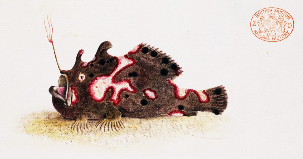
Ted Pietsch: A member of a family of bony fishes, containing 52 species, all of which are highly camouflaged and whose feeding strategy consists of mimicking the immobile, inert, and benign appearance of a sponge or an algae-encrusted rock, while wiggling a highly conspicuous lure to attract prey.
This is a fish that “walks” and “hops” across the sea bottom, and clambers about over rocks and coral like a four-legged terrestrial animal but, at the same time, can jet-propel itself through open water. Some lay their eggs encapsulated in a complex, floating, mucus mass, called an “egg raft,” while some employ elaborate forms of parental care, carrying their eggs around until they hatch.
They are among the most colorful of nature’s productions, existing in nearly every imaginable color and color pattern, with an ability to completely alter their color and pattern in a matter of days or seconds. All these attributes combined make them one of the most intriguing groups of aquatic vertebrates for the aquarist, diver, and underwater photographer as well as the professional zoologist.
…
I couldn’t resist the ‘frog’ reference and I’m glad since this is a good read with a number of fascinating photographs and illustrations.,
An illustration of the frogfish Antennarius pictus, published by George Shaw in 1794. From a new book by Ted Pietsch, UW professor of emeritus of aquatic and fishery sciences. Courtesy: University of Washington (state)
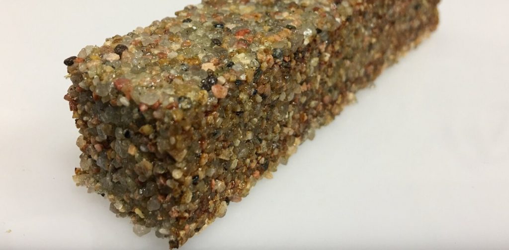
A block of sand particles held together by living cells. Credit: The University of Colorado Boulder College of Engineering and Applied Science
A March 24, 2020 news item on phys.org features the future of building construction as perceived by synthetic biologists,
Buildings are not unlike a human body. They have bones and skin; they breathe. Electrified, they consume energy, regulate temperature and generate waste. Buildings are organisms—albeit inanimate ones.
But what if buildings—walls, roofs, floors, windows—were actually alive—grown, maintained and healed by living materials? Imagine architects using genetic tools that encode the architecture of a building right into the DNA of organisms, which then grow buildings that self-repair, interact with their inhabitants and adapt to the environment.
…
A March 23, 2020 essay by Wil Srubar (Professor of Architectural Engineering and Materials Science, University of Colorado Boulder), which originated the news item, provides more insight,
Living architecture is moving from the realm of science fiction into the laboratory as interdisciplinary teams of researchers turn living cells into microscopic factories. At the University of Colorado Boulder, I lead the Living Materials Laboratory. Together with collaborators in biochemistry, microbiology, materials science and structural engineering, we use synthetic biology toolkits to engineer bacteria to create useful minerals and polymers and form them into living building blocks that could, one day, bring buildings to life.
In our most recent work, published in Matter, we used photosynthetic cyanobacteria to help us grow a structural building material – and we kept it alive. Similar to algae, cyanobacteria are green microorganisms found throughout the environment but best known for growing on the walls in your fish tank. Instead of emitting CO2, cyanobacteria use CO2 and sunlight to grow and, in the right conditions, create a biocement, which we used to help us bind sand particles together to make a living brick.
By keeping the cyanobacteria alive, we were able to manufacture building materials exponentially. We took one living brick, split it in half and grew two full bricks from the halves. The two full bricks grew into four, and four grew into eight. Instead of creating one brick at a time, we harnessed the exponential growth of bacteria to grow many bricks at once – demonstrating a brand new method of manufacturing materials.
Researchers have only scratched the surface of the potential of engineered living materials. Other organisms could impart other living functions to material building blocks. For example, different bacteria could produce materials that heal themselves, sense and respond to external stimuli like pressure and temperature, or even light up. If nature can do it, living materials can be engineered to do it, too.
It also take less energy to produce living buildings than standard ones. Making and transporting today’s building materials uses a lot of energy and emits a lot of CO2. For example, limestone is burned to make cement for concrete. Metals and sand are mined and melted to make steel and glass. The manufacture, transport and assembly of building materials account for 11% of global CO2 emissions. Cement production alone accounts for 8%. In contrast, some living materials, like our cyanobacteria bricks, could actually sequester CO2.
…
The field of engineered living materials is in its infancy, and further research and development is needed to bridge the gap between laboratory research and commercial availability. Challenges include cost, testing, certification and scaling up production. Consumer acceptance is another issue. For example, the construction industry has a negative perception of living organisms. Think mold, mildew, spiders, ants and termites. We’re hoping to shift that perception. Researchers working on living materials also need to address concerns about safety and biocontamination.
The [US] National Science Foundation recently named engineered living materials one of the country’s key research priorities. Synthetic biology and engineered living materials will play a critical role in tackling the challenges humans will face in the 2020s and beyond: climate change, disaster resilience, aging and overburdened infrastructure, and space exploration.
…
If you have time and interest, this is fascinating. Strubar is a little exuberant and, at this point, I welcome it.
Fitness
The Lithuanians are here for us. Scientists from the Kaunas University of Technology have just published a paper on better exercises for lower back pain in our increasingly sedentary times, from a March 23, 2020 Kaunas University of Technology press release (also on EurekAlert) Note: There are a few minor grammatical issues,
With the significant part of the global population forced to work from home, the occurrence of lower back pain may increase. Lithuanian scientists have devised a spinal stabilisation exercise programme for managing lower back pain for people who perform a sedentary job. After testing the programme with 70 volunteers, the researchers have found that the exercises are not only efficient in diminishing the non-specific lower back pain, but their effect lasts 3 times longer than that of a usual muscle strengthening exercise programme.
According to the World Health Organisation, lower back pain is among the top 10 diseases and injuries that are decreasing the quality of life across the global population. It is estimated that non-specific low back pain is experienced by 60% to 70% of people in industrialised societies. Moreover, it is the leading cause of activity limitation and work absence throughout much of the world. For example, in the United Kingdom, low back pain causes more than 100 million workdays lost per year, in the United States – an estimated 149 million.
Chronic lower back pain, which starts from long-term irritation or nerve injury affects the emotions of the afflicted. Anxiety, bad mood and even depression, also the malfunctioning of the other bodily systems – nausea, tachycardia, elevated arterial blood pressure – are among the conditions, which may be caused by lower back pain.
During the coronavirus disease (COVID-19) outbreak, with a significant part of the global population working from home and not always having a properly designed office space, the occurrence of lower back pain may increase.
“Lower back pain is reaching epidemic proportions. Although it is usually clear what is causing the pain and its chronic nature, people tend to ignore these circumstances and are not willing to change their lifestyle. Lower back pain usually comes away itself, however, the chances of the recurring pain are very high”, says Dr Irina Klizienė, a researcher at Kaunas University of Technology (KTU) Faculty of Social Sciences, Humanities and Arts.
Dr Klizienė, together with colleagues from KTU and from Lithuanian Sports University has designed a set of stabilisation exercises aimed at strengthening the muscles which support the spine at the lower back, i.e. lumbar area. The exercise programme is based on Pilates methodology.
According to Dr Klizienė, the stability of lumbar segments is an essential element of body biomechanics. Previous research evidence shows that in order to avoid the lower back pain it is crucial to strengthen the deep muscles, which are stabilising the lumbar area of the spine. One of these muscles is multifidus muscle.
“Human central nervous system is using several strategies, such as preparing for keeping the posture, preliminary adjustment to the posture, correcting the mistakes of the posture, which need to be rectified by specific stabilising exercises. Our aim was to design a set of exercises for this purpose”, explains Dr Klizienė.
The programme, designed by Dr Klizienė and her colleagues is comprised of static and dynamic exercises, which train the muscle strength and endurance. The static positions are to be held from 6 to 20 seconds; each exercise to be repeated 8 to 16 times.
Caption: The static positions are to be held from 6 to 20 seconds; each exercise to be repeated 8 to 16 times. Credit: KTU
The previous set is a little puzzling but perhaps you’ll find these ones below easier to follow,
Caption: The exercises are aimed at strengthening the muscles which support the spine at the lower back. Credit: KTU
I think more pictures of intervening moves would have been useful. Now. getting back to the press release,
In order to check the efficiency of the programme, 70 female volunteers were randomly enrolled either to the lumbar stabilisation exercise programme or to a usual muscle strengthening exercise programme. Both groups were exercising twice a week for 45 minutes for 20 weeks. During the experiment, ultrasound scanning of the muscles was carried out.
As soon as 4 weeks in lumbar stabilisation programme, it was observed that the cross-section area of the multifidus muscle of the subjects of the stabilisation group has increased; after completing the programme, this increase was statistically significant (p < 0,05). This change was not observed in the strengthening group.
Moreover, although both sets of exercises were efficient in eliminating lower back pain and strengthening the muscles of the lower back area, the effect of stabilisation exercises lasted 3 times longer – 12 weeks after the completion of the stabilisation programme against 4 weeks after the completion of the muscle strengthening programme.
“There are only a handful of studies, which have directly compared the efficiency of stabilisation exercises against other exercises in eliminating lower back pain”, says Dr Klizienė, “however, there are studies proving that after a year, lower back pain returned only to 30% of people who have completed a stabilisation exercise programme, and to 84% of people who haven’t taken these exercises. After three years these proportions are 35% and 75%.”
According to her, research shows that the spine stabilisation exercises are more efficient than medical intervention or usual physical activities in curing the lower back pain and avoiding the recurrence of the symptoms in the future.
[downloaded from https://sp.spiber.jp/en/tnfsp/mp/]
Adele Peters in her October 31, 2019 article for Fast Company describes the technology used to make this jacket,
A typical waterproof winter jacket is made with nylon—a material that, like other plastics, is made from petroleum. But a new limited-edition jacket from The North Face Japan uses something called “brewed protein” instead. It’s a material inspired by spider silk that is fermented in giant vats, the same way that breweries make beer.
It’s one of the first uses of a material produced by the Japanese startup Spiber, a company that has spent more than a decade developing a new process to make high-performance textiles and other products that don’t rely on fossil fuels, animals, or natural fibers like cotton, all of which have environmental issues. …
The company designs genes that code for a specific protein—the first was an exact replica of natural spider silk, known for its extreme strength—and then introduces the genes into microorganisms that can produce the protein efficiently. Inside giant tanks, the microorganisms are fed sugar, grow and multiply, and produce the protein through fermentation. …
Spiber first started collaborating with Goldwin, a Japanese outdoor brand that owns the Japanese rights to The North Face, in 2015, and created an early prototype of a jacket then. But it quickly realized that an exact replica of spider silk wouldn’t work well for the application; the material sucks up water, and the jacket needed to be waterproof.
“We spent the last four years going back to the drawing board, redesigning our protein molecule—the very order of the amino acids in the molecule,” says Meyer [Daniel Meyer, Spiber’s head of corporate global marketing]. “And we created our own hydrophobic [water repellent] version of spider silk. It’s inspired by natural spider silk, but we have made our own design changes such that it would be more hydrophobic and meet the performance requirements of The North Face Japan.”
…
The jacket is available for purchase but only by a lottery, which has now closed. According to Peters, a large, commercial production facility is being built in Thailand so that at some point a Moon Parka will be affordable. For reference, the lottery jackets were priced at ¥150,000 (about $1,377 US).
You can find Spiber here in mid-March [2020] according to the homepage.
An October 9, 2019 news item on ScienceDaily provides some insight into the latest US Army research into robots,
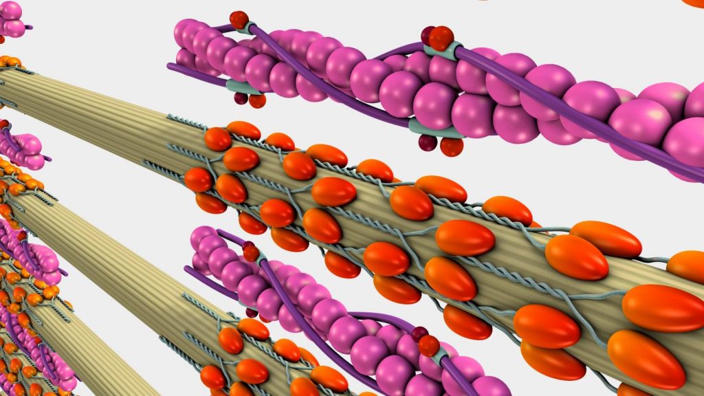
In an effort to make robots more effective and versatile teammates for Soldiers in combat, Army researchers are on a mission to understand the value of the molecular living functionality of muscle, and the fundamental mechanics that would need to be replicated in order to artificially achieve the capabilities arising from the proteins responsible for muscle contraction.
Caption: Army researchers are on a mission to understand the value of the molecular ‘living’ functionality of muscle, and the fundamental mechanics that would need to be replicated in order to artificially achieve the capabilities arising from the proteins responsible for muscle contraction. Credit: US Army-Shutterstock
Bionanomotors, like myosins that move along actin networks, are responsible for most methods of motion in all life forms. Thus, the development of artificial nanomotors could be game-changing in the field of robotics research.
Researchers from the U.S. Army Combat Capabilities Development Command’s [CCDC] Army Research Laboratory [ARL] have been looking to identify a design that would allow the artificial nanomotor to take advantage of Brownian motion, the property of particles to agitatedly move simply because they are warm.
The CCDC ARL researchers believe understanding and developing these fundamental mechanics are a necessary foundational step toward making informed decisions on the viability of new directions in robotics involving the blending of synthetic biology, robotics, and dynamics and controls engineering.
…
“By controlling the stiffness of different geometrical features of a simple lever-arm design, we found that we could use Brownian motion to make the nanomotor more capable of reaching desirable positions for creating linear motion,” said Dean Culver, a researcher in CCDC ARL’s Vehicle Technology Directorate. “This nano-scale feature translates to more energetically efficient actuation at a macro scale, meaning robots that can do more for the warfighter over a longer amount of time.”
According to Culver, the descriptions of protein interactions in muscle contraction are typically fairly high-level. More specifically, rather than describing the forces that act on an individual protein to seek its counterpart, prescribed or empirical rate functions that dictate the conditions under which a binding or a release event occurs have been used by the research community to replicate this biomechanical process.
“These widely accepted muscle contraction models are akin to a black-box understanding of a car engine,” Culver said. “More gas, more power. It weighs this much and takes up this much space. Combustion is involved. But, you can’t design a car engine with that kind of surface-level information. You need to understand how the pistons work, and how finely injection needs to be tuned. That’s a component-level understanding of the engine. We dive into the component-level mechanics of the built-up protein system and show the design and control value of living functionality as well as a clearer understanding of design parameters that would be key to synthetically reproducing such living functionality.”
Culver stated that the capacity for Brownian motion to kick a tethered particle from a disadvantageous elastic position to an advantageous one, in terms of energy production for a molecular motor, has been illustrated by ARL at a component level, a crucial step in the design of artificial nanomotors that offer the same performance capabilities as biological ones.
“This research adds a key piece of the puzzle for fast, versatile robots that can perform autonomous tactical maneuver and reconnaissance functions,” Culver said. “These models will be integral to the design of distributed actuators that are silent, low thermal signature and efficient – features that will make these robots more impactful in the field.”
Culver noted that they are silent because the muscles don’t make a lot of noise when they actuate, especially compared to motors or servos, cold because the amount of heat generation in a muscle is far less than a comparable motor, and efficient because of the advantages of the distributed chemical energy model and potential escape via Brownian motion.
According to Culver, the breadth of applications for actuators inspired by the biomolecular machines in animal muscles is still unknown, but many of the existing application spaces have clear Army applications such as bio-inspired robotics, nanomachines and energy harvesting.
“Fundamental and exploratory research in this area is therefore a wise investment for our future warfighter capabilities,” Culver said.
Moving forward, there are two primary extensions of this research.
“First, we need to better understand how molecules, like the tethered particle discussed in our paper, interact with each other in more complicated environments,” Culver said. “In the paper, we see how a tethered particle can usefully harness Brownian motion to benefit the contraction of the muscle overall, but the particle in this first model is in an idealized environment. In our bodies, it’s submerged in a fluid carrying many different ions and energy-bearing molecules in solution. That’s the last piece of the puzzle for the single-motor, nano-scale models of molecular motors.”
The second extension, stated Culver, is to repeat this study with a full 3-D model, paving the way to scaling up to practical designs.
Also notable is the fact that because this research is so young, ARL researchers used this project to establish relationships with other investigators in the academic community.
“Leaning on their expertise will be critical in the years to come, and we’ve done a great job of reaching out to faculty members and researchers from places like the University of Washington, Duke University and Carnegie Mellon University,” Culver said.
According to Culver, taking this research project into the next steps with help from collaborative partners will lead to tremendous capabilities for future Soldiers in combat, a critical requirement considering the nature of the ever-changing battlefield.
Here’s a link to and a citation for the paper,
A Dynamic Escape Problem of Molecular Motors by Dean Culver, Bryan Glaz, Samuel Stanton. J Biomech Eng. Paper No: BIO-18-1527 https://doi.org/10.1115/1.4044580 Published Online: August 1, 2019
An August 5, 2019 news item on Nanowerk announces a new technology for detecting killer bacteria (Note: A link has been removed),
A combination of off-the-shelf quantum dots and a smartphone camera soon could allow doctors to identify antibiotic-resistant bacteria in just 40 minutes, potentially saving patient lives.
Staphylococcus aureus (golden staph), is a common form of bacterium that causes serious and sometimes fatal conditions such as pneumonia and heart valve infections. Of particular concern is a strain that does not respond to methicillin, the antibiotic of first resort, and is known as methicillin-resistant S. aureus, or MRSA.
Recent reports estimate that 700 000 deaths globally could be attributed to antimicrobial resistance, such as methicillin-resistance. Rapid identification of MRSA is essential for effective treatment, but current methods make it a challenging process, even within well-equipped hospitals.
Soon, however, that may change, using nothing except existing technology.
Researchers from Macquarie University and the University of New South Wales, both in Australia, have demonstrated a proof-of-concept device that uses bacterial DNA to identify the presence of Staphylococcus aureus positively in a patient sample – and to determine if it will respond to frontline antibiotics.
In a paper published in the international peer-reviewed journal Sensors and Actuators B: Chemical the Macquarie University team of Dr Vinoth Kumar Rajendran, Professor Peter Bergquist and Associate Professor Anwar Sunna with Dr Padmavathy Bakthavathsalam (UNSW) reveal a new way to confirm the presence of the bacterium, using a mobile phone and some ultra-tiny semiconductor particles known as quantum dots.
“Our team is using Synthetic Biology and NanoBiotechnology to address biomedical challenges. Rapid and simple ways of identifying the cause of infections and starting appropriate treatments are critical for treating patients effectively,” says Associate Professor Anwar Sunna, head of the Sunna Lab at Macquarie University.
“This is true in routine clinical situations, but also in the emerging field of personalised medicine.”
The researchers’ approach identifies the specific strain of golden staph by using a method called convective polymerase chain reaction (or cPCR). This is a derivative of a widely -employed technique in which a small segment of DNA is copied thousands of times, creating multiple samples suitable for testing.
Vinoth Kumar and colleagues then subject the DNA copies to a process known as lateral flow immunoassay – a paper-based diagnostic tool used to confirm the presence or absence of a target biomarker. The researchers use probes fitted with quantum dots to detect two unique genes, that confirms the presence of methicillin resistance in golden staph
A chemical added at the PCR stage to the DNA tested makes the sample fluoresce when the genes are detected by the quantum dots – a reaction that can be captured easily using the camera on a mobile phone.
The result is a simple and rapid method of detecting the presence of the bacterium, while simultaneously ruling first-line treatment in or out.
Although currently at proof-of-concept stage, the researchers say their system which is powered by a simple battery is suitable for rapid detection in different settings.
“We can see this being used easily not only in hospitals, but also in GP clinics and at patient bedsides,” says lead author, Macquarie’s Vinoth Kumar Rajendran.
This* story actually started in 2018 with an August 1, 2018 Harvard University news release (h/t Aug. 1, 2018 news item on phys.org) by Leslie Brownell announcing molecular and synthetic biology educational kits that been tested in the classroom. (In 2019, a new kit was released but more about that later.)
As biologists have probed deeper into the molecular and genetic underpinnings of life, K-12 schools have struggled to provide a curriculum that reflects those advances. Hands-on learning is known to be more engaging and effective for teaching science to students, but even the most basic molecular and synthetic biology experiments require equipment far beyond an average classroom’s budget, and often involve the use of bacteria and other substances that can be difficult to manage outside a controlled lab setting.
Now, a collaboration between the Wyss Institute at Harvard University, MIT [Massachusetts Institute of Technology], and Northwestern University has developed BioBits, new educational biology kits that use freeze-dried cell-free (FD-CF) reactions to enable students to perform a range of simple, hands-on biological experiments. The BioBits kits introduce molecular and synthetic biology concepts without the need for specialized lab equipment, at a fraction of the cost of current standard experimental designs. The kits are described in two papers published in Science Advances [2018].
“The main motivation in developing these kits was to give students fun activities that allow them to actually see, smell, and touch the outcomes of the biological reactions they’re doing at the molecular level,” said Ally Huang, a co-first author on both papers who is an MIT graduate student in the lab of Wyss Founding Core Faculty member Jim Collins, Ph.D. “My hope is that they will inspire more kids to consider a career in STEM [science, technology, engineering, and math] and, more generally, give all students a basic understanding of how biology works, because they may one day have to make personal or policy decisions based on modern science.”
Synthetic and molecular biology frequently make use of the cellular machinery found in E. coli bacteria to produce a desired protein. But this system requires that the bacteria be kept alive and contained for an extended period of time, and involves several complicated preparation and processing steps. The FD-CF reactions pioneered in Collins’ lab for molecular manufacturing, when combined with innovations from the lab of Michael Jewett, Ph.D. at Northwestern University, offer a solution to this problem by removing bacteria from the equation altogether.
“You can think of it like opening the hood of a car and taking the engine out: we’ve taken the ‘engine’ that drives protein production out of a bacterial cell and given it the fuel it needs, including ribosomes and amino acids, to create proteins from DNA outside of the bacteria itself,” explained Jewett, who is the Charles Deering McCormick Professor of Teaching Excellence at Northwestern University’s McCormick School of Engineering and co-director of Northwestern’s Center for Synthetic Biology, and co-corresponding author of both papers. This collection of molecular machinery is then freeze-dried into pellets so that it becomes shelf-stable at room temperature. To initiate the transcription of DNA into RNA and the translation of that RNA into a protein, a student just needs to add the desired DNA and water to the freeze-dried pellets.
An expansion of the BioBits Bright kit, called BioBits Explorer, includes experiments that engage the senses of smell and touch and allow students to probe their environment using designer synthetic biosensors. In the first experiment, the FD-CF reaction pellets contain a gene that drives the conversion of isoamyl alcohol to isoamyl acetate, a compound that produces a strong banana odor. In the second experiment, the FD-CF reactions contain a gene coding for the enzyme sortase, which recognizes and links specific segments of proteins in a liquid solution together to form a squishy, semi-solid hydrogel, which the students can touch and manipulate. The third module uses another Wyss technology, the toehold switch sensor, to identify DNA extracted from a banana or a kiwi. The sensors are hairpin-shaped RNA molecules designed such that when they bind to a “trigger” RNA, they spring open and reveal a genetic sequence that produces a fluorescent protein. When fruit DNA is added to the sensor-containing FD-CF pellets, only the sensors that are designed to open in the presence of each fruit’s RNA will produce the fluorescent protein.
The researchers tested their BioBits kits in the Chicago Public School system, and demonstrated that students and teachers were able to perform the experiments in the kits with the same success as trained synthetic biology researchers. In addition to refining the kits’ design so that they can one day provide them to classrooms around the world, the authors hope to create an open-source online database where teachers and students can share their results and ideas for ways to modify the kits to explore different biological questions.
“Synthetic biology is going to be one of the defining technologies of the century, and yet it has been challenging to teach the fundamental concepts of the field in K-12 classrooms given that such efforts often require expensive, complicated equipment,” said Collins, who is a co-corresponding author of both papers and also the Termeer Professor of Medical Engineering & Science at MIT. “We show that it is possible to use freeze-dried, cell-free extracts along with freeze-dried synthetic biology components to conduct innovative educational experiments in classrooms and other low-resource settings. The BioBits kits enable us to expose young kids, older kids, and even adults to the wonders of synthetic biology and, as a result, are poised to transform science education and society.
“All scientists are passionate about what they do, and we are frustrated by the difficulty our educational system has had in inciting a similar level of passion in young people. This BioBits project demonstrates the kind of out-of-the-box thinking and refusal to accept the status quo that we value and cultivate at the Wyss Institute, and we all hope it will stimulate young people to be intrigued by science,” said Wyss Institute Founding Director Donald Ingber, M.D., Ph.D., who is also the Judah Folkman Professor of Vascular Biology at Harvard Medical School (HMS) and the Vascular Biology Program at Boston Children’s Hospital, as well as Professor of Bioengineering at Harvard’s John A. Paulson School of Engineering and Applied Sciences (SEAS). “It’s exciting to see this project move forward and become available to biology classrooms worldwide and, hopefully some of these students will pursue a path in science because of their experience.”
Additional authors of the papers include Peter Nguyen, Ph.D., Nina Donghia, and Tom Ferrante from the Wyss Institute; Melissa Takahashi, Ph.D. and Aaron Dy from MIT; Karen Hsu and Rachel Dubner from Northwestern University; Keith Pardee, Ph.D., Assistant Professor at the University of Toronto; and a number of teachers and students in the Chicago school system including: Mary Anderson, Ada Kanapskyte, Quinn Mucha, Jessica Packett, Palak Patel, Richa Patel, Deema Qaq, Tyler Zondor, Julie Burke, Tom Martinez, Ashlee Miller-Berry, Aparna Puppala, Kara Reichert, Miriam Schmid, Lance Brand, Lander Hill, Jemima Chellaswamy, Nuhie Faheem, Suzanne Fetherling, Elissa Gong, Eddie Marie Gonzales, Teresa Granito, Jenna Koritsaris, Binh Nguyen, Sujud Ottman, Christina Palffy, Angela Patel, Sheila Skweres, Adriane Slaton, and TaRhonda Woods.
This research was supported by the Army Research Office, the National Science Foundation, the Air Force Research Laboratory Center of Excellence Grant, The Defense Threat Reduction Agency Grant, the David and Lucile Packard Foundation, the Camille Dreyfus Teacher-Scholar Program, the Wyss Institute at Harvard University, the Paul G. Allen Frontiers Group, The Air Force Office of Scientific Research, and the Natural Sciences and Engineering Council of Canada. [emphases mine]
Well, that list of funding agencies is quite interesting. The US Army and Air Force but not the Navy? As for what the Natural Sciences and Engineering Council of Canada is doing on that list, I can only imagine why.
This is what they were doing in 2018,
Now for the latest update, a May 7, 2019 news item on phys.org announces the BioBits Kits have been expanded,
How can high school students learn about a technology as complex and abstract as CRISPR? It’s simple: just add water.
A Northwestern University-led team has developed BioBits, a suite of hands-on educational kits that enable students to perform a range of biological experiments by adding water and simple reagents to freeze-dried cell-free reactions. The kits link complex biological concepts to visual, fluorescent readouts, so students know—after a few hours and with a single glance—the results of their experiments.
After launching BioBits last summer, the researchers are now expanding the kit to include modules for CRISPR [clustered regularly interspaced short palindromic repeats] and antibiotic resistance. A small group of Chicago-area teachers and high school students just completed the first pilot study for these new modules, which include interactive experiments and supplementary materials exploring ethics and strategies.
“After we unveiled the first kits, we next wanted to tackle current topics that are important for society,” said Northwestern’s Michael Jewett, principal investigator of the study. “That led us to two areas: antibiotic resistance and gene editing.”
Called BioBits Health, the new kits and pilot study are detailed in a paper published today (May 7 [2019]) in the journal ACS Synthetic Biology.
Jewett is a professor of chemical and biological engineering in Northwestern’s McCormick School of Engineering and co-director of Northwestern’s Center for Synthetic Biology. Jessica Stark, a graduate student in Jewett’s laboratory, led the study.
Test in a tube
Instead of using live cells, the BioBits team removed the essential cellular machinery from inside the cells and freeze-dried them for shelf stability. Keeping cells alive and contained for an extended period of time involves several complicated, time-consuming preparation and processing steps as well as expensive equipment. Freeze-dried cell-free reactions bypass those complications and costs.
“These are essentially test-tube biological reactions,” said Stark, a National Science Foundation graduate research fellow. “We break the cells open and use their guts, which still contain all of the necessary biological machinery to carry out a reaction. We no longer need living cells to demonstrate biology.”
This method to harness biological systems without intact, living cells became possible over the last two decades thanks to multiple innovations, including many in cell-free synthetic biology by Jewett’s lab. Not only are these experiments doable in the classroom, they also only cost pennies compared to standard high-tech experimental designs.
“I’m hopeful that students get excited about engineering biology and want to learn more,” Jewett said.
Conquering CRISPR
One of the biggest scientific breakthroughs of the past decade, CRISPR (pronounced “crisper”) stands for Clustered Regularly Interspaced Short Palindromic Repeats. The powerful gene-editing technology uses enzymes to cut DNA in precise locations to turn off or edit targeted genes. It could be used to halt genetic diseases, develop new medicines, make food more nutritious and much more.
BioBits Health uses three components required for CRISPR: an enzyme called the Cas9 protein, a target DNA sequence encoding a fluorescent protein and an RNA molecule that targets the fluorescent protein gene. When students add all three components — and water — to the freeze-dried cell-free system, it creates a reaction that edits, or cuts, the DNA for the fluorescent protein. If the DNA is cut, the system does not glow. If the DNA is not cut, the fluorescent protein is made, and the system glows fluorescent.
“We have linked this abstract, really advanced biological concept to the presence or absence of a fluorescent protein,” Stark said. “It’s something students can see, something they can visually understand.”
The curriculum also includes activities that challenge students to consider the ethical questions and dilemmas surrounding the use of gene-editing technologies.
“There is a lot of excitement about being able to edit genomes with these technologies,” Jewett said. “BioBits Health calls attention to a lot of important questions — not only about how CRISPR technology works but about ethics that society should be thinking about. We hope that this promotes a conversation and dialogue about such technologies.”
Reducing resistance
Jewett and Stark are both troubled by a prediction that, by the year 2050, drug-resistant bacterial infections could outpace cancer as a leading cause of death. This motivated them to help educate the future generation of scientists about how antibiotic resistance emerges and inspire them to take actions that could help limit the emergence of resistant bacteria. In this module, students run two sets of reactions to produce a glowing fluorescent protein — one set with an antibiotic resistance gene and one set without. Students then add antibiotics. If the experiment glows, the fluorescent protein has been made, and the reaction has become resistant to antibiotics. If the experiment does not glow, then the antibiotic has worked.
“Because we’re using cell-free systems rather than organisms, we can demonstrate drug resistance in a way that doesn’t create drug-resistant bacteria,” Stark explained. “We can demonstrate these concepts without the risks.”
A supporting curriculum piece challenges students to brainstorm and research strategies for slowing the rate of emerging antibiotic resistant strains.
Part of something cool
After BioBits was launched in summer 2018, 330 schools from around the globe requested prototype kits for their science labs. The research team, which includes members from Northwestern and MIT, has received encouraging feedback from teachers, students and parents.
“The students felt like scientists and doctors by touching and using the laboratory materials provided during the demo,” one teacher said. “Even the students who didn’t seem engaged were secretly paying attention and wanted to take their turn pipetting. They knew they were part of something really cool, so we were able to connect with them in a way that was new to them.”
“My favorite part was using the equipment,” a student said. “It was a fun activity that immerses you into what top scientists are currently doing.”
###
The study, “BioBits Health: Classroom activities exploring engineering, biology and human health with fluorescent readouts,” was supported by the Army Research Office (award number W911NF-16-1-0372), the National Science Foundation (grant numbers MCB-1413563 and MCB-1716766), the Air Force Research Laboratory Center of Excellence (grant number FA8650-15-2-5518), the Defense Threat Reduction Agency (grant number HDTRA1-15-10052/P00001), the Department of Energy (grant number DE-SC0018249), the Human Frontiers Science Program (grant number RGP0015/2017), the David and Lucile Packard Foundation, the Office of Energy Efficiency and Renewable Energy (grant number DE-EE008343) and the Camille Dreyfus Teacher-Scholar Program. [emphases mine]
This is an image you’ll find in the abstract for the 2019 paper,
[downloaded from https://pubs.acs.org/doi/10.1021/acssynbio.8b00381]
Here are links and citations for the 2018 papers and the 2019 paper,
BioBits™ Explorer: A modular synthetic biology education kit by Ally Huang, Peter Q. Nguyen, Jessica C. Stark, Melissa K. Takahashi, Nina Donghia, Tom Ferrante, Aaron J. Dy, Karen J. Hsu, Rachel S. Dubner, Keith Pardee, Michael C. Jewett, and James J. Collins. Science Advances 01 Aug 2018: Vol. 4, no. 8, eaat5105 DOI: 10.1126/sciadv.aat5105
BioBits™ Bright: A fluorescent synthetic biology education kit by Jessica C. Stark, Ally Huang, Peter Q. Nguyen, Rachel S. Dubner, Karen J. Hsu, Thomas C. Ferrante, Mary Anderson, Ada Kanapskyte, Quinn Mucha, Jessica S. Packett, Palak Patel, Richa Patel, Deema Qaq, Tyler Zondor, Julie Burke, Thomas Martinez, Ashlee Miller-Berry, Aparna Puppala, Kara Reichert, Miriam Schmid, Lance Brand, Lander R. Hill, Jemima F. Chellaswamy, Nuhie Faheem, Suzanne Fetherling, Elissa Gong, Eddie Marie Gonzalzles, Teresa Granito, Jenna Koritsaris, Binh Nguyen, Sujud Ottman, Christina Palffy, Angela Patel, Sheila Skweres, Adriane Slaton, TaRhonda Woods, Nina Donghia, Keith Pardee, James J. Collins, and Michael C. Jewett. Science Advances 01 Aug 2018: Vol. 4, no. 8, eaat5107 DOI: 10.1126/sciadv.aat5107
BioBits Health: Classroom Activities Exploring Engineering, Biology, and Human Health with Fluorescent Readouts by Jessica C. Stark, Ally Huang, Karen J. Hsu, Rachel S. Dubner, Jason Forbrook, Suzanne Marshalla, Faith Rodriguez, Mechelle Washington, Grant A. Rybnicky, Peter Q. Nguyen, Brenna Hasselbacher, Ramah Jabri, Rijha Kamran, Veronica Koralewski, Will Wightkin, Thomas Martinez, and Michael C. Jewett. ACS Synth. Biol., Article ASAP DOI: 10.1021/acssynbio.8b00381 Publication Date (Web): March 29, 2019
Both of the 2018 papers appear to be open access while the 2019 paper is behind a paywall.
Should you be interested in acquiring a BioBits kit, you can check out the BioBits website. As for ‘conguering’ CRISPR, do we really need to look at it that way? Maybe a more humble appraoch could work just as well or even better, eh?
They’re not calling this synthetic biology but I’ m pretty sure that altering a virus gene so the virus can spin gold (Rumpelstiltskin anyone?) qualifies. From an August 24, 2018 news item on ScienceDaily,
The race is on to find manufacturing techniques capable of arranging molecular and nanoscale objects with precision.
Engineers at the University of California, Riverside, have altered a virus to arrange gold atoms into spheroids measuring a few nanometers in diameter. The finding could make production of some electronic components cheaper, easier, and faster.
“Nature has been assembling complex, highly organized nanostructures for millennia with precision and specificity far superior to the most advanced technological approaches,” said Elaine Haberer, a professor of electrical and computer engineering in UCR’s Marlan and Rosemary Bourns College of Engineering and senior author of the paper describing the breakthrough. “By understanding and harnessing these capabilities, this extraordinary nanoscale precision can be used to tailor and build highly advanced materials with previously unattainable performance.”
Viruses exist in a multitude of shapes and contain a wide range of receptors that bind to molecules. Genetically modifying the receptors to bind to ions of metals used in electronics causes these ions to “stick” to the virus, creating an object of the same size and shape. This procedure has been used to produce nanostructures used in battery electrodes, supercapacitors, sensors, biomedical tools, photocatalytic materials, and photovoltaics.
The virus’ natural shape has limited the range of possible metal shapes. Most viruses can change volume under different scenarios, but resist the dramatic alterations to their basic architecture that would permit other forms.
The M13 bacteriophage, however, is more flexible. Bacteriophages are a type of virus that infects bacteria, in this case, gram-negative bacteria, such as Escherichia coli, which is ubiquitous in the digestive tracts of humans and animals. M13 bacteriophages genetically modified to bind with gold are usually used to form long, golden nanowires.
Studies of the infection process of the M13 bacteriophage have shown the virus can be converted to a spheroid upon interaction with water and chloroform. Yet, until now, the M13 spheroid has been completely unexplored as a nanomaterial template.
Haberer’s group added a gold ion solution to M13 spheroids, creating gold nanobeads that are spiky and hollow.
“The novelty of our work lies in the optimization and demonstration of a viral template, which overcomes the geometric constraints associated with most other viruses,” Haberer said. “We used a simple conversion process to make the M13 virus synthesize inorganic spherical nanoshells tens of nanometers in diameter, as well as nanowires nearly 1 micron in length.”
The researchers are using the gold nanobeads to remove pollutants from wastewater through enhanced photocatalytic behavior.
The work enhances the utility of the M13 bacteriophage as a scaffold for nanomaterial synthesis. The researchers believe the M13 bacteriophage template transformation scheme described in the paper can be extended to related bacteriophages.
The conference itself will be held from May 22 – 24, 2019 at Arizona State University (ASU) and the deadline for abstracts is January 31, 2019. Here’s the news straight from the January 8, 2019 email announcement,
The Seventh Annual Conference on Governance of Emerging Technologies & Science (GETS)
May 22-24, 2019 / ASU / Sandra Day O’Connor College of Law 111 E. Taylor St., Phoenix, AZ The conference will consist of plenary and session presentations and discussions on regulatory, governance, legal, policy, social and ethical aspects of emerging technologies, including nanotechnology, synthetic biology, gene editing, biotechnology, genomics, personalized medicine, digital health, human enhancement, artificial intelligence, virtual reality, internet of things (IoT), blockchain and much, much more! Submit Your Abstract Here: 2019 Abstract or Conference Website
Call for abstracts:
The co-sponsors invite submission of abstracts for proposed presentations. Submitters of abstracts need not provide a written paper, although provision will be made for posting and possible post-conference publication of papers for those who are interested. Abstracts are invited for any aspect or topic relating to the governance of emerging technologies, including any of the technologies listed above.
· Abstracts should not exceed 500 words and must contain your name and email address. · Abstracts must be submitted by January 31, 2019 to be considered. · The sponsors will pay for the conference registration (including all conference meals and events) for one presenter for each accepted abstract. In addition, we will have limited funds available for travel subsidies (application included in submission form). For more informationcontact our Executive Director Josh Abbott at Josh.Abbott@asu.edu.