Monthly Archives: March 2016

The extinct dodo bird in 3D

This piece is to honour my father who was from Mauritius, former habitat of the now extinct dodo bird.

A March 25, 2016 news item on ScienceDaily announces the first 3D atlas for the dodo,

The dodo represents one of the best-known examples of extinction caused by humans, yet we know surprisingly little about this flightless pigeon from a scientific perspective. Now, for the first time since its extinction, a 3-D atlas of the skeletal anatomy of the dodo has been created, based upon two exceptional dodo skeletons that have remained unstudied for over a century. This atlas, published as the fifteenth Memoir of the Society of Vertebrate Paleontology, represents the culmination of nearly five years of work and thousands of man-hours of digital investigation on the only two associated, near-complete skeletons of the dodo in existence.

A March 25, 2016 Society of Vertebrate Paleontology news release on EurekAlert, which originated the news item, expands on the theme,

The dodo (Raphus cucullatus), an extinct, giant flightless pigeon once endemic to the island of Mauritius, may arguably be the most widely known animal species to have gone extinct in human history. However, despite its prominence in popular culture, surprisingly little is known of the anatomy and biology of this animal. The dodo was extinct by 1693, less than one hundred years after the discovery and colonization of Mauritius by the Dutch. There is not a single complete specimen that exists from 17th century collections, only a few fragments remain; a single desiccated head, a skull, a beak, and a foot. There are also a few genuine but often contradictory contemporary written accounts and drawings. It was not until the discovery of a mid-Holocene fossil concentration-Lagerstätte on Mauritius in 1865, the Mare aux Songes (MAS), that scientists, most notably Sir Richard Owen, were able to reconstruct the dodo’s skeletal anatomy by constructing composite, partially incomplete skeletons. Surprisingly, only few additions to our knowledge of dodo anatomy, paleoecology and extinction have been made since Owen’s 1866 seminal publication, a vast library of semi-popular works on the dodo notwithstanding.

The fossil discoveries made by barber and amateur naturalist Etienne Thirioux between 1899 and 1910 include some of the best dodo remains existing today, including the only complete skeleton known from a single bird (housed in the Natural History Museum in Port Louis, Mauritius), and another largely complete skeleton (housed in the Durban Natural Science Museum in South Africa). Sadly, Thirioux’s discoveries never received the attention they deserved. Our anatomical atlas of the Thirioux skeletons, produced using modern techniques such as 3D laser surface scanning, opens a new window into the life of this famous extinct bird.

Society of Vertebrate Paleontology Memoir number fifteen is the first complete, comprehensive treatise on dodo skeletal anatomy ever produced and only the third monograph on dodo skeletal anatomy; the last one dating from 150 years ago. It represents years of collaborative efforts from a large team of international scientists, with a substantial contribution from undergraduate student researchers in the 3-D laser surface scanning 3-D of the Thirioux skeletons.

Here’s a link to and a citation for the fifteenth memoir,

ANATOMY OF THE DODO (RAPHUS CUCULLATUS L., 1758): AN OSTEOLOGICAL STUDY OF THE THIRIOUX SPECIMENS by Leon P. A. M. Claessens, Hanneke J. M. Meijer, Julian P. Hume, and Kenneth F. Rijsdijk (Editors). Society of Vertebrate Paleontology Memoir 15, Journal of Vertebrate Paleontology Vol. 35, Supplement to No. 6.

This atlas is behind a paywall.

4th USA Science & Engineering Festival on April 16 and 17, 2016

Thanks to David Bruggeman and a March 23, 2016 posting on his Pasco Phronesis blog for reminding me of this event (Note: Links have been removed),

The Fourth USA Science and Engineering Festival will return [if memory serves this festival takes place every two years] to Washington D.C. next month.  The big Expo is April 16th and 17th [2016] at the Walter Washington Convention Center and open to all.  There is a Sneak Peek taking place on the 15th, which is open to school groups (including homeschooled children) and military families.  Registration is required and now open.

Here’s more from the USA Science and Engineering Festival’s homepage,

Attendees will have access to over 3000 hands-on, interactive activities offered by more than 1000 leading science organizations from around the nation. Imagine chatting with Albert Einstein, flying a simulated jet fighter, being a crime scene investigator, building an underwater robot, taking a vacation in space or watching a science magician. …

The 2016 sneak peek mentioned in the excerpt from David’s post is shown here in a video of 2014 festival sneak peek event,

There aren’t many details about the 2016 programme but I did find this in a March 16, 2016 posting on the USA Science & Engineering festival blog,

Tracking Sharks with Chris Fischer on Stage at the Festival

OCEARCH is a recognized world leader in generating critical scientific data related to tracking (telemetry) and biological studies of keystone marine species such as great white sharks, in conjunction with conservation outreach and education at a measurable global scale. In a collaborative environment established by Founding Chairman and Expedition Leader Chris Fischer, OCEARCH shares real-time data through OCEARCH’s Global Shark Tracker, inspires current and future generations of explorers, scientists, and stewards of the ocean, and enables leading researchers and institutions to generate previously unattainable data. OCEARCH has completed 22 expeditions as of September 2015; by 2016, a total of 26 will be completed.

Meet Chris Fischer as he speaks about his expeditions and efforts to track white sharks and other ocean giants at the USA Science & Engineering Festival. …

The festival itself is free.

You may want to read David’s March 23, 2016 posting in its entirety as he highlights different aspects of the festival.

US Nanotechnology Initiative for water sustainability

Wednesday, March 23, 2016 was World Water Day and to coincide with that event the US National Nanotechnology Initiative (NNI) in collaboration with several other agencies announced a new ‘signature initiative’. From a March 24, 2016 news item on Nanowerk (Note: A link has been removed),

As a part of the White House Water Summit held yesterday on World Water Day, the Federal agencies participating in the National Nanotechnology Initiative (NNI) announced the launch of a Nanotechnology Signature Initiative (NSI), Water Sustainability through Nanotechnology: Nanoscale Solutions for a Global-Scale Challenge.

A March 23, 2016 NNI news release provides more information about why this initiative is important,

Access to clean water remains one of the world’s most pressing needs. As today’s White House Office of Science and Technology blog post explains, “the small size and exceptional properties of engineered nanomaterials are particularly promising for addressing the key technical challenges related to water quality and quantity.”

“One cannot find an issue more critical to human life and global security than clean, plentiful, and reliable water sources,” said Dr. Michael Meador, Director of the National Nanotechnology Coordination Office (NNCO). “Through the NSI mechanism, NNI member agencies will have an even greater ability to make meaningful strides toward this initiative’s thrust areas: increasing water availability, improving the efficiency of water delivery and use, and enabling next-generation water monitoring systems.”

A March 23, 2016 US White House blog posting by Lloyd Whitman and Lisa Friedersdorf describes the efforts in more detail (Note: A link has been removed),

The small size and exceptional properties of engineered nanomaterials are particularly promising for addressing the pressing technical challenges related to water quality and quantity. For example, the increased surface area—a cubic centimeter of nanoparticles has a surface area larger than a football field—and reactivity of nanometer-scale particles can be exploited to create catalysts for water purification that do not require rare or precious metals. And composites incorporating nanomaterials such as carbon nanotubes might one day enable stronger, lighter, and more durable piping systems and components. Under this NSI, Federal agencies will coordinate and collaborate to more rapidly develop nanotechnology-enabled solutions in three main thrusts: [thrust 1] increasing water availability; [thrust 2] improving the efficiency of water delivery and use; and [thrust 3] enabling next-generation water monitoring systems.

A technical “white paper” released by the agencies this week highlights key technical challenges for each thrust, identifies key objectives to overcome those challenges, and notes areas of research and development where nanotechnology promises to provide the needed solutions. By shining a spotlight on these areas, the new NSI will increase Federal coordination and collaboration, including with public and private stakeholders, which is vital to making progress in these areas. The additional focus and associated collective efforts will advance stewardship of water resources to support the essential food, energy, security, and environment needs of all stakeholders.

We applaud the commitment of the Federal agencies who will participate in this effort—the Department of Commerce/National Institute of Standards and Technology, Department of Energy, Environmental Protection Agency, National Aeronautics and Space Administration, National Science Foundation, and U.S. Department of Agriculture/National Institute of Food and Agriculture. As made clear at this week’s White House Water Summit, the world’s water systems are under tremendous stress, and new and emerging technologies will play a critical role in ensuring a sustainable water future.

The white paper (12 pp.) is titled: Water Sustainability through Nanotechnology: Nanoscale Solutions for a Global-Scale Challenge and describes the thrusts in more detail.

A March 22, 2016 US White House fact sheet lays out more details including funding,

Click here to learn more about all of the commitments and announcements being made today. They include:

  • Nearly $4 billion in private capital committed to investment in a broad range of water-infrastructure projects nationwide. This includes $1.5 billion from Ultra Capital to finance decentralized and scalable water-management solutions, and $500 million from Sustainable Water to develop water reclamation and reuse systems.
  • More than $1 billion from the private sector over the next decade to conduct research and development into new technologies. This includes $500 million from GE to fuel innovation, expertise, and global capabilities in advanced water, wastewater, and reuse technologies.
  • A Presidential Memorandum and supporting Action Plan on building national capabilities for long-term drought resilience in the United States, including by setting drought resilience policy goals, directing specific drought resilience activities to be completed by the end of the year, and permanently establishing the National Drought Resilience Partnership as an interagency task force responsible for coordinating drought-resilience, response, and recovery efforts.
  • Nearly $35 million this year in Federal grants from the Environmental Protection Agency, the National Oceanic and Atmospheric Administration, the National Science Foundation, and the U.S. Department of Agriculture to support cutting-edge water science;
  • The release of a new National Water Model that will dramatically enhance the Nation’s river-forecasting capabilities by delivering forecasts for approximately 2.7 million locations, up from 4,000 locations today (a 700-fold increase in forecast density).

This seems promising and hopefully other countries will follow suit.

Listening to bacteria for superior organic nanowires

Researchers at Michigan State University (MSU; US) claim to have  discovered organic nanowires that are superior to the engineered kind according to a March 24, 2016 news item on ScienceDaily,

A microbial protein fiber discovered by a Michigan State University scientist transports charges at rates high enough to be applied in humanmade nanotechnologies.

The discovery, featured in the current issue of Scientific Reports, describes the high-speed protein fiber produced by uranium-reducing Geobacter bacteria. The fibers are hair-like protein filaments called “pili” that have the unique property of transporting charges at speeds of 1 billion electrons per second.

A March 24, 2016 MSU news release, which originated the news item, provides more information,

“This microbial nanowire is made of but a single peptide subunit,” said Gemma Reguera, lead author and MSU microbiologist. “Being made of protein, these organic nanowires are biodegradable and biocompatible. This discovery thus opens many applications in nanoelectronics such as the development of medical sensors and electronic devices that can be interfaced with human tissues.”

Since existing nanotechnologies incorporate exotic metals into their designs, the cost of organic nanowires is much more cost effective as well, she added.

How the nanowires function in nature is comparable to breathing. Bacterial cells, like humans, have to breathe. The process of respiration involves moving electrons out of an organism. Geobacter bacteria use the protein nanowires to bind and breathe metal-containing minerals such as iron oxides and soluble toxic metals such as uranium. The toxins are mineralized on the nanowires’ surface, preventing the metals from permeating the cell.

Reguera’s team purified their protein fibers, which are about 2 nanometers in diameter. Using the same toolset of nanotechnologists, the scientists were able to measure the high velocities at which the proteins were passing electrons.

“They are like power lines at the nanoscale,” Reguera said. “This also is the first study to show the ability of electrons to travel such long distances — more than a 1,000 times what’s been previously proven — along proteins.”

The researchers also identified metal traps on the surface of the protein nanowires that bind uranium with great affinity and could potentially trap other metals. These findings could provide the basis for systems that integrate protein nanowires to mine gold and other precious metals, scrubbers that can be deployed to immobilize uranium at remediation sites and more.

Reguera’s nanowires also can be modified to seek out other materials in which to help them breathe.

“The Geobacter cells are making these protein fibers naturally to breathe certain metals. We can use genetic engineering to tune the electronic and biochemical properties of the nanowires and enable new functionalities. We also can mimic the natural manufacturing process in the lab to mass-produce them in inexpensive and environmentally friendly processes,” Reguera said. “This contrasts dramatically with the manufacturing of humanmade inorganic nanowires, which involve high temperatures, toxic solvents, vacuums and specialized equipment.”

This discovery came from truly listening to bacteria, Reguera said.

“The protein is getting the credit, but we can’t forget to thank the bacteria that invented this,” she said. “It’s always wise to go back and ask bacteria what else they can teach us. In a way, we are eavesdropping on microbial conversations. It’s like listening to our elders, learning from their wisdom and taking it further.”

Asking what else bacteria can teach us? That’s a lovely thought and  different from the still common ‘let’s wipe them all out’ approach to bacteria. It suggests scientific research that is more amenable to sharing the planet with all forms of life.

Here’s a link to and a citation for the paper,

Thermally activated charge transport in microbial protein nanowires by Sanela Lampa-Pastirk, Joshua P. Veazey, Kathleen A. Walsh, Gustavo T. Feliciano, Rebecca J. Steidl, Stuart H. Tessmer, & Gemma Reguera. Scientific Reports 6, Article number: 23517 (2016) doi:10.1038/srep23517 Published online: 24 March 2016

This paper is open access.

Clothes washers and dryers begone! Nano-enhanced textiles can self-clean

It will be a while yet even it this technique proves to be viable commercially, still, the possibilities tantalize: self-cleaning textiles. A March 22, 2016 news item on ScienceDaily announced research in Australia that may, one day, change your life,

A spot of sunshine is all it could take to get your washing done, thanks to pioneering nano research into self-cleaning textiles.

Researchers at RMIT University in Melbourne, Australia, have developed a cheap and efficient new way to grow special nanostructures — which can degrade organic matter when exposed to light — directly onto textiles.

The work paves the way towards nano-enhanced textiles that can spontaneously clean themselves of stains and grime simply by being put under a light bulb or worn out in the sun.

A March 22, 2016 RMIT media release (also on EurekAlert), which originated the news item, expands on the theme,

Dr Rajesh Ramanathan said the process developed by the team had a variety of applications for catalysis-based industries such as agrochemicals, pharmaceuticals and natural products, and could be easily scaled up to industrial levels.

“The advantage of textiles is they already have a 3D structure so they are great at absorbing light, which in turn speeds up the process of degrading organic matter,” he said.

“There’s more work to do to before we can start throwing out our washing machines, but this advance lays a strong foundation for the future development of fully self-cleaning textiles.”

The researchers from the Ian Potter NanoBioSensing Facility and NanoBiotechnology Research Lab at RMIT worked with copper and silver-based nanostructures, which are known for their ability to absorb visible light.

When the nanostructures are exposed to light, they receive an energy boost that creates “hot electrons”. These “hot electrons” release a burst of energy [emphasis mine] that enables the nanostructures to degrade organic matter.

The challenge for researchers has been to bring the concept out of the lab by working out how to build these nanostructures on an industrial scale and permanently attach them to textiles.

The RMIT team’s novel approach was to grow the nanostructures directly onto the textiles by dipping them into a few solutions, resulting in the development of stable nanostructures within 30 minutes.

When exposed to light, it took less than six minutes for some of the nano-enhanced textiles to spontaneously clean themselves.

“Our next step will be to test our nano-enhanced textiles with organic compounds that could be more relevant to consumers, to see how quickly they can handle common stains like tomato sauce or wine,” Ramanathan said.

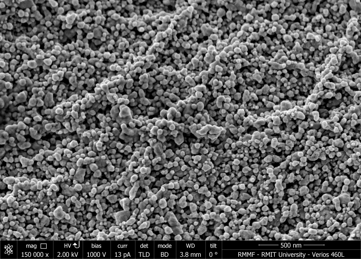
I wonder if these “hot electrons” mean that when they release “a burst of energy” your clothing will heat up when exposed to light? This image supplied by the researchers does not help to answer the question but it is intriguing,

Caption: Close-up of the nanostructures grown on cotton textiles by RMIT University researchers. Image magnified 150,000 times. Credit: RMIT University

Caption: Close-up of the nanostructures grown on cotton textiles by RMIT University researchers. Image magnified 150,000 times. Credit: RMIT University

Here’s a link to and a citation for the paper,

Robust Nanostructured Silver and Copper Fabrics with Localized Surface Plasmon Resonance Property for Effective Visible Light Induced Reductive Catalysis by Samuel R. Anderson, Mahsa Mohammadtaheri, Dipesh Kumar, Anthony P. O’Mullane, Matthew R. Field, Rajesh Ramanathan, and Vipul Bansal. Advanced Materials Interfaces DOI: 10.1002/admi.201500632 Article first published online: 7 JAN 2016

© 2016 WILEY-VCH Verlag GmbH & Co. KGaA, Weinheim

This paper is behind a paywall.

A new Shrinky Dinks story: super-wrinkled and super-crumpled graphene for self-cleaning surfaces and other applications

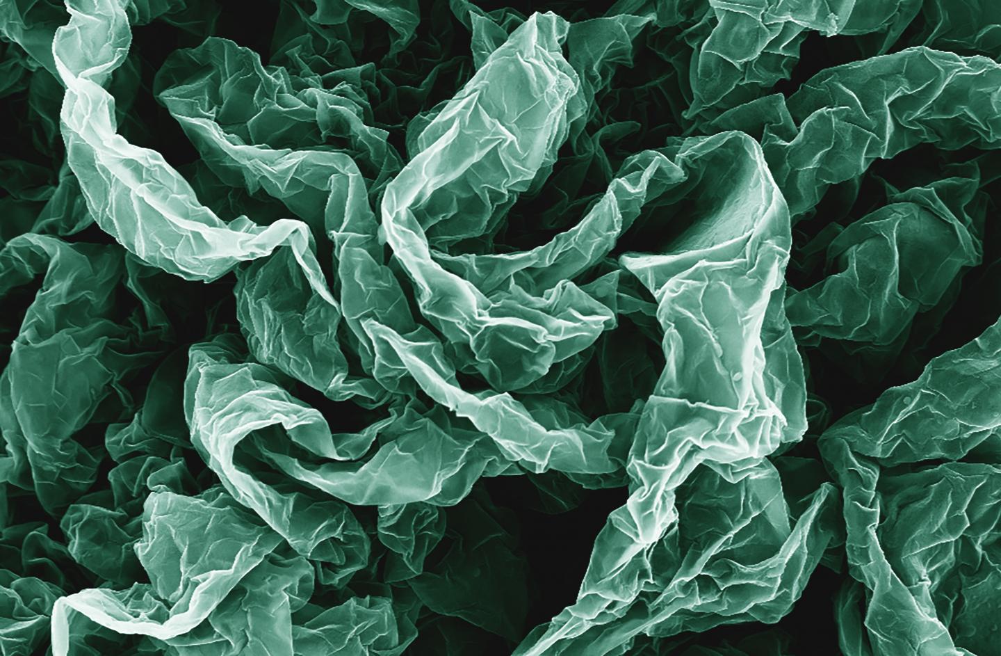
Caption: Wrinkles and crumples, introduced by placing graphene on shrinky polymers, can enhance graphene's properties. Credit: Hurt and Wong Labs / Brown University

Caption: Wrinkles and crumples, introduced by placing graphene on shrinky polymers, can enhance graphene’s properties. Credit: Hurt and Wong Labs / Brown University

A March 21, 2016 news item on ScienceDaily describes how Brown University (US) researchers developed super-wrinkled and super-crumpled graphene,

Crumple a piece of paper and it’s probably destined for the trash can, but new research shows that repeatedly crumpling sheets of the nanomaterial graphene can actually enhance some of its properties. In some cases, the more crumpled the better.

The research by engineers from Brown University shows that graphene, wrinkled and crumpled in a multi-step process, becomes significantly better at repelling water–a property that could be useful in making self-cleaning surfaces. Crumpled graphene also has enhanced electrochemical properties, which could make it more useful as electrodes in batteries and fuel cells.

A March 21, 2016 Brown University news release (also on EurekAlert), which originated the news item, provides more detail about the current and previous research,

This new research builds on previous work done by Robert Hurt and Ian Wong, from Brown’s School of Engineering. The team had previously showed that by introducing wrinkles into graphene, they could make substrates for culturing cells that were more similar to the complex environments in which cells grow in the body. For this latest work, the researchers led by Po-Yen Chen, a Hibbit postdoctoral fellow, wanted to build more complex architectures incorporating both wrinkles and crumples. “I wanted to see if there was a way to create higher-generational structures,” Chen said.

To do that, the researchers deposited layers of graphene oxide onto shrink films–polymer membranes that shrink when heated (kids may know these as Shrinky Dinks [emphasis mine]). As the films shrink, the graphene on top is compressed, causing it to wrinkle and crumple. To see what kind of structures they could create, the researchers compressed same graphene sheets multiple times. After the first shrink, the film was dissolved away, and the graphene was placed in a new film to be shrunk again.

The researchers experimented with different configurations in the successive generations of shrinking. For example, sometimes they clamped opposite ends of the films, which caused them to shrink only along one axis. Clamped films yielded graphene sheets with periodic, basically parallel wrinkles across its surface. Unclamped films shrank in two dimensions, both length- and width-wise, creating a graphene surface that was crumpled in random shapes.

The team experimented with those different modes of shrinking over three successive generations. For example, they might shrink the same graphene sheet on a clamped film, then an unclamped film, then clamped again; or unclamped, clamped, unclamped. They also rotated the graphene in different configurations between shrinkings, sometimes placing the sheet perpendicular to its original orientation.

The team found that the multi-generational approach could substantially compress the graphene sheets, making them as small as one-fortieth their original size. They also showed that successive generations could create interesting patterns along the surface–wrinkles and crumples that were superimposed onto each other, for example.

“As you go deeper into the generations you tend to get larger wavelength structures with the original, smaller wavelength structure from earlier generations built into them,” said Robert Hurt, a professor of engineering at Brown and one of the paper’s corresponding authors.

A sheet that was shrunk clamped, unclamped, and then clamped looked different from ones that were unclamped, clamped, unclamped, for example.

“The sequence matters,” said Wong, also a corresponding author on the paper. “It’s not like multiplication where 2 times 3 is the same as 3 times 2. The material has a ‘memory’ and we get different results when we wrinkle or crumple in a different order.”

The researchers generated a kind of taxonomy of structures born from different shrinking configurations. They then tested several of those structures to see how they altered the properties of the graphene sheets.

Enhanced properties

They showed that a highly crumpled graphene surface becomes superhydrophobic–able to resist wetting by water. When water touches a hydrophobic surface, it beads up and rolls off. When the contact angle of those water beads with an underlying surface exceeds 160 degrees–meaning very little of the water bead’s surface touches the material–the material is said to be superhydrophobic. The researchers showed that they could make superhydrophobic graphene with three unclamped shrinks.

The team also showed that crumpling could enhance the electrochemical behaviors of graphene, which could be useful in next-generation energy storage and generation. The research showed that crumpled graphene used as a battery electrode had as much as a 400 percent increase in electrochemical current density over flat graphene sheets. That increase in current density could make for vastly more efficient batteries.

“You don’t need a new material to do it,” Chen said. “You just need to crumple the graphene.”

In additional to batteries and water resistant coatings, graphene compressed in this manner might also be useful in stretchable electronics–a wearable sensor, for example.

The group plans to continue experimenting with different ways of generating structures on graphene and other nanomaterials.

“There are many new two-dimensional nanomaterials that have interesting properties, not just graphene,” Wong said. “So other materials or combinations of materials may also organize into interesting structures with unexpected functionalities.”

Here’s a link to and a citation for the paper,

Multiscale Graphene Topographies Programmed by Sequential Mechanical Deformation by Po-Yen Chen, Jaskiranjeet Sodhi, Yang Qiu, Thomas M. Valentin, Ruben Spitz Steinberg, Zhongying Wang, Robert H. Hurt, and Ian Y. Wong. Advanced Materials DOI: 10.1002/adma.201506194 Article first published online: 21 MAR 2016

© 2016 WILEY-VCH Verlag GmbH & Co. KGaA, Weinheim

This paper is behind a paywall.

As for Shrinky Dinks, I first featured this material and its use in science research in an Aug. 16, 2010 posting about Shrinky Dinks and nanopatterning. It was originally developed by Betty J. Morris as craft material for children. Both she and the scientist kindly answered some followup questions inspired by the original news release and published in the 2010 post.

Wearable device to monitor and control diabetes is based on graphene

The research comes from Korea’s Institute of Basic Science and was announced in a March 22, 2016 news article by Lee Chi-dong for Yonhap News Agency,

A team of South Korean scientists announced Tuesday [March 22, 2016] that they have developed a wearable device, based on nanotechnology, for more convenient diabetes monitoring and therapy.

The graphene-using “smart patch” has improved the accuracy of blood sugar level measurements as it checks not only glucose in sweat but also temperature and acidity, according to the Institute for Basic Science (IBS) located in Daejeon, some 160 kilometers south of Seoul.

Existing smart patches gauge blood sugar merely in sweat.

Google is working on “smart contact lens” with an ultra-tiny super sensitive glucose sensor for tear fluid. Its accuracy remains a question amid concerns about adverse effects on eye health.

A March 21, 2016 IBS press release on EurekAlert provides more details about the work,

A scientific team from the Center for Nanoparticle Research at IBS has created a wearable GP [graphene]-based patch that allows accurate diabetes monitoring and feedback therapy by using human sweat. The researchers improved the device’s detecting capabilities by integrating electrochemically active and soft functional materials on the hybrid of gold-doped graphene and a serpentine-shape gold mesh. The device’s pH and temperature monitoring functions enable systematic corrections of sweat glucose measurements as the enzyme-based glucose sensor is affected by pH (blood acidity levels) and temperature.

Diabetes and regulating glucose levels

Insulin is produced in the pancreas and regulates the use of glucose, maintaining a balance in blood sugar levels. Diabetes causes an imbalance: insufficient amounts of insulin results in high blood glucose levels, known as hyperglycemia. Type 2 diabetes is the most common form of diabetes with no known cure. It affects some 3 million Koreans with the figure increasing due to dietary patterns and an aging society. The current treatments available to diabetics are painful, inconvenient and costly; regular visits to a doctor and home testing kits are needed to record glucose levels. Patients also have to inject uncomfortable insulin shots to regulate glucose levels. There is a significant need for non-invasive, painless, and stress-free monitoring of important markers of diabetes using multifunctional wearable devices. The IBS device facilitates this and thereby reduces the lengthy and expensive cycles of visiting doctors and pharmacies.

Components of the graphene-based wearable device

KIM Dae-Hyeong, a scientist from the Center for Nanoparticle Research, describes the vast array of components: “Our wearable GP-based device is capable of not only sweat-based glucose and pH monitoring but also controlled transcutaneous drug delivery through temperature-responsive microneedles. Precise measurement of sweat glucose concentrations are used to estimate the levels of glucose in the blood of a patient. The device retains its original sensitivity after multiple uses, thereby allowing for multiple treatments. The connection of the device to a portable/ wireless power supply and data transmission unit enables the point-of-care treatment of diabetes.” The professor went on to describe how the device works, “The patch is applied to the skin where sweat-based glucose monitoring begins on sweat generation. The humidity sensor monitors the increase in relative humidity (RH). It takes an average of 15 minutes for the sweat-uptake layer of the patch to collect sweat and reach a RH over 80% at which time glucose and pH measurements are initiated.”

Merits of the device and drug administration

The device shows dramatic advances over current treatment methods by allowing non-invasive treatments. During the team’s research, two healthy males participated in tests to demonstrate the sweat-based glucose sensing of the device. Glucose and pH levels of both subjects were recorded; a statistical analysis confirmed the reliable correlation between sweat glucose data from the diabetes patch and those from commercial glucose tests. If abnormally high levels of glucose are detected, a drug is released into a patient’s bloodstream via drug loaded microneedles. The malleable, semi-transparent skin-like appearance of the GP device provides easy and comfortable contact with human skin, allowing the sensors to remain unaffected by any skin deformations. This enables stable sensing and efficient drug delivery.

The scientific team also demonstrated the therapeutic effects by experimenting on diabetic (db/db) mice. Treatment began by applying the device near the abdomen of the db mouse. Microneedles pierced the skin of the mouse and released Metformin, an insulin regulating drug, into the bloodstream. The group treated with microneedles showed a significant suppression of blood glucose concentrations with respect to control groups. “One can easily replace the used microneedles with new ones. Treatment with Metformin through the skin is more efficient than that through the digestive system because the drug is directly introduced into metabolic circulation through the skin,” commented KIM Dae-Hyeong. He went on: “These advances using nanomaterials and devices provide new opportunities for the treatment of chronic diseases like diabetes.”

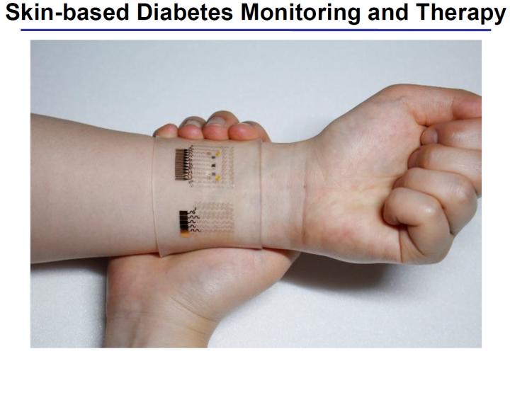
The researchers have made an image illustrating their work available,

Caption: Optical image of the GP-hybrid electrochemical device array on the human skin Credit: IBS

Caption: Optical image of the GP-hybrid electrochemical device array on the human skin Credit: IBS

Here’s a link to and a citation for the paper,

A graphene-based electrochemical device with thermoresponsive microneedles for diabetes monitoring and therapy by Hyunjae Lee, Tae Kyu Choi, Young Bum Lee, Hye Rim Cho, Roozbeh Ghaffari, Liu Wang, Hyung Jin Choi, Taek Dong Chung, Nanshu Lu, Taeghwan Hyeon, Seung Hong Choi, & Dae-Hyeong Kim. Nature Nanotechnology (2016) doi:10.1038/nnano.2016.38 Published online 21 March 2016

This paper is behind a paywall.

Explaining research into matching plasmonic nanoantenna resonances with atoms, molecules, and quantum dots

There’s a very nice explanation of the difficulties associeated with using plasmonic nanoantennas as sensors in a March 21, 2016 news item on phys.org,

Plasmonic nanoantennas are among the hot topics in science at the moment because of their ability to interact strongly with light, which for example makes them useful for different kinds of sensing. But matching their resonances with atoms, molecules or so called quantum dots has been difficult so far because of the very different length scales involved. Thanks to a grant from the Engkvist foundation, Timur Shegai, assistant professor at Chalmers University of Technology, hopes to find a way to do this and by that open doors for applications such as safe long distance communication channels.

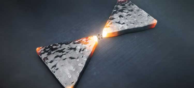
A molecule being illuminated by two gold nanoantennas. By: Alexander Ericson Courtesy: Chalmers University of Technology

A molecule being illuminated by two gold nanoantennas. By: Alexander Ericson Courtesy: Chalmers University of Technology

The image, looking like a stylized butterfly or bow tie, above accompanies Karin Weijdegård’s March ??, 2016 Chalmers University of Technology press release, which originated the news item, expands on the research theme,

The diffraction limit makes it very hard for light to interact with the very smallest particles or so called quantum systems such as atoms, molecules or quantum dots. The size of such a particle is simply so much smaller than the wavelength of light that there cannot be a strong interaction between the two. But by using plasmonic nanoantennas, which can be described as metallic nanostructures that are able to focus light very strongly and in wavelengths smaller than those of the visible light, one can build a bridge between the light and the atom, molecule or quantum dot and that is what Timur Shegai is working on.

“Plasmonic nanostructures are themselves smaller than wavelengths of light, but because they have a lot of free electrons they can store the electromagnetic energy in a volume which is actually a lot smaller than the diffraction limit, which helps to bridge the gap between really small objects such as molecules and the larger wavelengths of light,” he says.

Matching the harmonic with the un-harmonic

This might sound easy enough, but the problem with combining the two is that they behave in very different ways. The behaviour of plasmonic nanostructures is very linear, like a harmonic oscillator it will regularly move from side to side no matter how much energy or in other words how many excitations are stored in it. On the other hand, so called quantum systems like atoms, molecules or quantum dots are very much the opposite – their optical properties are highly un-harmonic. Here it makes a big difference if you excite the system with one or two or hundreds of photons.

“Now imagine that you couple together this un-harmonic resonator and a harmonic resonator, and add the possibility to interact with light much stronger than the un-harmonic system alone would have allowed. That opens up very interesting possibilities for quantum technologies and for non-linear optics for example. But as opposed to previous attempts that have been done at very low temperatures and in a vacuum, we will do it at room temperature.”

Communication channels impossible to hack

One possible application where this technology could be useful in the future is to create channels for long distance communications that are impossible to hack. With the current technology this kind of safe communication is only possible if the persons communicating is within a distance of about one hundred kilometres from each other, because that is the maximum distance that an individual photon can run in fibres before it scatters and the signal is lost.

“The kind of ultra small and ultra fast technology we want to develop could be useful in a so called quantum repeater, a device that could be installed across the line from for example New York to London, that would repeat the photon every time it is about to be scattered,” says Timur Shegai.

At the moment though, it is the fundamental aspects of merging plasmons with quantum systems that interest Timur Shegai. To be able to experimentally prove that the there can be interactions between the two systems, he first of all needs to fabricate model systems at the nano level. This is a big challenge, but with the grant of 1,6 million SEK over a period of two years that he just received from the Engkvist foundation, the chances of success have improved.

“Since I am a researcher at the beginning of my career every person is a huge improvement and now I can hire a post doc to work with my group. This means that the project can be divided into sub parts and together we will be able to explore more possibilities about this new technology.”

Thank you Karin Weijdegård for the explanation.

Using Google Glass to monitor organs-on-chips

Google Glass : Explorer version Credit: Dan Leveille, twitter.com/danlev Courtesty: Wikimedi

Google Glass: Explorer Edition Credit: Dan Leveille, twitter.com/danlev Courtesy: Wikimedia

Researchers have introduced Google Glass as a new application for monitoring and controlling organs-on-chips according to a March 18, 2016 news item on ScienceDaily,

Investigators from Brigham and Women’s Hospital (BWH [Boston, Massachussetts]) have developed hardware and software to remotely monitor and control devices that mimic the human physiological system. Devices known as organs-on-chips allow researchers to test drug compounds and predict physiological responses with high accuracy in a laboratory setting. But monitoring the results of such experiments from a conventional desktop computer has several limitations, especially when results must be monitored over the course of hours, days or even weeks.

Google Glass, one of the newest forms of wearable technology, offers researchers a hands-free and flexible monitoring system. To make Google Glass work for their purposes, Zhang et al. custom developed hardware and software that takes advantage of voice control command (“ok glass”) and other features in order to not only monitor but also remotely control their liver- and heart-on-a-chip systems. Using valves remotely activated by the Glass, the team introduced pharmaceutical compounds on liver organoids and collected the results. …

A March 18, 2016 BWH press release on EurekAlert, which originated the news item, describes the hopes for this new combined platform,

“We believe such a platform has widespread applications in biomedicine, and may be further expanded to health care settings where remote monitoring and control could make things safer and more efficient,” said senior author Ali Khademhosseini, PhD, Director of the Biomaterials Innovation Research Center at BWH.

“This may be of particular importance in cases where experimental conditions threaten human life – such as work involving highly pathogenic bacteria or viruses or radioactive compounds,” said leading author, Shrike Zhang, PhD, also of BWH’s Biomedical Division.

Here’s a link to and a citation for the paper,

Google Glass-Directed Monitoring and Control of Microfluidic Biosensors and Actuators by Yu Shrike Zhang, Fabio Busignani, João Ribas, Julio Aleman, Talles Nascimento Rodrigues, Seyed Ali Mousavi Shaegh, Solange Massa, Camilla Baj Rossi, Irene Taurino, Su-Ryon Shin, Giovanni Calzone, Givan Mark Amaratunga, Douglas Leon Chambers, Saman Jabari, Yuxi Niu, Vijayan Manoharan, Mehmet Remzi Dokmeci, Sandro Carrara, Danilo Demarchi, & Ali Khademhossein. Scientific Reports 6, Article number: 22237 (2016) doi:10.1038/srep22237 Published online: 01 March 2016

This paper is open access.