Monthly Archives: June 2016

Ukidama-structured nanoparticles discovered

The researchers discovered a new nanoparticle structure that resemble the ukidama, glass fishing floats, used regularly by Japanese fishermen. The nanoparticle has a core of one element (copper) and is surrounded by a “cage” of another element (silver). The silver does not cover certain areas of the copper core, which is very similar to the rope that surrounds the glass float. Courtesy: Okinawa Institute of Science and Technology (OIST)

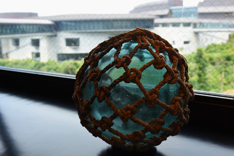
The researchers discovered a new nanoparticle structure that resemble the ukidama, glass fishing floats, used regularly by Japanese fishermen. The nanoparticle has a core of one element (copper) and is surrounded by a “cage” of another element (silver). The silver does not cover certain areas of the copper core, which is very similar to the rope that surrounds the glass float. Courtesy: Okinawa Institute of Science and Technology (OIST)

What a beautiful image to illustrate the new ukidama nanoparticle structure! Here’s the announcement in a June 13, 2016 news item on ScienceDaily,

Sometimes it is the tiny things in the world that can make an incredible difference. One of these things is the nanoparticle. Nanoparticles may be small, but they have a variety of important applications in areas such as, medicine, manufacturing, and energy. A team of researchers from Okinawa Institute of Science and Technology Graduate University (OIST) recently discovered a unique copper-silver nanoparticle structure that has a core of one element surrounded by a “cage” of the other element. However, the cage does not cover certain areas of the core, which very much resembles the Japanese glass fishing floats traditionally covered with rope called ukidama.

This previously undiscovered ukidama structure may have properties that can help the team on their mission for optimal nanotechnology. …

A June 13, 2016 OIST press release by Rebecca Holland (also on EurekAlert; the June 12, 2016 publication date discrepancy is likely due to timezone issues), which originated the news item, provides more insight into the research team’s workings,

“The ukidama is a unique structure, which means that it can likely give us unique properties,” said Panagiotis Grammatikopoulos, first author and group leader of the OIST Nanoparticles by Design Unit. “The idea is that now that we know about this structure we may be able to fine tune it to our applications.”

The OIST researchers are continually working to create and design nanoparticles that can be used in biomedical technology. Specifically, the team works to design the optimal nanoparticles for technologies like smart gas sensors that can send information about what is going on inside your body to your smart phone for better diagnoses. Another application is the label free biosensor, a device that can detect chemical substances without the hindrance of fluorescent or radioactive labels. The identification of the ukidama structure is important in this endeavour because having a new structure increases the possibilities for technological advancements.

“The more parameters that we can control the more flexibility we have in our applications and devices,” Prof. Mukhles Sowwan, author and head of OIST’s Nanoparticles by Design Unit said. “Therefore, we need to optimize many properties of these nanoparticles: the size, chemical composition, crystallinity, shape, and structure.”

The discovery of the ukidama structure was found through sputtering copper and silver atoms simultaneously, but independently, through a magnetron-sputtering system at high temperatures. When the atoms began to cool they combined into bi-metallic nanoparticles. During the sputtering process, researchers could control the ratio of silver to copper, with the rate of power with which the atoms were sputtered. They found that the ukidama structure was possible, especially when the copper was the dominant element, since silver atoms have a higher tendency to diffuse on the nanoparticle surface. From their experimental findings, the team was able to create simulations that can clearly show how the ukidama nanoparticles form.

The team is now looking to see if this structure can be recreated in other types of nanoparticles, which could be an even bigger step in the optimization of nanoparticles for biomedical application and nanotechnology.

“We design and optimize nanoparticles for biomedical devices and nanotechnology,” Sowwan said. “Because the ukidama is a new structure, it may have properties that could be utilized in our applications.”

Co-author, Antony Galea, formerly of the Nanoparticles by Design Unit, was responsible for the experimental portion of this study and has since moved to OIST’s Technology and Licensing Section to help research – like this work being done with nanoparticles that can be utilized in applications – move into the market.

“Our aim is to take research created by OIST from the lab to the real world,” Galea said. “This is a way that work done at OIST, such as by the Nanoparticles by Design Unit, can benefit society.”

Here’s a link to and a citation for the paper,

Kinetic trapping through coalescence and the formation of patterned Ag–Cu nanoparticles by Panagiotis Grammatikopoulos, Joseph Kioseoglou, Antony Galea, Jerome Vernieres, Maria Benelmekki, Rosa E. Diaz, Mukhles Sowwan. Nanoscale, 2016; 8 (18): 9780 DOI: 10.1039/C5NR08256K

I believe this paper is behind a paywall.

X-rays reveal memristor workings

A June 14, 2016 news item on ScienceDaily focuses on memristors. (It’s been about two months since my last memristor posting on April 22, 2016 regarding electronic synapses and neural networks). This piece announces new insight into how memristors function at the atomic scale,

In experiments at two Department of Energy national labs — SLAC National Accelerator Laboratory and Lawrence Berkeley National Laboratory — scientists at Hewlett Packard Enterprise (HPE) [also referred to as HP Labs or Hewlett Packard Laboratories] have experimentally confirmed critical aspects of how a new type of microelectronic device, the memristor, works at an atomic scale.

This result is an important step in designing these solid-state devices for use in future computer memories that operate much faster, last longer and use less energy than today’s flash memory. …

“We need information like this to be able to design memristors that will succeed commercially,” said Suhas Kumar, an HPE scientist and first author on the group’s technical paper.

A June 13, 2016 SLAC news release, which originated the news item, offers a brief history according to HPE and provides details about the latest work,

The memristor was proposed theoretically [by Dr. Leon Chua] in 1971 as the fourth basic electrical device element alongside the resistor, capacitor and inductor. At its heart is a tiny piece of a transition metal oxide sandwiched between two electrodes. Applying a positive or negative voltage pulse dramatically increases or decreases the memristor’s electrical resistance. This behavior makes it suitable for use as a “non-volatile” computer memory that, like flash memory, can retain its state without being refreshed with additional power.

Over the past decade, an HPE group led by senior fellow R. Stanley Williams has explored memristor designs, materials and behavior in detail. Since 2009 they have used intense synchrotron X-rays to reveal the movements of atoms in memristors during switching. Despite advances in understanding the nature of this switching, critical details that would be important in designing commercially successful circuits  remained controversial. For example, the forces that move the atoms, resulting in dramatic resistance changes during switching, remain under debate.

In recent years, the group examined memristors made with oxides of titanium, tantalum and vanadium. Initial experiments revealed that switching in the tantalum oxide devices could be controlled most easily, so it was chosen for further exploration at two DOE Office of Science User Facilities – SLAC’s Stanford Synchrotron Radiation Lightsource (SSRL) and Berkeley Lab’s Advanced Light Source (ALS).

At ALS, the HPE researchers mapped the positions of oxygen atoms before and after switching. For this, they used a scanning transmission X-ray microscope and an apparatus they built to precisely control the position of their sample and the timing and intensity of the 500-electronvolt ALS X-rays, which were tuned to see oxygen.

The experiments revealed that even weak voltage pulses create a thin conductive path through the memristor. During the pulse the path heats up, which creates a force that pushes oxygen atoms away from the path, making it even more conductive. Reversing the voltage pulse resets the memristor by sucking some of oxygen atoms back into the conducting path, thereby increasing the device’s resistance. The memristor’s resistance changes between 10-fold and 1 million-fold, depending on operating parameters like the voltage-pulse amplitude. This resistance change is dramatic enough to exploit commercially.

To be sure of their conclusion, the researchers also needed to understand if the tantalum atoms were moving along with the oxygen during switching. Imaging tantalum required higher-energy, 10,000-electronvolt X-rays, which they obtained at SSRL’s Beam Line 6-2. In a single session there, they determined that the tantalum remained stationary.

“That sealed the deal, convincing us that our hypothesis was correct,” said HPE scientist Catherine Graves, who had worked at SSRL as a Stanford graduate student. She added that discussions with SLAC experts were critical in guiding the HPE team toward the X-ray techniques that would allow them to see the tantalum accurately.

Kumar said the most promising aspect of the tantalum oxide results was that the scientists saw no degradation in switching over more than a billion voltage pulses of a magnitude suitable for commercial use. He added that this knowledge helped his group build memristors that lasted nearly a billion switching cycles, about a thousand-fold improvement.

“This is much longer endurance than is possible with today’s flash memory devices,” Kumar said. “In addition, we also used much higher voltage pulses to accelerate and observe memristor failures, which is also important in understanding how these devices work. Failures occurred when oxygen atoms were forced so far away that they did not return to their initial positions.”

Beyond memory chips, Kumar says memristors’ rapid switching speed and small size could make them suitable for use in logic circuits. Additional memristor characteristics may also be beneficial in the emerging class of brain-inspired neuromorphic computing circuits.

“Transistors are big and bulky compared to memristors,” he said. “Memristors are also much better suited for creating the neuron-like voltage spikes that characterize neuromorphic circuits.”

The researchers have provided an animation illustrating how memristors can fail,

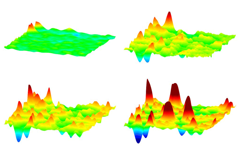
This animation shows how millions of high-voltage switching cycles can cause memristors to fail. The high-voltage switching eventually creates regions that are permanently rich (blue pits) or deficient (red peaks) in oxygen and cannot be switched back. Switching at lower voltages that would be suitable for commercial devices did not show this performance degradation. These observations allowed the researchers to develop materials processing and operating conditions that improved the memristors’ endurance by nearly a thousand times. (Suhas Kumar) Courtesy: SLAC

This animation shows how millions of high-voltage switching cycles can cause memristors to fail. The high-voltage switching eventually creates regions that are permanently rich (blue pits) or deficient (red peaks) in oxygen and cannot be switched back. Switching at lower voltages that would be suitable for commercial devices did not show this performance degradation. These observations allowed the researchers to develop materials processing and operating conditions that improved the memristors’ endurance by nearly a thousand times. (Suhas Kumar) Courtesy: SLAC

Here’s a link to and a citation for the paper,

Direct Observation of Localized Radial Oxygen Migration in Functioning Tantalum Oxide Memristors by Suhas Kumar, Catherine E. Graves, John Paul Strachan, Emmanuelle Merced Grafals, Arthur L. David Kilcoyne3, Tolek Tyliszczak, Johanna Nelson Weker, Yoshio Nishi, and R. Stanley Williams. Advanced Materials, First published: 2 February 2016; Print: Volume 28, Issue 14 April 13, 2016 Pages 2772–2776 DOI: 10.1002/adma.201505435

This paper is behind a paywall.

Some of the ‘memristor story’ is contested and you can find a brief overview of the discussion in this Wikipedia memristor entry in the section on ‘definition and criticism’. There is also a history of the memristor which dates back to the 19th century featured in my May 22, 2012 posting.

Animal technology: a touchscreen for your dog, sonar lunch orders for dolphins, and more

A rather unexpected (for ignorant folks like me) approach to animal technology has been taken by Ilyena Hirskyj-Douglas in her June 17, 2016 piece on phys.org,

Imagine leaving your dog at home while it turns on the smart TV and chooses a programme to watch. Meanwhile you visit a zoo where you play interactive touchscreen games with the apes and watch the dolphins using sonar to order their lunch. In the field behind you, a farmer is stroking his flock of chickens virtually, leaving the drones to collect sheep while the cows milk themselves. Welcome to the unusual world of animal technology.

Hirskyj-Douglas’s piece was originally published as a June 15, 2016 essay  about animal-computer interaction (ACI) and some of the latest work being done in the field on The Conversation website (Note: Links have been removed),

Animals have interacted with technology for a long time, from tracking devices for conservation research to zoos with early touchscreen computers. But more recently, the field of animal-computer interaction (ACI) has begun to explore in more detail exactly how animals use technology like this. The hope is that better understanding animals’ relationship with technology will means we can use it to monitor and improve their welfare.

My own research involves building intelligent tracking devices for dogs that let them interact with media on a screen so we can study how dogs use TV and what they like to watch (if anything). Perhaps unsurprisingly, I’ve found that dogs like to watch videos of other dogs. This has led me to track dogs dogs’ gaze across individual and multiple screens and attempts to work out how best to make media just for dogs.

Eventually I hope to make an interactive system that allows a dog to pick what they want to watch and that evolves by learning what media they like. This isn’t to create a toy for indulgent pet owners. Dogs are often left at home alone during the day or isolated in kennels. So interactive media technology could improve the animals’ welfare by providing a stimulus and a source of entertainment. …

This 2014 video (embedded in Hirskyj-Douglas’s essay) illustrates how touchscreens are used by great apes,

It’s all quite intriguing and I encourage you to read the essay in it entirety.

If you find the great apes project interesting, you can find  out more about it (I believe it’s in the Primate Research category) and others at the Atlanta Zoo’s research webpage.

A human user manual—for robots

Researchers from the Georgia Institute of Technology (Georgia Tech), funded by the US Office of Naval Research (ONR), have developed a program that teaches robots to read stories and more in an effort to educate them about humans. From a June 16, 2016 ONR news release by Warren Duffie Jr. (also on EurekAlert),

With support from the Office of Naval Research (ONR), researchers at the Georgia Institute of Technology have created an artificial intelligence software program named Quixote to teach robots to read stories, learn acceptable behavior and understand successful ways to conduct themselves in diverse social situations.

“For years, researchers have debated how to teach robots to act in ways that are appropriate, non-intrusive and trustworthy,” said Marc Steinberg, an ONR program manager who oversees the research. “One important question is how to explain complex concepts such as policies, values or ethics to robots. Humans are really good at using narrative stories to make sense of the world and communicate to other people. This could one day be an effective way to interact with robots.”

The rapid pace of artificial intelligence has stirred fears by some that robots could act unethically or harm humans. Dr. Mark Riedl, an associate professor and director of Georgia Tech’s Entertainment Intelligence Lab, hopes to ease concerns by having Quixote serve as a “human user manual” by teaching robots values through simple stories. After all, stories inform, educate and entertain–reflecting shared cultural knowledge, social mores and protocols.

For example, if a robot is tasked with picking up a pharmacy prescription for a human as quickly as possible, it could: a) take the medicine and leave, b) interact politely with pharmacists, c) or wait in line. Without value alignment and positive reinforcement, the robot might logically deduce robbery is the fastest, cheapest way to accomplish its task. However, with value alignment from Quixote, it would be rewarded for waiting patiently in line and paying for the prescription.

For their research, Riedl and his team crowdsourced stories from the Internet. Each tale needed to highlight daily social interactions–going to a pharmacy or restaurant, for example–as well as socially appropriate behaviors (e.g., paying for meals or medicine) within each setting.

The team plugged the data into Quixote to create a virtual agent–in this case, a video game character placed into various game-like scenarios mirroring the stories. As the virtual agent completed a game, it earned points and positive reinforcement for emulating the actions of protagonists in the stories.

Riedl’s team ran the agent through 500,000 simulations, and it displayed proper social interactions more than 90 percent of the time.

“These games are still fairly simple,” said Riedl, “more like ‘Pac-Man’ instead of ‘Halo.’ However, Quixote enables these artificial intelligence agents to immerse themselves in a story, learn the proper sequence of events and be encoded with acceptable behavior patterns. This type of artificial intelligence can be adapted to robots, offering a variety of applications.”

Within the next six months, Riedl’s team hopes to upgrade Quixote’s games from “old-school” to more modern and complex styles like those found in Minecraft–in which players use blocks to build elaborate structures and societies.

Riedl believes Quixote could one day make it easier for humans to train robots to perform diverse tasks. Steinberg notes that robotic and artificial intelligence systems may one day be a much larger part of military life. This could involve mine detection and deactivation, equipment transport and humanitarian and rescue operations.

“Within a decade, there will be more robots in society, rubbing elbows with us,” said Riedl. “Social conventions grease the wheels of society, and robots will need to understand the nuances of how humans do things. That’s where Quixote can serve as a valuable tool. We’re already seeing it with virtual agents like Siri and Cortana, which are programmed not to say hurtful or insulting things to users.”

This story brought to mind two other projects: RoboEarth (an internet for robots only) mentioned in my Jan. 14, 2014 which was an update on the project featuring its use in hospitals and RoboBrain, a robot learning project (sourcing the internet, YouTube, and more for information to teach robots) was mentioned in my Sept. 2, 2014 posting.

The ‘Lasso of Truth’ and the lie detector have a common origin

There’s a fascinating June 17, 2016 article about Wonder Woman’s seventy-fifth anniversary (points to anyone who her recognized her ‘Lasso of Truth’) by Susan Karlin for Fast Company,

William Moulton Marston—an attorney and psychologist who invented a systolic blood pressure deception test, the precursor to the modern polygraph—created Wonder Woman as a new type of superhero who, beyond her strength, used wisdom and compassion as weapons against evil—not to mention a magic golden lasso to compel people to tell the truth.

“Marston recognized not only the thereto untapped commercial market for a strong female superhero, but also the powerful potential for comic books to educate and inspire. He understood that education and entertainment need not be mutually exclusive,” says Vasilis Pozios, a forensic psychiatrist who cofounded [Broadcast Thought; mental health-and-media think tank with three forensic psychiatrists – H. Eric Bender, M.D., Praveen Kambam, M.D., and Pozios], which uses media and comic convention panels to educate about mental illness, and author of Aura, an award-winning comic about bipolar disorder.

The article has various versions of Wonder Woman images embedded throughout and it includes a few nuggets like this about her and her originator,

Wonder Woman is the only female comic book character to have her own stories continuously published for the past three-quarters of a century, spawning numerous other incarnations, including the hit 1975-1979 TV series starring Lynda Carter, and finally a big-screen introduction in this year’s [2016] Batman v Superman: Dawn of Justice.

Marston, who was strongly influenced by the women’s suffrage movement, devised that WW’s would lose her strength if men bound her in chains. Initially controversial due to a look inspired by pinup art and bondage intimations, she emerged as a symbol of equality and female empowerment—gracing Ms. magazine’s inaugural cover in 1972—that resonates today.

I gather this Wonder Woman 75th anniversary is going to be celebrated over a two year period with DC Comics hosting a 2016 Wonder Woman 75 San Diego Comic-Con panel and costume display and then, releasing the first (and fortuitously timed) Wonder Woman feature film starring Gal Gadot on June 2, 2017.

Do read Karlin’s if only to catch sight of the images. I have written about Wonder Woman before notably in a July 1, 2010 (Canada Day) posting featuring a then new makeover,

wonder_woman_makeover

I wasn’t thrilled by the makeover and was not alone in my opinion although reasons for the ‘lack of thrill’ varied from mine.

Oil spill cleanups with supergelators

Researchers in Singapore have proposed a new technology for cleaning up oil spills, according to a June 17, 2016 news item on Nanowerk,

Large-scale oil spills, where hundreds of tons of petroleum products are accidentally released into the oceans, not only have devastating effects on the environment, but have significant socio-economic impact as well [1].

Current techniques of cleaning up oil spills are not very efficient and may even cause further pollution or damage to the environment. These methods, which include the use of toxic detergent-like compounds called dispersants or burning of the oil slick, result in incomplete removal of the oil. The oil molecules remain in the water over long periods and may even be spread over a larger area as they are carried by wind and waves. Further, burning can only be applied to fresh oil slicks of at least 3 millimeters thick, and this process would also cause secondary environmental pollution.

In a bid to improve the technology utilized by cleanup crews to manage and contain such large spills, researchers from the Institute of Bioengineering and Nanotechnology (IBN) of A*STAR [located in Singapore] have invented a smart oil-scavenging material or supergelators that could help clean up oil spills efficiently and rapidly to prevent secondary pollution.

These supergelators are derived from highly soluble small organic molecules, which instantly self-assemble into nanofibers to form a 3D net that traps the oil molecules so that they can be removed easily from the surface of the water.

A June 17, 2016 IBN A*STAR media release, which originated the news item, provides more detail,

“Marine oil spills have a disastrous impact on the environment and marine life, and result in an enormous economic burden on society. Our rapid-acting supergelators offer an effective cleanup solution that can help to contain the severe environmental damage and impact of such incidents in the future,” said IBN Executive Director Professor Jackie Y. Ying.

Motivated by the urgent need for a more effective oil spill control solution, the IBN researchers developed new compounds that dissolve easily in environmentally friendly solvents and gel rapidly upon contact with oil. The supergelator molecules arrange themselves into a 3D network, entangling the oil molecules into clumps that can then be easily skimmed off the water’s surface.

“The most interesting and useful characteristic of our molecules is their ability to stack themselves on top of each other. These stacked columns allow our researchers to create and test different molecular constructions, while finding the best structure that will yield the desired properties,” said IBN Team Leader and Principal Research Scientist Dr Huaqiang Zeng. (Animation: Click to see how the supergelators stack themselves into columns.)

IBN’s supergelators have been tested on various types of weathered and unweathered crude oil in seawater, and have been found to be effective in solidifying all of them. The supergelators take only minutes to solidify the oil at room temperature for easy removal from water. In addition, tests carried out by the research team showed that the supergelator was not toxic to human cells, as well as zebrafish embryos and larvae. The researchers believe that these qualities would make the supergelators suitable for use in large oil spill areas.

The Institute is looking for industrial partners to further develop its technology for commercial use. [emphasis mine]

Video: Click to watch the supergelators in action

  1. The well documented BP Gulf of Mexico oil well accident in 2010 was a catastrophe on an unprecedented scale, with damages amounting to hundreds of billions of dollars. Its wide-ranging effects on the marine ecosystem, as well as the fishing and tourism industries, can still be felt six years on.

Here’s a link to and a citation for the paper,

Instant Room-Temperature Gelation of Crude Oil by Chiral Organogelators by Changliang Ren, Grace Hwee Boon Ng, Hong Wu, Kiat-Hwa Chan, Jie Shen, Cathleen Teh, Jackie Y. Ying, and Huaqiang Zeng. Chem. Mater., 2016, 28 (11), pp 4001–4008 DOI: 10.1021/acs.chemmater.6b01367 Publication Date (Web): May 10, 2016

Copyright © 2016 American Chemical Society

This paper is behind a paywall.

I have featured other nanotechnology-enabled oil spill cleanup solutions here. One of the more recent pieces is my Dec. 7, 2015 post about boron nitride sponges. The search terms: ‘oil spill’ and ‘oil spill cleanup’ will help you unearth more.

There have been some promising possibilities and I hope one day these clean up technologies will be brought to market.

Biohackers (also known as bodyhackers or grinders) become more common?

Stephen Melendez’s June 11, 2016 story about biohackers/bodyhackers/grinders for Fast Company sports a striking image in the banner, an x-ray of a pair hands featuring some mysterious additions to the webbing between thumbs and forefingers (Note: Links have been removed),

Tim Shank can guarantee he’ll never leave home without his keys. Why? His house keys are located inside his body.

Shank, the president of the Minneapolis futurist group TwinCities+, has a chip installed in his hand that can communicate electronically with his front door and tell it to unlock itself. His wife has one, too.

In fact, Shank has several chips in his hand, including a near field communication (NFC) chip like the ones used in Apple Pay and similar systems, which stores a virtual business card with contact information for TwinCities+. “[For] people with Android phones, I can just tap their phone with my hand, right over the chip, and it will send that information to their phone,” he says. In the past, he’s also used a chip to store a bitcoin wallet.

Shank is one of a growing number of “biohackers” who implant hardware ranging from microchips to magnets inside their bodies.

Certainly the practice seems considerably more developed since the first time it was mentioned here in a May 27, 2010 posting about a researcher who’d implanted a chip into his body which he then contaminated with a computer virus. In the comments, you’ll find Amal Grafstraa who’s mentioned in the Melendez article at some length, from the Melendez article (Note: Links have been removed),

Some biohackers use their implants in experimental art projects. Others who have disabilities or medical conditions use them to improve their quality of life, while still others use the chips to extend the limits of human perception. …

Experts sometimes caution that the long-term health risks of the practice are still unknown. But many biohackers claim that, if done right, implants can be no more dangerous than getting a piercing or tattoo. In fact, professional body piercers are frequently the ones tasked with installing these implants, given that they possess the training and sterilization equipment necessary to break people’s skin safely.

“When you talk about things like risk, things like putting it in your body, the reality is the risk of having one of these installed is extremely low—it’s even lower than an ear piercing,” claims Amal Graafstra, the founder of Dangerous Things, a biohacking supply company.

Graafstra, who is also the author of the book RFID Toys, says he first had an RFID chip installed in his hand in 2005, which allowed him to unlock doors without a key. When the maker movement took off a few years later, and as more hackers began to explore what they could put inside their bodies, he founded Dangerous Things with the aim of ensuring these procedures were done safely.

“I decided maybe it’s time to wrap a business model around this and make sure that the things people are trying to put in their bodies are safe,” he says. The company works with a network of trained body piercers and offers online manuals and videos for piercers looking to get up to speed on the biohacking movement.

At present, these chips are capable of verifying users’ identities and opening doors. And according to Graafstra, a next-generation chip will have enough on-board cryptographic power to potentially work with credit card terminals securely.

“The technology is there—we can definitely talk to payment terminals with it—but we don’t have the agreements in place with banks [and companies like] MasterCard to make that happen,” he says.

Paying for goods with an implantable chip might sound unusual for consumers and risky for banks, but Graafstra thinks the practice will one day become commonplace. He points to a survey released by Visa last year that found that 25% of Australians are “at least slightly interested” in paying for purchases through a chip implanted in their bodies.

Melendez’s article is fascinating and well worth reading in its entirety. It’s not all keys and commerce as this next and last excerpt shows,

Other implantable technology has more of an aesthetic focus: Pittsburgh biohacking company Grindhouse Wetware offers a below-the-skin, star-shaped array of LED lights called Northstar. While the product was inspired by the on-board lamps of a device called Circadia that Grindhouse founder Tim Cannon implanted to send his body temperature to a smartphone, the commercially available Northstar features only the lights and is designed to resemble natural bioluminescence.

“This particular device is mainly aesthetic,” says Grindhouse spokesman Ryan O’Shea. “It can backlight tattoos or be used in any kind of interpretive dance, or artists can use it in various ways.”

The lights activate in the presence of a magnetic field—one that is often provided by magnets already implanted in the same user’s fingertips. Which brings up another increasingly common piece of bio-hardware: magnetic finger implants. ….

There are other objects that can be implanted in bodies. In one case, an artist, Wafaa Bilal had a camera implanted into the back of his head for a 3rd eye. I mentioned the Iraqi artist in my April 13, 2011 posting titled: Blood, memristors, cyborgs plus brain-controlled computers, prosthetics, and art (scroll down about 75% of the way). Bilal was unable to find a doctor who would perform the procedure so he went to a body-piercing studio. Unfortunately, the posting chronicles his infection and subsequent removal of the camera (h/t Feb. 11, 2011 BBC [British Broadcasting Corporation] news online article).

Observations

It’s been a while since I’ve written about bodyhacking and I’d almost forgotten about the practice relegating it to the category of “one of those trendy ideas that get left behind as interest shifts.” My own interest had shifted more firmly to neuroprosthetics (the integration of prostheses into the nervous system).

I had coined a tag for bodyhacking and neuroprostheses: machine/flesh which covers both those topics and more (e.g. cyborgs) as we continue to integrate machines into our bodies.

Final note

I was reminded of Wafaa Bilal recently when checking out a local arts magazine, Preview: the gallery guide, June/July/August 2016 issue. His work (the 168:01show) is being shown in Calgary, Alberta, Canada at the Esker Foundation from May 27 to August 28, 2016,

168:01 is a major solo exhibition of new and recent work by Iraqi-born, New York-based artist Wafaa Bilal, renowned for his online performances and technologically driven encounters that speak to the impact of international politics on individual lives.

In 168:01, Bilal takes the Bayt al-Hikma, or House of Wisdom, as a starting point for a sculptural installation of a library. The Bayt al-Hikma was a major academic center during the Islamic Golden Age where Muslim, Jewish, and Christian scholars studied the humanities and science. By the middle of the Ninth Century, the House of Wisdom had accumulated the largest library in the world. Four centuries later, a Mongol siege laid waste to all the libraries of Baghdad along with the House of Wisdom. According to some accounts, the library was thrown into the Tigris River to create a bridge of books for the Mongol army to cross. The pages bled ink into the river for seven days – or 168 hours, after which the books were drained of knowledge. Today, the Bayt al-Hikma represents one of the most well-known examples of historic cultural loss as a casualty of wartime.

For this exhibition, Bilal has constructed a makeshift library filled with empty white books. The white books symbolize the priceless cultural heritage destroyed at Bayt al-Hikma as well as the libraries, archives, and museums whose systematic decimation by occupying forces continues to ravage his homeland. Throughout the duration of the exhibition, the white books will slowly be replaced with visitor donations from a wishlist compiled by The College of Fine Arts at the University of Baghdad, whose library was looted and destroyed in 2003. At the end of each week a volunteer unpacks the accumulated shipments, catalogues each new book by hand, and places the books on the shelves. At the end of the exhibition, all the donated books will be sent to the University of Baghdad to help rebuild their library. This exchange symbolizes the power of individuals to rectify violence inflicted on cultural spaces that are meant to preserve and store knowledge for future generations.

In conjunction with the library, Bilal presents a powerful suite of photographs titled The Ashes Series that brings the viewer closer to images of violence and war in the Middle East. In an effort to foster empathy and humanize the onslaught of violent images that inundate Western media during wartime, Bilal has reconstructed journalistic images of the destruction caused by the Iraq War. He writes, “Reconstructing the destructed spaces is a way to exist in them, to share them with an audience, and to provide a layer of distance, as the original photographs are too violent and run the risk of alienating the viewer. It represents an attempt to make sense of the destruction and to preserve the moment of serenity after the dust has settled, to give the ephemeral moment extended life in a mix of beauty and violence.” In the photograph Al-Mutanabbi Street from The Ashes Series, the viewer encounters dilapidated historic and modern buildings on a street covered with layers upon layers of rubble and fragments of torn books. Bilal’s images emanate a slowness that deepens engagement between the viewer and the image, thereby inviting them to share the burden of obliterated societies and reimagine a world built on the values of peace and hope.

The House of Wisdom has been mentioned here a few times perhaps most comprehensively and in the context of the then recent opening of the King Abdullah University for Science and Technology (KAUST; located in Saudi Arabia) in this Sept. 24, 2009 posting (scroll down about 45% of the way).

Anyone interested in hacking their own body?


I expect you can find out more Amal Grafstraa’s website.
DNA as a framework for rationally designed nanostructures

After publishing a June 15, 2016 post about taking DNA (deoxyribonucleic acid) beyond genetics, it seemed like a good to publish a companion piece featuring a more technical explanation of at least one way DNA might provide the base for living computers and robots. From a June 13, 2016 BrookHaven National Laboratory news release (also on EurekAlert),

A cube, an octahedron, a prism–these are among the polyhedral structures, or frames, made of DNA that scientists at the U.S. Department of Energy’s (DOE) Brookhaven National Laboratory have designed to connect nanoparticles into a variety of precisely structured three-dimensional (3D) lattices. The scientists also developed a method to integrate nanoparticles and DNA frames into interconnecting modules, expanding the diversity of possible structures.

These achievements, described in papers published in Nature Materials and Nature Chemistry, could enable the rational design of nanomaterials with enhanced or combined optical, electric, and magnetic properties to achieve desired functions.

“We are aiming to create self-assembled nanostructures from blueprints,” said physicist Oleg Gang, who led this research at the Center for Functional Nanomaterials (CFN), a DOE Office of Science User Facility at Brookhaven. “The structure of our nanoparticle assemblies is mostly controlled by the shape and binding properties of precisely designed DNA frames, not by the nanoparticles themselves. By enabling us to engineer different lattices and architectures without having to manipulate the particles, our method opens up great opportunities for designing nanomaterials with properties that can be enhanced by precisely organizing functional components. For example, we could create targeted light-absorbing materials that harness solar energy, or magnetic materials that increase information-storage capacity.”

The news release goes on to describe the frames,

Gang’s team has previously exploited DNA’s complementary base pairing–the highly specific binding of bases known by the letters A, T, G, and C that make up the rungs of the DNA double-helix “ladder”–to bring particles together in a precise way. Particles coated with single strands of DNA link to particles coated with complementary strands (A binds with T and G binds with C) while repelling particles coated with non-complementary strands.

They have also designed 3D DNA frames whose corners have single-stranded DNA tethers to which nanoparticles coated with complementary strands can bind. When the scientists mix these nanoparticles and frames, the components self-assemble into lattices that are mainly defined by the shape of the designed frame. The Nature Materials paper describes the most recent structures achieved using this strategy.

“In our approach, we use DNA frames to promote the directional interactions between nanoparticles such that the particles connect into specific configurations that achieve the desired 3D arrays,” said Ye Tian, lead author on the Nature Materials paper and a member of Gang’s research team. “The geometry of each particle-linking frame is directly related to the lattice type, though the exact nature of this relationship is still being explored.”

So far, the team has designed five polyhedral frame shapes–a cube, an octahedron, an elongated square bipyramid, a prism, and a triangular bypyramid–but a variety of other shapes could be created.

“The idea is to construct different 3D structures (buildings) from the same nanoparticle (brick),” said Gang. “Usually, the particles need to be modified to produce the desired structures. Our approach significantly reduces the structure’s dependence on the nature of the particle, which can be gold, silver, iron, or any other inorganic material.”

Nanoparticles (yellow balls) capped with short single-stranded DNA (blue squiggly lines) are mixed with polyhedral DNA frames (from top to bottom): cube, octahedron, elongated square bipyramid, prism, and triangular bipyramid. The frames' vertices are encoded with complementary DNA strands for nanoparticle binding. When the corresponding frames and particles mix, they form a framework. Courtesy of Brookhaven National Laboratory

Nanoparticles (yellow balls) capped with short single-stranded DNA (blue squiggly lines) are mixed with polyhedral DNA frames (from top to bottom): cube, octahedron, elongated square bipyramid, prism, and triangular bipyramid. The frames’ vertices are encoded with complementary DNA strands for nanoparticle binding. When the corresponding frames and particles mix, they form a framework. Courtesy of Brookhaven National Laboratory

There’s also a discussion about how DNA origami was used to design the frames,

To design the frames, the team used DNA origami, a self-assembly technique in which short synthetic strands of DNA (staple strands) are mixed with a longer single strand of biologically derived DNA (scaffold strand). When the scientists heat and cool this mixture, the staple strands selectively bind with or “staple” the scaffold strand, causing the scaffold strand to repeatedly fold over onto itself. Computer software helps them determine the specific sequences for folding the DNA into desired shapes.

The folding of the single-stranded DNA scaffold introduces anchoring points that contain free “sticky” ends–unpaired strings of DNA bases–where nanoparticles coated with complementary single-strand tethers can attach. These sticky ends can be positioned anywhere on the DNA frame, but Gang’s team chose the corners so that multiple frames could be connected.

For each frame shape, the number of DNA strands linking a frame corner to an individual nanoparticle is equivalent to the number of edges converging at that corner. The cube and prism frames have three strands at each corner, for example. By making these corner tethers with varying numbers of bases, the scientists can tune the flexibility and length of the particle-frame linkages.

The interparticle distances are determined by the lengths of the frame edges, which are tens of nanometers in the frames designed to date, but the scientists say it should be possible to tailor the frames to achieve any desired dimensions.

The scientists verified the frame structures and nanoparticle arrangements through cryo-electron microscopy (a type of microscopy conducted at very low temperatures) at the CFN and Brookhaven’s Biology Department, and x-ray scattering at the National Synchrotron Light Source II (NSLS-II), a DOE Office of Science User Facility at Brookhaven.

The team started with a relatively simple form (from the news release),

In the Nature Chemistry paper, Gang’s team described how they used a similar DNA-based approach to create programmable two-dimensional (2D), square-like DNA frames around single nanoparticles.

DNA strands inside the frames provide coupling to complementary DNA on the nanoparticles, essentially holding the particle inside the frame. Each exterior side of the frame can be individually encoded with different DNA sequences. These outer DNA strands guide frame-frame recognition and connection.

Gang likens these DNA-framed nanoparticle modules to Legos whose interactions are programmed: “Each module can hold a different kind of nanoparticle and interlock to other modules in different but specific ways, fully determined by the complementary pairing of the DNA bases on the sides of the frame.”

In other words, the frames not only determine if the nanoparticles will connect but also how they will connect. Programming the frame sides with specific DNA sequences means only frames with complementary sequences can link up.

Mixing different types of modules together can yield a variety of structures, similar to the constructs that can be generated from Lego pieces. By creating a library of the modules, the scientists hope to be able to assemble structures on demand.

Finally, the discussion turns to the assembly of multifuctional nanomaterials (from the news release),

The selectivity of the connections enables different types and sizes of nanoparticles to be combined into single structures.

The geometry of the connections, or how the particles are oriented in space, is very important to designing structures with desired functions. For example, optically active nanoparticles can be arranged in a particular geometry to rotate, filter, absorb, and emit light–capabilities that are relevant for energy-harvesting applications, such as display screens and solar panels.

By using different modules from the “library,” Gang’s team demonstrated the self-assembly of one-dimensional linear arrays, “zigzag” chains, square-shaped and cross-shaped clusters, and 2D square lattices. The scientists even generated a simplistic nanoscale model of Leonardo da Vinci’s Vitruvian Man.

“We wanted to demonstrate that complex nanoparticle architectures can be self-assembled using our approach,” said Gang.

Again, the scientists used sophisticated imaging techniques–electron and atomic force microscopy at the CFN and x-ray scattering at NSLS-II–to verify that their structures were consistent with the prescribed designs and to study the assembly process in detail.

“Although many additional studies are required, our results show that we are making advances toward our goal of creating designed matter via self-assembly, including periodic particle arrays and complex nanoarchitectures with freeform shapes,” said Gang. “Our approach is exciting because it is a new platform for nanoscale manufacturing, one that can lead to a variety of rationally designed functional materials.”

Here’s an image illustrating among other things da Vinci’s Vitruvian Man,

A schematic diagram (left) showing how a nanoparticle (yellow ball) is incorporated within a square-like DNA frame. The DNA strands inside the frame (blue squiggly lines) are complementary to the DNA strands on the nanoparticle; the colored strands on the outer edges of the frame have different DNA sequences that determine how the DNA-framed nanoparticle modules can connect. The architecture shown (middle) is a simplistic nanoscale representation of Leonardo da Vinci's Vitruvian Man, assembled from several module types. The scientists used atomic force microscopy to generate the high-magnification image of this assembly (right). Courtesy Brookhaven National Laboratory

A schematic diagram (left) showing how a nanoparticle (yellow ball) is incorporated within a square-like DNA frame. The DNA strands inside the frame (blue squiggly lines) are complementary to the DNA strands on the nanoparticle; the colored strands on the outer edges of the frame have different DNA sequences that determine how the DNA-framed nanoparticle modules can connect. The architecture shown (middle) is a simplistic nanoscale representation of Leonardo da Vinci’s Vitruvian Man, assembled from several module types. The scientists used atomic force microscopy to generate the high-magnification image of this assembly (right). Courtesy Brookhaven National Laboratory

I enjoy the overviews provided by various writers and thinkers in the field but it’s details such as these that are often most compelling to me.

Gold-144 is a polymorph

Au-144 (also known as Gold-144) is an iconic gold nanocluster according to a June 14, 2016 news item announcing its polymorphic nature on ScienceDaily,

Chemically the same, graphite and diamonds are as physically distinct as two minerals can be, one opaque and soft, the other translucent and hard. What makes them unique is their differing arrangement of carbon atoms.

Polymorphs, or materials with the same composition but different structures, are common in bulk materials, and now a new study in Nature Communications confirms they exist in nanomaterials, too. Researchers describe two unique structures for the iconic gold nanocluster Au144(SR)60, better known as Gold-144, including a version never seen before. Their discovery gives engineers a new material to explore, along with the possibility of finding other polymorphic nanoparticles.

A June 14, 2016 Columbia University news release (also on EurekAlert), which originated the news item, provides more insight into the work,

“This took four years to unravel,” said Simon Billinge, a physics professor at Columbia Engineering and a member of the Data Science Institute. “We weren’t expecting the clusters to take on more than one atomic arrangement. But this discovery gives us more handles to turn when trying to design clusters with new and useful properties.”

Gold has been used in coins and jewelry for thousands of years for its durability, but shrink it to a size 10,000 times smaller than a human hair [at one time one billionth of a meter or a nanometer was said to be 1/50,000, 1/60,000 or 1/100,000 of the diameter of a human hair], and it becomes wildly unstable and unpredictable. At the nanoscale, gold likes to split apart other particles and molecules, making it a useful material for purifying water, imaging and killing tumors, and making solar panels more efficient, among other applications.

Though a variety of nanogold particles and molecules have been made in the lab, very few have had their secret atomic arrangement revealed. But recently, new technologies are bringing these miniscule structures into focus.

Under one approach, high-energy x-ray beams are fired at a sample of nanoparticles. Advanced data analytics are used to interpret the x-ray scattering data and infer the sample’s structure, which is key to understanding how strong, reactive or durable the particles might be.

Billinge and his lab have pioneered a method, the atomic Pair Distribution Function (PDF) analysis, for interpreting this scattering data. To test the PDF method, Billinge asked chemists at the Colorado State University to make tiny samples of Gold-144, a molecule-sized nanogold cluster first isolated in 1995. Its structure had been theoretically predicted in 2009, and though never confirmed, Gold-144 has found numerous applications, including in tissue-imaging.

Hoping the test would confirm Gold-144’s structure, they analyzed the clusters at the European Synchrotron Radiation Source in Grenoble, and used the PDF method to infer their structure. To their surprise, they found an angular core, and not the sphere-like icosahedral core predicted. When they made a new sample and tried the experiment again, this time using synchrotrons at Brookhaven and Argonne national laboratories, the structure came back spherical.

“We didn’t understand what was going on, but digging deeper, we realized we had a polymorph,” said study coauthor Kirsten Jensen, formerly a postdoctoral researcher at Columbia, now a chemistry professor at the University of Copenhagen.

Further experiments confirmed the cluster had two versions, sometimes found together, each with a unique structure indicating they behave differently. The researchers are still unsure if Gold-144 can switch from one version to the other or, what exactly, differentiates the two forms.

To make their discovery, the researchers solved what physicists call the nanostructure inverse problem. How can the structure of a tiny nanoparticle in a sample be inferred from an x-ray signal that has been averaged over millions of particles, each with different orientations?

“The signal is noisy and highly degraded,” said Billinge. “It’s the equivalent of trying to recognize if the bird in the tree is a robin or a cardinal, but the image in your binoculars is too blurry and distorted to tell.”

“Our results demonstrate the power of PDF analysis to reveal the structure of very tiny particles,” added study coauthor Christopher Ackerson, a chemistry professor at Colorado State. “I’ve been trying, off and on, for more than 10 years to get the single-crystal x-ray structure of Gold-144. The presence of polymorphs helps to explain why this molecule has been so resistant to traditional methods.”

The PDF approach is one of several rival methods being developed to bring nanoparticle structure into focus. Now that it has proven itself, it could help speed up the work of describing other nanostructures.

The eventual goal is to design nanoparticles by their desired properties, rather than through trial and error, by understanding how form and function relate. Databases of known and predicted structures could make it possible to design new materials with a few clicks of a mouse.

The study is a first step.

“We’ve had a structure model for this iconic gold molecule for years and then this study comes along and says the structure is basically right but it’s got a doppelgänger,” said Robert Whetten, a professor of chemical physics at the University of Texas, San Antonio, who led the team that first isolated Gold-144. “It seemed preposterous, to have two distinct structures that underlie its ubiquity, but this is a beautiful paper that will persuade a lot of people.”

Here’s an image illustrating the two shapes,

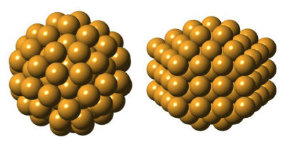
Setting out to confirm the predicted structure of Gold-144, researchers discovered an entirely unexpected atomic arrangement (right). The two structures, described in detail for the first time, each have 144 gold atoms, but are uniquely shaped, suggesting they also behave differently. (Courtesy of Kirsten Ørnsbjerg Jensen)

Setting out to confirm the predicted structure of Gold-144, researchers discovered an entirely unexpected atomic arrangement (right). The two structures, described in detail for the first time, each have 144 gold atoms, but are uniquely shaped, suggesting they also behave differently. (Courtesy of Kirsten Ørnsbjerg Jensen)

Here’s a link to and a citation for the paper,

Polymorphism in magic-sized Au144(SR)60 clusters by Kirsten M.Ø. Jensen, Pavol Juhas, Marcus A. Tofanelli, Christine L. Heinecke, Gavin Vaughan, Christopher J. Ackerson, & Simon J. L. Billinge.  Nature Communications 7, Article number: 11859  doi:10.1038/ncomms11859 Published 14 June 2016

This is an open access paper.

Turning gold into see-through rubber for an updated Rumpelstiltskin story

Rumpelstiltskin is a fairy tale whereby a young girl is trapped by her father’s lie that she can spin straw into gold. She is forced to spin gold by the King under pain of execution when an imp offers to help in exchange for various goods. As she succeeds each time, the King demands more until finally she has nothing left to trade for the imp’s help. Well, there is one last thing: her first-born child. She agrees to the bargain and she marries the King. On the birth of their first child, the imp reappears and under pressure of her pleas makes one last bargain. She must guess his name which she does, Rumplestiltskin. (The full story along with variants is here in this Wikipedia entry.)

With this latest research, we have a reverse Rumpelstiltskin story where gold is turned into something else according to a June 13, 2016 news item on Nanowerk (Note: A link has been removed),

Flexible solar panels that could be rolled up for easy transport and other devices would benefit from transparent metal electrodes that can conduct electricity, are stretchable, and resist damage following repeated stretching. Researchers found that topology and the adhesion between a metal nanomesh and the underlying substrate played key roles in creating such materials. The metal nanomesh can be stretched to three times its length while maintaining a transparency comparable to similar commercial materials used in solar cells and flat panel displays. Also, nanomeshes on pre-stretched slippery substrates led to electrodes that didn’t wear out, even after being stretched 50,000 times (Proceedings of the National Academy of Sciences, “Fatigue-free, superstretchable, transparent, and biocompatible metal electrodes”).

Tuning topology and adhesion of metal nanomeshes has led to super stretchable, transparent electrodes that don’t wear out. The scanning electron microscopy image shows the structure of a gold mesh created with a special lithographic technique that controlled the dimensions of the mesh structure. Optimizing this structure and its adhesion to the substrate was key to achieving super stretchability and long lifetimes in use—nanomeshes on pre-stretched slippery substrates did not show signs of wear even after repeated stretching, up to 50,000 cycles.

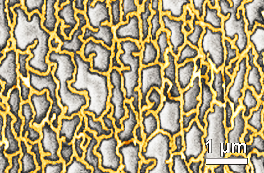
Tuning topology and adhesion of metal nanomeshes has led to super stretchable, transparent electrodes that don’t wear out. The scanning electron microscopy image shows the structure of a gold mesh created with a special lithographic technique that controlled the dimensions of the mesh structure. Optimizing this structure and its adhesion to the substrate was key to achieving super stretchability and long lifetimes in use—nanomeshes on pre-stretched slippery substrates did not show signs of wear even after repeated stretching, up to 50,000 cycles.

A June 9, 2016 US Dept. of Energy news release,which originated the news item, provides more detail,

Next-generation flexible electronics require highly stretchable and transparent electrodes. Fatigue, structural damage due to repeated use, is deadly in metals as it leads to poor conductivity and it commonly occurs in metals with repeated stretching—even with short elongations. However, few electronic conductors are transparent and stretchable, even fewer can be cyclically stretched to a large strain without causing fatigue. Now researchers led by the University of Houston found that optimizing topology of a metal nanomesh and its adhesion to an underlying substrate improved stretchability and eliminated fatigue, while maintaining transparency. A special lithographic technique called “grain boundary lithography” controlled the dimensions of the mesh structure. The metal nanomesh remained transparent after being stretched to three times its length. Gold nanomeshes on prestretched slippery substrates impressively showed no wear when stretched 50,000 times. The slippery surface advantageously allowed the structure of the nanomesh to reorient to relax the stress. Such electrically conductive, flexible, and transparent electrodes could lead to next-generation flexible electronics such as advanced solar cells.  The nanomesh electrodes are also promising for implantable electronics because the nanomeshes are biocompatible.

Here’s a link to and a citation for the paper,

Fatigue-free, superstretchable, transparent, and biocompatible metal electrodes by Chuan Fei Guo, Qihan Liu, Guohui Wang, Yecheng Wang, Zhengzheng Shi, Zhigang Suo, Ching-Wu Chu, and Zhifeng Ren. Proceedings of the National Academy of Sciences, vol. 112 no. 40,  12332–12337, doi: 10.1073/pnas.1516873112

This paper appears to be open access.