Monthly Archives: November 2018

‘One health in the 21st century’ event and internship opportunities at the Woodrow Wilson Center

One health

This event at the Woodrow Wilson International Center for Scholars (Wilson Center) is the first that I’ve seen of its kind (from a November 2, 2018 Wilson Center Science and Technology Innovation Program [STIP] announcement received via email; Note: Logistics such as date and location follow directly after),

One Health in the 21st Century Workshop

The  One Health in the 21st Century workshop will serve as a snapshot of government, intergovernmental organization and non-governmental organization innovation as it pertains to the expanding paradigm of One Health. One Health being the umbrella term for addressing animal, human, and environmental health issues as inextricably linked [emphasis mine], each informing the other, rather than as distinct disciplines.

This snapshot, facilitated by a partnership between the Wilson Center, World Bank, and EcoHealth Alliance, aims to bridge professional silos represented at the workshop to address the current gaps and future solutions in the operationalization and institutionalization of One Health across sectors. With an initial emphasis on environmental resource management and assessment as well as federal cooperation, the One Health in the 21st Century Workshop is a launching point for upcoming events, convenings, and products, sparked by the partnership between the hosting organizations. RSVP today.

Agenda:

1:00pm — 1:15pm: Introductory Remarks

1:15pm — 2:30pm: Keynote and Panel: Putting One Health into Practice

Larry Madoff — Director of Emerging Disease Surveillance; Editor, ProMED-mail
Lance Brooks — Chief, Biological Threat Reduction Department at DoD
Further panelists TBA

2:30pm — 2:40pm: Break

2:40pm — 3:50pm: Keynote and Panel: Adding Seats at the One Health Table: Promoting the Environmental Backbone at Home and Abroad

Assaf Anyamba — NASA Research Scientist
Jonathan Sleeman — Center Director for the U.S. Geological Survey’s National Wildlife Health Center
Jennifer Orme-Zavaleta — Principal Deputy Assistant Administrator for Science for the Office of Research and Development and the EPA Science Advisor
Further panelists TBA

3:50pm — 4:50pm: Breakout Discussions and Report Back Panel

4:50pm — 5:00pm: Closing Remarks

5:00pm — 6:00pm: Networking Happy Hour

Co-Hosts:

Sponsor Logos

You can register/RSVP here.

Logistics are:

November 26
1:00pm – 5:00pm
Reception to follow
5:00pm – 6:00pm

Flom Auditorium, 6th floor

Directions

Wilson Center
Ronald Reagan Building and
International Trade Center
One Woodrow Wilson Plaza
1300 Pennsylvania, Ave., NW
Washington, D.C. 20004

Phone: 202.691.4000

stip@wilsoncenter.org

Privacy Policy

Internships

The Woodrow Wilson Center is gearing up for 2019 although the deadline for a Spring 2019  November 15, 2018. (You can find my previous announcement for internships in a July 23, 2018 posting). From a November 5, 2018 Wilson Center STIP announcement (received via email),

Internships in DC for Science and Technology Policy

Deadline for Fall Applicants November 15

The Science and Technology Innovation Program (STIP) at the Wilson Center welcomes applicants for spring 2019 internships. STIP focuses on understanding bottom-up, public innovation; top-down, policy innovation; and, on supporting responsible and equitable practices at the point where new technology and existing political, social, and cultural processes converge. We recommend exploring our blog and website first to determine if your research interests align with current STIP programming.

We offer two types of internships: research (open to law and graduate students only) and a social media and blogging internship (open to undergraduates, recent graduates, and graduate students). Research internships might deal with one of the following key objectives:

  • Artificial Intelligence
  • Citizen Science
  • Cybersecurity
  • One Health
  • Public Communication of Science
  • Serious Games Initiative
  • Science and Technology Policy

Additionally, we are offering specific internships for focused projects, such as for our Earth Challenge 2020 initiative.

Special Project Intern: Earth Challenge 2020

Citizen science involves members of the public in scientific research to meet real world goals.  In celebration of the 50th anniversary of Earth Day, Earth Day Network (EDN), The U.S. Department of State, and the Wilson Center are launching Earth Challenge 2020 (EC2020) as the world’s largest ever coordinated citizen science campaign.  EC2020 will collaborate with existing citizen science projects as well as build capacity for new ones as part of a larger effort to grow citizen science worldwide.  We will become a nexus for collecting billions of observations in areas including air quality, water quality, biodiversity, and human health to strengthen the links between science, the environment, and public citizens.

We are seeking a research intern with a specialty in topics including citizen science, crowdsourcing, making, hacking, sensor development, and other relevant topics.

This intern will scope and implement a semester-long project related to Earth Challenge 2020 deliverables. In addition to this the intern may:

  • Conduct ad hoc research on a range of topics in science and technology innovation to learn while supporting department priorities.
  • Write or edit articles and blog posts on topics of interest or local events.
  • Support meetings, conferences, and other events, gaining valuable event management experience.
  • Provide general logistical support.

This is a paid position available for 15-20 hours a week.  Applicants from all backgrounds will be considered, though experience conducting cross and trans-disciplinary research is an asset.  Ability to work independently is critical.

Interested applicants should submit a resume, cover letter describing their interest in Earth Challenge 2020 and outlining relevant skills, and two writing samples. One writing sample should be formal (e.g., a class paper); the other, informal (e.g., a blog post or similar).

For all internships, non-degree seeking students are ineligible. All internships must be served in Washington, D.C. and cannot be done remotely.

Full application process outlined on our internship website.

I don’t see a specific application deadline for the special project (Earth Challenge 2010) internship. In any event, good luck with all your applications.

Nanoplastics accumulating in marine organisms

I’m starting to have a collection of postings related to plastic nanoparticles and aquatic life (I have a listing below). The latest originates in Singapore (from a May 31, 2018 news item on ScienceDaily),

Plastic nanoparticles — these are tiny pieces of plastic less than 1 micrometre in size — could potentially contaminate food chains, and ultimately affect human health, according to a recent study by scientists from the National University of Singapore (NUS). They discovered that nanoplastics are easily ingested by marine organisms, and they accumulate in the organisms over time, with a risk of being transferred up the food chain, threatening food safety and posing health risks.

A May 31, 2018 NUS press release (also on EurekAlert), which originated the news item, expands on the theme,

Ocean plastic pollution is a huge and growing global problem. It is estimated that the oceans may already contain over 150 million tonnes of plastic, and each year, about eight million tonnes of plastic will end up in the ocean. Plastics do not degrade easily. In the marine environment, plastics are usually broken down into smaller pieces by the sun, waves, wind and microbial action. These micro- and nanoplastic particles in the water may be ingested by filter-feeding marine organisms such as barnacles, tube worms and sea-squirts.

Using the acorn barnacle Amphibalanus amphitrite as a model organism, the NUS research team demonstrated for the first time that nanoplastics consumed during the larval stage are retained and accumulated inside the barnacle larvae until they reach adulthood.

“We opted to study acorn barnacles as their short life cycle and transparent bodies made it easy to track and visualise the movement of nanoplastics in their bodies within a short span of time,” said Mr Samarth Bhargava, a PhD student from the Department of Chemistry at the NUS Faculty of Science, who is the first author of the research paper.

“Barnacles can be found in all of the world’s oceans. This accumulation of nanoplastics within the barnacles is of concern. Further work is needed to better understand how they may contribute to longer term effects on marine ecosystems,” said Dr Serena Teo, Senior Research Fellow from the Tropical Marine Science Institute at NUS, who co-supervised the research.

Studying the fate of nanoplastics in marine organisms

The NUS research team incubated the barnacle larvae in solutions of their regular feed coupled with plastics that are about 200 nanometres in size with green fluorescent tags. The larvae were exposed to two different treatments: ‘acute’ and ‘chronic’.

Under the ‘acute’ treatment, the barnacle larvae were kept for three hours in a solution that contained 25 times more nanoplastics than current estimates of what is present in the oceans. On the other hand, under the ‘chronic’ treatment, the barnacle larvae were exposed to a solution containing low concentrations of nanoplastics for up to four days.

The larvae were subsequently filtered from the solution, and examined under the microscope. The distribution and movement of the nanoplastics were monitored by examining the fluorescence from the particles present within the larvae over time.

“Our results showed that after exposing the barnacle larvae to nanoplastics in both treatments, the larvae had not only ingested the plastic particles, but the tiny particles were found to be distributed throughout the bodies of the larvae,” said Ms Serina Lee from the Tropical Marine Science Institute at NUS, who is the second author of the paper.

Even though the barnacles’ natural waste removal pathways of moulting and excretion resulted in some removal of the nanoplastics, the team detected the continued presence of nanoplastics inside the barnacles throughout their growth until they reached adulthood.

“Barnacles may be at the lower levels of the food chain, but what they consume will be transferred to the organisms that eat them. In addition, plastics are capable of absorbing pollutants and chemicals from the water. These toxins may be transferred to the organisms if the particles of plastics are consumed, and can cause further damage to marine ecosystems and human health,” said marine biologist Dr Neo Mei Lin from the Tropical Marine Science Institute at NUS, who is one of the authors of the paper.

The team’s research findings were first published online in the journal ACS Sustainable Chemistry & Engineering in March 2018. The study was funded under the Marine Science Research and Development Programme of the National Research Foundation Singapore.

Next steps

The NUS research team seeks to further their understanding of the translocation of nanoparticles within the marine organisms and potential pathways of transfer in the marine ecosystem.

“The life span and fate of plastic waste materials in marine environment is a big concern at the moment owing to the large amounts of plastic waste and its potential impact on marine ecosystem and food security around the world. The team would like to explore such topics in the near future and possibly to come up with pathways to address such problems,” explained Associate Professor Suresh Valiyaveettil from the Department of Chemistry at the NUS Faculty of Science, who co-supervised the research.

The team is currently examining how nanoplastics affect other invertebrate model organisms to understand the impact of plastics on marine ecosystems.

Here’s a link to and a citation for the paper,

Fate of Nanoplastics in Marine Larvae: A Case Study Using Barnacles, Amphibalanus amphitrite by Samarth Bhargava, Serina Siew Chen Lee, Lynette Shu Min Ying, Mei Lin Neo, Serena Lay-Ming Teo, and Suresh Valiyaveettil. ACS Sustainable Chem. Eng., 2018, 6 (5), pp 6932–6940 DOI: 10.1021/acssuschemeng.8b00766 Publication Date (Web): March 21, 2018

Copyright © 2018 American Chemical Society

This paper is behind a paywall.

Other plastic nanoparticle postings:

While this doesn’t relate directly to aquatic life, the research focuses on how plastic degrades into plastic nanoparticles,

That’s it for now.

Colo(u)r-changing bandage for better compression

This is a structural colo(u)r story, from a May 29, 2018 news item on Nanowerk,

Compression therapy is a standard form of treatment for patients who suffer from venous ulcers and other conditions in which veins struggle to return blood from the lower extremities. Compression stockings and bandages, wrapped tightly around the affected limb, can help to stimulate blood flow. But there is currently no clear way to gauge whether a bandage is applying an optimal pressure for a given condition.

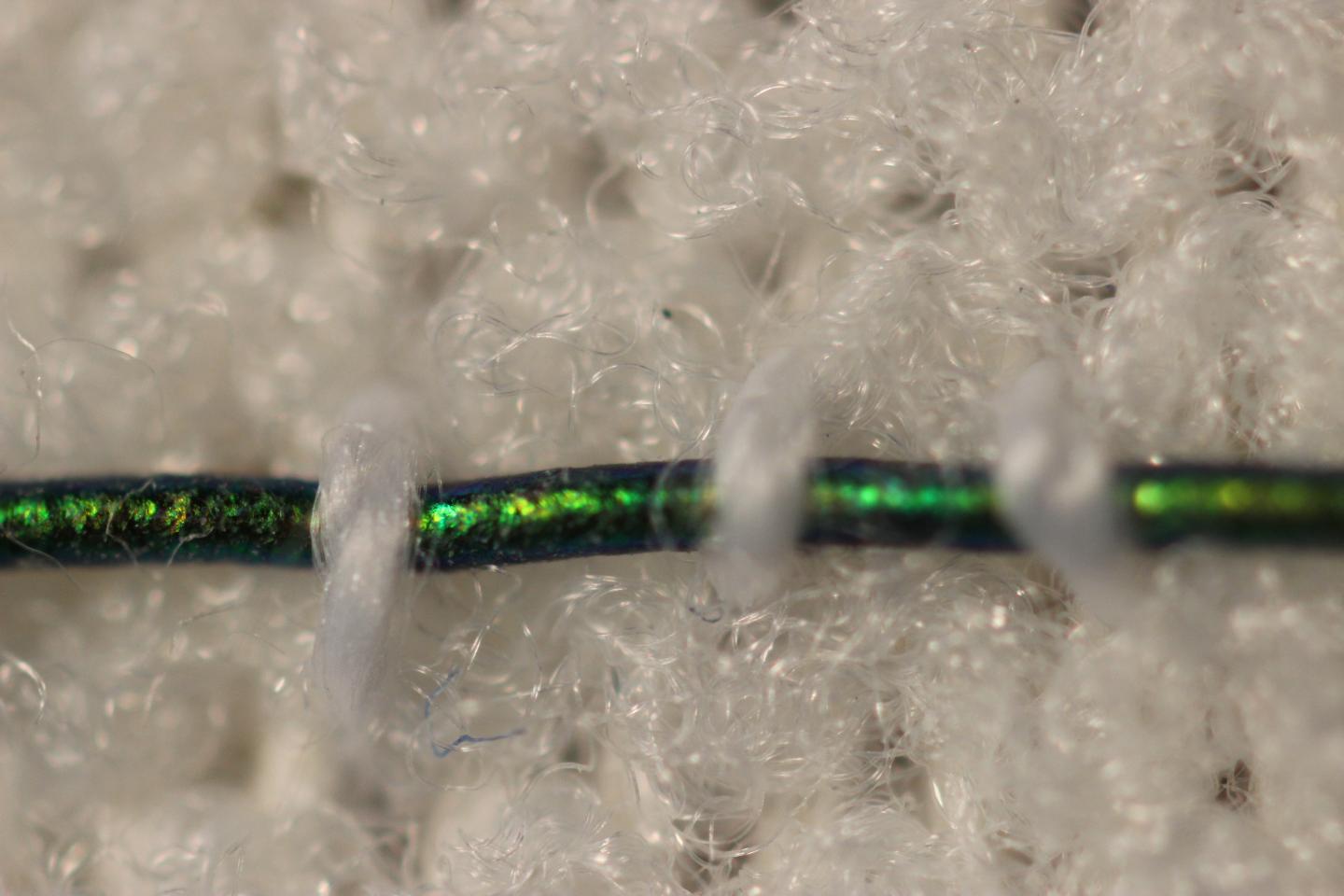
Now engineers at MIT {Massachusetts Institute of Technology] have developed pressure-sensing photonic fibers that they have woven into a typical compression bandage. As the bandage is stretched, the fibers change color. Using a color chart, a caregiver can stretch a bandage until it matches the color for a desired pressure, before, say, wrapping it around a patient’s leg.

The photonic fibers can then serve as a continuous pressure sensor — if their color changes, caregivers or patients can use the color chart to determine whether and to what degree the bandage needs loosening or tightening.

A May 29, 2018 MIT news release (also on EurekAlert), which originated the news item, provides more detail,

“Getting the pressure right is critical in treating many medical conditions including venous ulcers, which affect several hundred thousand patients in the U.S. each year,” says Mathias Kolle, assistant professor of mechanical engineering at MIT. “These fibers can provide information about the pressure that the bandage exerts. We can design them so that for a specific desired pressure, the fibers reflect an easily distinguished color.”

Kolle and his colleagues have published their results in the journal Advanced Healthcare Materials. Co-authors from MIT include first author Joseph Sandt, Marie Moudio, and Christian Argenti, along with J. Kenji Clark of the Univeristy of Tokyo, James Hardin of the United States Air Force Research Laboratory, Matthew Carty of Brigham and Women’s Hospital-Harvard Medical School, and Jennifer Lewis of Harvard University.

Natural inspiration

The color of the photonic fibers arises not from any intrinsic pigmentation, but from their carefully designed structural configuration. Each fiber is about 10 times the diameter of a human hair. The researchers fabricated the fiber from ultrathin layers of transparent rubber materials, which they rolled up to create a jelly-roll-type structure. Each layer within the roll is only a few hundred nanometers thick.

In this rolled-up configuration, light reflects off each interface between individual layers. With enough layers of consistent thickness, these reflections interact to strengthen some colors in the visible spectrum, for instance red, while diminishing the brightness of other colors. This makes the fiber appear a certain color, depending on the thickness of the layers within the fiber.

“Structural color is really neat, because you can get brighter, stronger colors than with inks or dyes just by using particular arrangements of transparent materials,” Sandt says. “These colors persist as long as the structure is maintained.”

The fibers’ design relies upon an optical phenomenon known as “interference,” in which light, reflected from a periodic stack of thin, transparent layers, can produce vibrant colors that depend on the stack’s geometric parameters and material composition. Optical interference is what produces colorful swirls in oily puddles and soap bubbles. It’s also what gives peacocks and butterflies their dazzling, shifting shades, as their feathers and wings are made from similarly periodic structures.

“My interest has always been in taking interesting structural elements that lie at the origin of nature’s most dazzling light manipulation strategies, to try recreating and employing them in useful applications,” Kolle says.

A multilayered approach

The team’s approach combines known optical design concepts with soft materials, to create dynamic photonic materials.

While a postdoc at Harvard in the group of Professor Joanna Aizenberg, Kolle was inspired by the work of Pete Vukusic, professor of biophotonics at the University of Exeter in the U.K., on Margaritaria nobilis, a tropical plant that produces extremely shiny blue berries. The fruits’ skin is made up of cells with a periodic cellulose structure, through which light can reflect to give the fruit its signature metallic blue color.

Together, Kolle and Vukusic sought ways to translate the fruit’s photonic architecture into a useful synthetic material. Ultimately, they fashioned multilayered fibers from stretchable materials, and assumed that stretching the fibers would change the individual layers’ thicknesses, enabling them to tune the fibers’ color. The results of these first efforts were published in Advanced Materials in 2013.

When Kolle joined the MIT faculty in the same year, he and his group, including Sandt, improved on the photonic fiber’s design and fabrication. In their current form, the fibers are made from layers of commonly used and widely available transparent rubbers, wrapped around highly stretchable fiber cores. Sandt fabricated each layer using spin-coating, a technique in which a rubber, dissolved into solution, is poured onto a spinning wheel. Excess material is flung off the wheel, leaving a thin, uniform coating, the thickness of which can be determined by the wheel’s speed.

For fiber fabrication, Sandt formed these two layers on top of a water-soluble film on a silicon wafer. He then submerged the wafer, with all three layers, in water to dissolve the water-soluble layer, leaving the two rubbery layers floating on the water’s surface. Finally, he carefully rolled the two transparent layers around a black rubber fiber, to produce the final colorful photonic fiber.

Reflecting pressure

The team can tune the thickness of the fibers’ layers to produce any desired color tuning, using standard optical modeling approaches customized for their fiber design.

“If you want a fiber to go from yellow to green, or blue, we can say, ‘This is how we have to lay out the fiber to give us this kind of [color] trajectory,'” Kolle says. “This is powerful because you might want to have something that reflects red to show a dangerously high strain, or green for ‘ok.’ We have that capacity.”

The team fabricated color-changing fibers with a tailored, strain-dependent color variation using the theoretical model, and then stitched them along the length of a conventional compression bandage, which they previously characterized to determine the pressure that the bandage generates when it’s stretched by a certain amount.

The team used the relationship between bandage stretch and pressure, and the correlation between fiber color and strain, to draw up a color chart, matching a fiber’s color (produced by a certain amount of stretching) to the pressure that is generated by the bandage.

To test the bandage’s effectiveness, Sandt and Moudio enlisted over a dozen student volunteers, who worked in pairs to apply three different compression bandages to each other’s legs: a plain bandage, a bandage threaded with photonic fibers, and a commercially-available bandage printed with rectangular patterns. This bandage is designed so that when it is applying an optimal pressure, users should see that the rectangles become squares.

Overall, the bandage woven with photonic fibers gave the clearest pressure feedback. Students were able to interpret the color of the fibers, and based on the color chart, apply a corresponding optimal pressure more accurately than either of the other bandages.

The researchers are now looking for ways to scale up the fiber fabrication process. Currently, they are able to make fibers that are several inches long. Ideally, they would like to produce meters or even kilometers of such fibers at a time.

“Currently, the fibers are costly, mostly because of the labor that goes into making them,” Kolle says. “The materials themselves are not worth much. If we could reel out kilometers of these fibers with relatively little work, then they would be dirt cheap.”

Then, such fibers could be threaded into bandages, along with textiles such as athletic apparel and shoes as color indicators for, say, muscle strain during workouts. Kolle envisions that they may also be used as remotely readable strain gauges for infrastructure and machinery.

“Of course, they could also be a scientific tool that could be used in a broader context, which we want to explore,” Kolle says.

Here’s what the bandage looks like,

Caption: Engineers at MIT have developed pressure-sensing photonic fibers that they have woven into a typical compression bandage. Credit Courtesy of the researchers

Here’s a link to and a citation for the paper,

Stretchable Optomechanical Fiber Sensors for Pressure Determination in Compressive Medical Textiles by Joseph D. Sandt, Marie Moudio, J. Kenji Clark, James Hardin, Christian Argenti, Matthew Carty, Jennifer A. Lewis, Mathias Kolle. Advanced Healthcare Materials https://doi.org/10.1002/adhm.201800293 First published: 29 May 2018

This paper is behind a paywall.

ArtSci Salon visits the Society for Literature, Science & the Arts 2018 Meeting in Toronto (Canada) while Vancouver’s Curiosity Collider provides a November 2018 update

I have two art/sci (or sciart) announcements, one concerns an event and the other is a news update.

Toronto’s ArtSci Salon and the Society of Literature, Science & the Arts (SLSA) 2018 Meeting

How could I not have stumbled across SLSA until now? Better late than never but the 2018 meeting/conference in Toronto, Canada is the 32nd of this annual event. (sigh)

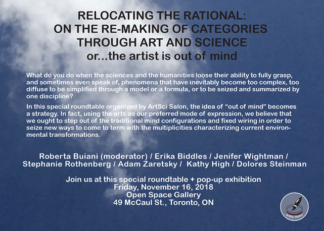
Getting on to the important points, the ArtSci Salon is hosting a special roundtable as part of the conference (from a November 14, 2018 announcement received via email),

ArtSci Salon has organized a special roundtable at the annual SLSA
(Society for Science Literature and the Arts) which will take place in
Toronto this week.

The roundtable is public and will be held at OCADU [Ontario College of Art and Design University] in the gallery on 49 McCaul Street.

Re-locating the rational: on the re-making of categories through art and science (or: the artist is out of mind!)

A roundtable and a mobile/pop-up exhibition organized by ArtSci Salon

The world always exceeds our conception of it (Shotwell, 2016)

Coinciding with this year’s SLSA “Out of Mind” conference in Toronto, ArtSci Salon is proposing a panel/roundtable on “out-of-mindedness” as a way to re-think categories, and to disrupt the disciplinary and methodological status quo through which we normally see science and the humanities unfolding in academic contexts. We plan to do it through a pop-up exhibition featuring the works of local artists and members of SLSA.

What to do when the sciences and the humanities loose [sic] their ability to fully grasp, and sometimes even speak of, phenomena that have inevitably become too complex, too diffuse to be simplified through a model or a formula, or to be seized and summarized by one discipline?

This initiative is not designed to propose a set of new categories, but to pose a series of open questions, highlighting the necessity to conduct collaborative research between artistic practices and scientific research. We interpret the idea of “out of mind” as a strategy. In fact, using the arts as our preferred mode of expression, we believe that we ought to step out of the traditional mind configurations and fixed wiring in order to seize new ways to come to term with the multiplicities characterizing current environmental transformations. These occurrences have proved to be connected with nature, culture, and society in too many intricate ways, to the extent that neither science, nor technological methods are able to fully comprehend them.

Roundtable Participants:

Roberta Buiani (Chair)

Erika Biddles

Jenifer Wightman

Stephanie Rothenberg

Adam Zaretsky

Kathy High

Dolores Steinman

Here’s the poster:

One more logistical detail,

[T]he roundtable will be at 10:30-12:00 noon [Friday, November 16, 2018] followed by a small tour of the mobile pop-up exhibition[.]

For the curious, here’s the SLSA website and the SLSA 2018 [Meeting]—Out of Your Mind website. Unexpectedly, the University of Toronto is not one of the conference hosts, instead we have the University of Waterloo [Waterloo, Ontario] and York University [Toronto, Ontario] as joint hosts with OCAD University—Canada’s oldest art and design institution—partnering with the Rochester Institute of Technology (New York state, US).

Vancouver’s Curiosity Collider

Coincidentally on the same day I received the ArtSci Salon event information, I received a November 14, 2018 update for Vancouver’s art/sci (or sciart) organization, Curiosity Collider. From the update received via email,

Collider Update

Next events (save-the-date), call for submissions, and other art+science in town

Collisions Festival:
Meet Up & Hang Out

Are you an artist working in the sci/art genre? A scientist interested in collaborating with artists? Or one who wears both hats?

In the fall of 2019, the Curiosity Collider will be hosting our inaugural Sci-Art festival The Collisions Festival; the first theme will be Invasive Systems. The call for submission will be open in spring, 2019. The theme is meant to be broad in scope and not limited to any specific scientific subject/discipline; participants are encouraged to suggest various interpretation of the theme.

We would like to invite all artists and scientists who are interested in participating or potentially submitting a proposal to join us at this meet up event, chat about possible collaborations, and learn more about projects and details on “collaborative work” proposals we are looking for.

RSVP now so we know how many to expect.. This is a casual drop in event; feel free to stay, or just stop by and say hi!

Notice that RSVP? Taken with the next announcement, something becomes evident,

Join the Collider Team!

Are you passionate about art and science? Want to be part of the awesome Curiosity Collider team to help create new ways to experience science? 

We are now inviting applications for the following positions:

Read more on our volunteer page. Feel free to contact us if you have any questions!

In the old days a ‘development director’ was a ‘fundraiser’. That RSVP? Likely, they’re trying to establish the size of their potential audience so they can get government grants. Audience size is important to corporate or ‘other’ funders but if you want a government grant you need numbers.

Getting back to the update, this is a grouping of Curiosity Collider’s latest hits,

#ColliderCafe: Art. Science. Cadence.

Did you miss our most recent Collider Cafe event? You can now chek out the talks by Singer-songwriter Devon More, Biologists Wayne Maddison and David Maddison, as well as Integrated Media Artist Victoria Gibson on our YouTube Channel.

Check out the talks now.

Et al 3: Collaboration Process for Quantum Futures

Nerd Nite, Science Slam, and Curiosity Collider joined forces for the 3rd edition of Et al: the ultimate bar science night event. During the event, Quantum Physicist James Day and our Creative Managing Director Char Hoyt gave attendees an overview of the collaboration process that made Night shift: Quantum Futures, an event curated by CC and hosted at the Museum of Anthropology, possible.

Missed the show? Watch the presentation on our YouTube channel now.

While they don’t seem to have any details, there is a date for the next Collider Cafe,

Save the Date:
Next Collider Cafe

Our next Collider Cafe will be on Wednesday, January 23 at Cafe Deux Soleils. #ColliderCafe is a space for artists, scientists, makers, and anyone interested in art+science. Meet. Discover. Connect. Create.

Are you curious? Join us to explore how art and science intersect in the exploration of curiosity.

Finally, a miscellaneous listing of related events being held in Vancouver, mostly, this November,

Looking for more art+science in town?

  • November 17 (Victoria) Science Writers and Communicators of Canada is hosting a workshop on science writing in an age of reconciliation: What science writers can learn from indigenous community members about better representation and relationships. Only a few spots left! Register now.
  • November 15-18 CC friend Dzee Louise will open her studio during the East Side Cultural Crawl! Drop by at studio #5 just at the top of the stairs of the William Clark Building at 1310 William Street (on the corner of Clark).
  • November 21 Natural History (Paleoart) Illustrator Julius Csotonyi will present a public lecture at the Vancouver Public Library (Kits branch) on the mutually beneficial affair between science and art.
  • November 21 Our friends at Nerd Nite Vancouver is hosting another awesome event next week, including a presentation by artist Michael Markowsky who will talk about how he ends up “Painting on the Moon”. Get your tickets now!
  • Until December 15 Vancouver Biennale’s CURIOUS IMAGININGS continues…check out the exhibition that will “challenge us to explore the social impacts of emerging biotechnology and our ethical limits in an age where genetic engineering and digital technologies are already pushing the boundaries of humanity.”

For more Vancouver art+science events, visit the Curiosity Collider events calendar. Let us know about your art+science events by emailing info@curiositycollider.org.

I did write a preview (June 18, 2018) for the last event on the list, Curious Imaginings, which included some of the latest science on xenotransplantation and chimeras (i.e., examples of  emerging biotechnology). That’s all folks!

Wooden supercapacitors: a cellulose nanofibril story

A May 24, 2018 news item on Nanowerk announces a technique for making sustainable electrodes (Note: A link has been removed),

Carbon aerogels are ultralight, conductive materials, which are extensively investigated for applications in supercapacitor electrodes in electrical cars and cell phones. Chinese scientists have now found a way to make these electrodes sustainably. The aerogels can be obtained directly from cellulose nanofibrils, the abundant cell-wall material in wood, finds the study reported in the journal Angewandte Chemie (“Wood-Derived Ultrathin Carbon Nanofiber Aerogels”).

A May 24, 2018 Wiley Publications press release, which originated the news item, explains further,

Supercapacitors are capacitors that can take up and release a very large amount of energy in a very short time. Key requirements for supercapacitor electrodes are a large surface area and conductivity, combined with a simple production method. Another growing issue in supercapacitor production–mainly for smartphone and electric car technologies–is sustainability. However, sustainable and economical production of carbon aerogels as supercapacitor electrode materials is possible, propose Shu-Hong Yu and colleagues from the University of Science and Technology of China, Hefei, China.

Carbon aerogels are ultralight conductive materials with a very large surface area. They can be prepared by two production routes: the first and cheapest starts from mostly phenolic components and produces aerogels with improvable conductivity, while the second route is based on graphene- and carbon-nanotube precursors. The latter method delivers high-performance aerogels but is expensive and non-environmentally friendly. In their search for different precursors, Yu and colleagues have found an abundant, far less expensive, and sustainable source: wood pulp.

Well, not really wood pulp, but its major ingredient, nanocellulose. Plant cell walls are stabilized by fibrous nanocellulose, and this extractable material has very recently stimulated substantial research and technological development. It forms a highly porous, but very stable transparent network, and, with the help of a recent technique–oxidation with a radical scavenger called TEMPO–it forms a microporous hydrogel of highly oriented cellulose nanofibrils with a uniform width and length. As organic aerogels are produced from hydrogels by drying and pyrolysis, the authors attempted pyrolysis of supercritically or freeze-dried nanofibrillated cellulose hydrogel.

As it turns out, the method was not as straightforward as expected because ice crystal formation and insufficient dehydration hampered carbonization, according to the authors. Here, a trick helped. The scientists pyrolyzed the dried gel in the presence of the organic acid catalyst para-toluenesulfonic acid. The catalyst lowered the decomposition temperature and yielded a “mechanically stable and porous three-dimensional nanofibrous network” featuring a “large specific surface area and high electrical conductivity,” the authors reported.

The authors also demonstrated that their wood-derived carbon aerogel worked well as a binder-free electrode for supercapacitor applications. The material displayed electrochemical properties comparable to commercial electrodes. The method is an interesting and innovative way in which to fabricate sustainable materials suitable for use in high-performance electronic devices.

This is the first time I’ve seen work on wood-based nanocellulose from China. Cellulose according to its Wikipedia entry is: ” … the most abundant organic polymer on Earth.” For example, there’s more cellulose in cotton than there is wood. So, I find it interesting that in a country not known for its forests, nanocellulose (in this project anyway) is being derived from wood.

Here’s a link to and a citation for the paper,

Wood‐Derived Ultrathin Carbon Nanofiber Aerogels by Si‐Cheng Li, Bi‐Cheng Hu, Dr. Yan‐Wei Ding, Prof. Hai‐Wei Liang, Chao Li, Dr. Zi‐You Yu, Dr. Zhen‐Yu Wu, Prof. Wen‐Shuai Chen, Prof. Shu‐Hong Yu. Angewandt Chemie First published: 23 April 2018 DOI: https://doi.org/10.1002/anie.201802753

This paper is behind a paywall.

Electrode-filled elastic fiber for wearable electronics and robots

This work comes out of Switzerland. A May 25, 2018 École Polytechnique Fédérale de Lausanne (EPFL) press release (also on EurekAlert) announces their fibers,

EPFL scientists have found a fast and simple way to make super-elastic, multi-material, high-performance fibers. Their fibers have already been used as sensors on robotic fingers and in clothing. This breakthrough method opens the door to new kinds of smart textiles and medical implants.

It’s a whole new way of thinking about sensors. The tiny fibers developed at EPFL are made of elastomer and can incorporate materials like electrodes and nanocomposite polymers. The fibers can detect even the slightest pressure and strain and can withstand deformation of close to 500% before recovering their initial shape. All that makes them perfect for applications in smart clothing and prostheses, and for creating artificial nerves for robots.

The fibers were developed at EPFL’s Laboratory of Photonic Materials and Fiber Devices (FIMAP), headed by Fabien Sorin at the School of Engineering. The scientists came up with a fast and easy method for embedding different kinds of microstructures in super-elastic fibers. For instance, by adding electrodes at strategic locations, they turned the fibers into ultra-sensitive sensors. What’s more, their method can be used to produce hundreds of meters of fiber in a short amount of time. Their research has just been published in Advanced Materials.

Heat, then stretch
To make their fibers, the scientists used a thermal drawing process, which is the standard process for optical-fiber manufacturing. They started by creating a macroscopic preform with the various fiber components arranged in a carefully designed 3D pattern. They then heated the preform and stretched it out, like melted plastic, to make fibers of a few hundreds microns in diameter. And while this process stretched out the pattern of components lengthwise, it also contracted it crosswise, meaning the components’ relative positions stayed the same. The end result was a set of fibers with an extremely complicated microarchitecture and advanced properties.

Until now, thermal drawing could be used to make only rigid fibers. But Sorin and his team used it to make elastic fibers. With the help of a new criterion for selecting materials, they were able to identify some thermoplastic elastomers that have a high viscosity when heated. After the fibers are drawn, they can be stretched and deformed but they always return to their original shape.

Rigid materials like nanocomposite polymers, metals and thermoplastics can be introduced into the fibers, as well as liquid metals that can be easily deformed. “For instance, we can add three strings of electrodes at the top of the fibers and one at the bottom. Different electrodes will come into contact depending on how the pressure is applied to the fibers. This will cause the electrodes to transmit a signal, which can then be read to determine exactly what type of stress the fiber is exposed to – such as compression or shear stress, for example,” says Sorin.

Artificial nerves for robots

Working in association with Professor Dr. Oliver Brock (Robotics and Biology Laboratory, Technical University of Berlin), the scientists integrated their fibers into robotic fingers as artificial nerves. Whenever the fingers touch something, electrodes in the fibers transmit information about the robot’s tactile interaction with its environment. The research team also tested adding their fibers to large-mesh clothing to detect compression and stretching. “Our technology could be used to develop a touch keyboard that’s integrated directly into clothing, for instance” says Sorin.

The researchers see many other potential applications. Especially since the thermal drawing process can be easily tweaked for large-scale production. This is a real plus for the manufacturing sector. The textile sector has already expressed interest in the new technology, and patents have been filed.

There’s a video of the lead researcher discussing the work as he offers some visual aids,

Here’s a link to and a citation for the paper,

Superelastic Multimaterial Electronic and Photonic Fibers and Devices via Thermal Drawing by Yunpeng Qu, Tung Nguyen‐Dang, Alexis Gérald Page, Wei Yan, Tapajyoti Das Gupta, Gelu Marius Rotaru, René M. Rossi, Valentine Dominique Favrod, Nicola Bartolomei, Fabien Sorin. Advanced Materials First published: 25 May 2018 https://doi.org/10.1002/adma.201707251

This paper is behind a paywall.

Transparent graphene electrode technology and complex brain imaging

Michael Berger has written a May 24, 2018 Nanowerk Spotlight article about some of the latest research on transparent graphene electrode technology and the brain (Note: A link has been removed),

In new work, scientists from the labs of Kuzum [Duygu Kuzum, an Assistant Professor of Electrical and Computer Engineering at the University of California, San Diego {UCSD}] and Anna Devor report a transparent graphene microelectrode neural implant that eliminates light-induced artifacts to enable crosstalk-free integration of 2-photon microscopy, optogenetic stimulation, and cortical recordings in the same in vivo experiment. The new class of transparent brain implant is based on monolayer graphene. It offers a practical pathway to investigate neuronal activity over multiple spatial scales extending from single neurons to large neuronal populations.

Conventional metal-based microelectrodes cannot be used for simultaneous measurements of multiple optical and electrical parameters, which are essential for comprehensive investigation of brain function across spatio-temporal scales. Since they are opaque, they block the field of view of the microscopes and generate optical shadows impeding imaging.

More importantly, they cause light induced artifacts in electrical recordings, which can significantly interfere with neural signals. Transparent graphene electrode technology presented in this paper addresses these problems and allow seamless and crosstalk-free integration of optical and electrical sensing and manipulation technologies.

In their work, the scientists demonstrate that by careful design of key steps in the fabrication process for transparent graphene electrodes, the light-induced artifact problem can be mitigated and virtually artifact-free local field potential (LFP) recordings can be achieved within operating light intensities.

“Optical transparency of graphene enables seamless integration of imaging, optogenetic stimulation and electrical recording of brain activity in the same experiment with animal models,” Kuzum explains. “Different from conventional implants based on metal electrodes, graphene-based electrodes do not generate any electrical artifacts upon interacting with light used for imaging or optogenetics. That enables crosstalk free integration of three modalities: imaging, stimulation and recording to investigate brain activity over multiple spatial scales extending from single neurons to large populations of neurons in the same experiment.”

The team’s new fabrication process avoids any crack formation in the transfer process, resulting in a 95-100% yield for the electrode arrays. This fabrication quality is important for expanding this technology to high-density large area transparent arrays to monitor brain-scale cortical activity in large animal models or humans.

“Our technology is also well-suited for neurovascular and neurometabolic studies, providing a ‘gold standard’ neuronal correlate for optical measurements of vascular, hemodynamic, and metabolic activity,” Kuzum points out. “It will find application in multiple areas, advancing our understanding of how microscopic neural activity at the cellular scale translates into macroscopic activity of large neuron populations.”

“Combining optical techniques with electrical recordings using graphene electrodes will allow to connect the large body of neuroscience knowledge obtained from animal models to human studies mainly relying on electrophysiological recordings of brain-scale activity,” she adds.

Next steps for the team involve employing this technology to investigate coupling and information transfer between different brain regions.

This work is part of the US BRAIN (Brain Research through Advancing Innovative Neurotechnologies) initiative and there’s more than one team working with transparent graphene electrodes. John Hewitt in an Oct. 21, 2014 posting on ExtremeTech describes two other teams’ work (Note: Links have been removed),

The solution [to the problems with metal electrodes], now emerging from multiple labs throughout the universe is to build flexible, transparent electrode arrays from graphene. Two studies in the latest issue of Nature Communications, one from the University of Wisconsin-Madison and the other from Penn [University of Pennsylvania], describe how to build these devices.

The University of Wisconsin researchers are either a little bit smarter or just a little bit richer, because they published their work open access. It’s a no-brainer then that we will focus on their methods first, and also in more detail. To make the arrays, these guys first deposited the parylene (polymer) substrate on a silicon wafer, metalized it with gold, and then patterned it with an electron beam to create small contact pads. The magic was to then apply four stacked single-atom-thick graphene layers using a wet transfer technique. These layers were then protected with a silicon dioxide layer, another parylene layer, and finally molded into brain signal recording goodness with reactive ion etching.

PennTransparentelectrodeThe researchers went with four graphene layers because that provided optimal mechanical integrity and conductivity while maintaining sufficient transparency. They tested the device in opto-enhanced mice whose neurons expressed proteins that react to blue light. When they hit the neurons with a laser fired in through the implant, the protein channels opened and fired the cell beneath. The masterstroke that remained was then to successfully record the electrical signals from this firing, sit back, and wait for the Nobel prize office to call.

The Penn State group [Note: Every reearcher mentioned in the paper Hewitt linked to is from the University of Pennsylvania] in the  used a similar 16-spot electrode array (pictured above right), and proceeded — we presume — in much the same fashion. Their angle was to perform high-resolution optical imaging, in particular calcium imaging, right out through the transparent electrode arrays which simultaneously recorded in high-temporal-resolution signals. They did this in slices of the hippocampus where they could bring to bear the complex and multifarious hardware needed to perform confocal and two-photon microscopy. These latter techniques provide a boost in spatial resolution by zeroing in over narrow planes inside the specimen, and limiting the background by the requirement of two photons to generate an optical signal. We should mention that there are voltage sensitive dyes available, in addition to standard calcium dyes, which can almost record the fastest single spikes, but electrical recording still reigns supreme for speed.

What a mouse looks like with an optogenetics system plugged in

What a mouse looks like with an optogenetics system plugged in

One concern of both groups in making these kinds of simultaneous electro-optic measurements was the generation of light-induced artifacts in the electrical recordings. This potential complication, called the Becqueral photovoltaic effect, has been known to exist since it was first demonstrated back in 1839. When light hits a conventional metal electrode, a photoelectrochemical (or more simply, a photovoltaic) effect occurs. If present in these recordings, the different signals could be highly disambiguatable. The Penn researchers reported that they saw no significant artifact, while the Wisconsin researchers saw some small effects with their device. In particular, when compared with platinum electrodes put into the opposite side cortical hemisphere, the Wisconsin researchers found that the artifact from graphene was similar to that obtained from platinum electrodes.

Here’s a link to and a citation for the latest research from UCSD,

Deep 2-photon imaging and artifact-free optogenetics through transparent graphene microelectrode arrays by Martin Thunemann, Yichen Lu, Xin Liu, Kıvılcım Kılıç, Michèle Desjardins, Matthieu Vandenberghe, Sanaz Sadegh, Payam A. Saisan, Qun Cheng, Kimberly L. Weldy, Hongming Lyu, Srdjan Djurovic, Ole A. Andreassen, Anders M. Dale, Anna Devor, & Duygu Kuzum. Nature Communicationsvolume 9, Article number: 2035 (2018) doi:10.1038/s41467-018-04457-5 Published: 23 May 2018

This paper is open access.

You can find out more about the US BRAIN initiative here and if you’re curious, you can find out more about the project at UCSD here. Duygu Kuzum (now at UCSD) was at  the University of Pennsylvania in 2014 and participated in the work mentioned in Hewitt’s 2014 posting.

Creating cheap, small carbon nanotubes

The excitement fairly crackles off the video,

A May 24, 2018 news item on Nanowerk announces the research,

Imagine a box you plug into the wall that cleans your toxic air and pays you cash.

That’s essentially what Vanderbilt University researchers produced after discovering the blueprint for turning the carbon dioxide into carbon nanotubes with small diameters.

Carbon nanotubes are supermaterials that can be stronger than steel and more conductive than copper. The reason they’re not in every application from batteries to tires is that these amazing properties only show up in the tiniest nanotubes, which are extremely expensive. Not only did the Vanderbilt team show they can make these materials from carbon dioxide sucked from the air, but how to do this in a way that is much cheaper than any other method out there.

I’m not sure what ‘small’ means in this context. I’ve heard of long and short carbon nanotubes (CNTs) and also of single-walled, multi-walled, and double-walled CNTs. I wish there’d been an an explanation and measurements for ‘small diameter CNTs’. That nitpick aside, a May 23, 2018 Vanderbilt University news release by Heidi Hall adds a few more technical details,

These materials, which Assistant Professor of Mechanical Engineering Cary Pint calls “black gold,” could steer the conversation from the negative impact of emissions to how we can use them in future technology.

“One of the most exciting things about what we’ve done is use electrochemistry to pull apart carbon dioxide into elemental constituents of carbon and oxygen and stitch together, with nanometer precision, those carbon atoms into new forms of matter,” Pint said. “That opens the door to being able to generate really valuable products with carbon nanotubes.

“These could revolutionize the world.”

In a report published today in ACS [American Chemical Society] Applied Materials and Interfaces, Pint, interdisciplinary material science Ph.D. student Anna Douglas and their team describe how tiny nanoparticles 10,000 times smaller than a human hair can be produced from coatings on stainless steel surfaces. The key was making them small enough to be valuable.

“The cheapest carbon nanotubes on the market cost around $100-200 per kilogram,” Douglas said. “Our research advance demonstrates a pathway to synthesize carbon nanotubes better in quality than these materials with lower cost and using carbon dioxide captured from the air.”

But making small nanotubes is no small task. The research team showed that a process called Ostwald ripening — where the nanoparticles that grow the carbon nanotubes change in size to larger diameters — is a key contender against producing the infinitely more useful size. The team showed they could partially overcome this by tuning electrochemical parameters to minimize these pesky large nanoparticles.

side-by-side photos showing stainless steel plate becoming covered in carbon nanotubes (which look like lumps of ash or mud)
Small diameter carbon nanotubes grown on a stainless steel surface. (Pint Lab/Vanderbilt University)

This core technology led Pint and Douglas to co-found SkyNano LLC, a company focused on building upon the science of this process to scale up and commercialize products from these materials.

“What we’ve learned is the science that opens the door to now build some of the most valuable materials in our world, such as diamonds and single-walled carbon nanotubes, from carbon dioxide that we capture from air through our process,” Pint said.

Here’s a link to and a citation for the paper,

Toward Small-Diameter Carbon Nanotubes Synthesized from Captured Carbon Dioxide: Critical Role of Catalyst Coarsening by Anna Douglas, Rachel Carter, Mengya Li, and Cary L. Pint. ACS Appl. Mater. Interfaces, Article ASAP DOI: 10.1021/acsami.8b02834 Publication Date (Web): May 1, 2018

Copyright © 2018 American Chemical Society

This paper is behind a paywall.

Regarding the start-up, SkyNano, which Douglas and Pint have co-founded, it looks to be at a  very early stage.

Preserving art canvases (think Van Gogh, Picasso, Vermeer, and others) with nanomaterials

It has to be disconcerting to realize that your precious paintings are deteriorating day by day.  In a June 22, 2017 posting titled ‘Art masterpieces are turning into soap‘,

This piece of research has made a winding trek through the online science world. First it was featured in an April 20, 2017 American Chemical Society news release on EurekAlert,

A good art dealer can really clean up in today’s market, but not when some weird chemistry wreaks havoc on masterpieces [emphasis mine]. Art conservators started to notice microscopic pockmarks forming on the surfaces of treasured oil paintings that cause the images to look hazy. It turns out the marks are eruptions of paint caused, weirdly, by soap that forms via chemical reactions. Since you have no time to watch paint dry, we explain how paintings from Rembrandts to O’Keefes are threatened by their own compositions — and we don’t mean the imagery.

Here’s the video,


Now, for the latest: canavases are deteriorating too. A May 23, 2018 news item on Nanowerk announces the latest research on the ‘canvas issue’ (Note: A link has been removed),

Paintings by Vincent van Gogh, Pablo Picasso and Johannes Vermeer have been delighting art lovers for years. But it turns out that these works of art might be their own worst enemy — the canvases they were painted on can deteriorate over time.

In an effort to combat this aging process, one group is reporting in ACS Applied Nano Materials (“Combined Nanocellulose/Nanosilica Approach for Multiscale Consolidation of Painting Canvases”) that nanomaterials can provide multiple layers of reinforcement.

A May 23, 2018 American Chemical Society (ACS) news release (also on EurekAlert), which originated the news item,  expands on the theme,

One of the most important parts of a painting is the canvas, which is usually made from cellulose-based fibers. Over time, the canvas ages, resulting in discoloration, wrinkles, tears and moisture retention, all greatly affecting the artwork. To combat aging, painting conservators currently place a layer of adhesive and a lining on the back of a painting, but this treatment is invasive and difficult to reverse. In previous work, Romain Bordes and colleagues from Chalmers University of Technology, Sweden, investigated nanocellulose as a new way to strengthen painting canvases on their surfaces. In addition, together with Krzysztof Kolman, they showed that silica nanoparticles can strengthen individual paper and cotton fibers. So, they next wanted to combine these two methods to see if they could further strengthen aging canvas.

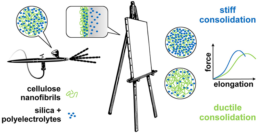
The team combined polyelectrolyte-treated silica nanoparticles (SNP) with cellulose nanofibrils (CNF) for a one-step treatment. The researchers first treated canvases with acid and oxidizing conditions to simulate aging. When they applied the SNP-CNF treatment, the SNP penetrated and strengthened the individual fibers of the canvas, making it stiffer compared to untreated materials. The CNF strengthened the surface of the canvas and increased the canvas’s flexibility. The team notes that this treatment could be a good alternative to conventional methods.

Here’s a link to and a citation for the paper,

Combined Nanocellulose/Nanosilica Approach for Multiscale Consolidation of Painting Canvases by Krzysztof Kolman, Oleksandr Nechyporchuk, Michael Persson, Krister Holmberg, and Romain Bordes. ACS Appl. Nano Mater., Article ASAP DOI: 10.1021/acsanm.8b00262 Publication Date (Web): April 26, 2018

Copyright © 2018 American Chemical Society

This image illustrating the researchers’ solution accompanies the article,

Courtesy: ACS

The European Union’s NanoRestART project was mentioned here before they’d put together this introductory video, which provides a good overview of the research,

For more details about the problems with contemporary and modern art, there’s my April 4, 2016 posting when the NanoRestART project was first mentioned here and there’s my Jan. 10, 2017 posting which details research into 3D-printed art and some of the questions raised by the use of 3D printing and other emerging technologies in the field of contemporary art.

ARPICO November 13, 2018 event in Vancouver (Canada): The Mysterious Dark-Side of the Universe: From Quarks to the Big Bang with Dark Matter

The Society of Italian Researchers and Professionals in Western Canada (ARPICO) is hosting a physics event for those of us who don’t have Phd’s in physics. From an October 24, 2018 ARPICO announcement (received via email),

The second event of ARPICO’s fall 2018 activity will take place on Tuesday, November 13th, 2018 at the Roundhouse Community Centre (Room B). Our speaker will be Dr. Pietro Giampa, a physicist who recently joined the ranks of the TRIUMF laboratories [Canada’s particle accelerator centre and, formerly, Canada’s National Laboratory for Particle and Nuclear Physics] here in Vancouver. Dr. Giampa will give us an intriguing and, importantly, layperson-intelligible overview on the state of our knowledge of the universe especially in regards to so-called dark matter, a chapter of physics that the most complete theoretical model to-date cannot explain. We will learn, among other things, about an ambitious experiment (set up in a Canadian mine!) [emphasis mine] to detect neutrinos, fundamental and very elusive particles of our  cosmos. You can read a summary of Pietro Giampa’s lecture as well as his short professional biography below.

We look forward to seeing everyone there.

The evening agenda is as follows:

  • 6:30 pm – Doors Open for Registration
  • 7:00 pm – Start of the evening event with introductions & lecture by Dr. Pietro Giampa
  • ~8:15 pm – Q & A Period
  • to follow – Mingling & Refreshments until about 9:30 pm

If you have not already done so, please register for the event by visiting the EventBrite link or RSVPing to info@arpico.ca.

Further details are also available at arpico.ca and Eventbrite.

More details from the email announcement,

The Mysterious Dark-Side of the Universe: From Quarks to the Big Bang with Dark Matter

Understanding the true nature of our universe is one of the most fundamental quests of our society. The path of knowledge acquisition in that quest has led us to the hypothesis of “dark matter”, that is, a large proportion of the mass of the universe which appears invisible. In this lecture, with minimal technical language we will journey through the structure and evolution of the universe, from subatomic particles to the big bang, which gave rise to our universe, in an ultimate research to describe the dark side of the universe called dark matter. We will review what we have learnt thus far about dark matter, and get an in-depth look at how scientists are searching for something that can not be seen.

Dr. Pietro Giampa originally completed his undergraduate in physics at Royal Holloway University of London in the UK, where he wrote a thesis on SuperSymmetry Searches with the ATLAS Detector (so LHC related). Following his undergraduate, he completed a Master Degree in particle physics at the same institute where he developed a novel technique for directional detection of neutrons. It was after his master that he moved to Canada to complete his Ph.D at Queen’s University in Particle Astrophysics, working on the DEAP-3600 Experiment with Nobel laureate Prof. Arthur McDonald. In the summer of 2017 he moved to TRIUMF, where he is currently the Otto Hausser Fellow. At TRIUMF he continues his research for new forms of physics, by studying Dark Matter and Ultra-Cold Neutrons.


WHEN: Tuesday, November 13th, 2018 at 7:00pm (doors open at 6:30pm)

WHERE: Roundhouse Community Centre, Room B – 181 Roundhouse Mews, Vancouver, BC, V6Z 2W3

RSVP: Please RSVP at EventBrite (https://mysteryofdarkmatter.eventbrite.ca/) or email info@arpico.ca


Tickets are Needed

  • Tickets are FREE, but all individuals are requested to obtain “free-admission” tickets on EventBrite site due to limited seating at the venue. Organizers need accurate registration numbers to manage wait lists and prepare name tags.
  • All ARPICO events are 100% staffed by volunteer organizers and helpers, however, room rental, stationery, and guest refreshments are costs incurred and underwritten by members of ARPICO. Therefore to be fair, all audience participants are asked to donate to the best of their ability at the door or via EventBrite to “help” defray costs of the event.

FAQs

  • Where can I contact the organizer with any questions? info@arpico.ca
  • Do I have to bring my printed ticket to the event? No, you do not. Your name will be on our Registration List at the Check-in Desk.
  • Is my registration/ticket transferrable? If you are unable to attend, another person may use your ticket. Please send us an email at info@arpico.ca of this substitution to correct our audience Registration List and to prepare guest name tags.
  • Can I update my registration information? Yes. If you have any questions, contact us at info@arpico.ca
  • I am having trouble using EventBrite and cannot reserve my ticket(s). Can someone at ARPICO help me with my ticket reservation? Of course, simply send your ticket request to us at info@arpico.ca so we help you.

What are my transport/parking options?

  • Bus/Train: The Canada Line Yaletown Skytrain station is a 1 minute walk from the Roundhouse Community Centre.
  • Parking: Pay Parking is underground at the community centre.  Access is available via Drake Street.

With regard to the Canadian mine and neutrino experiments, I hunted down a little more information (from an October 6, 2015 article by Kate Allen for thestar.com), Note: A link has been removed,

Canadian physicist Arthur B. McDonald has won the Nobel Prize for discoveries about the behaviour of a mysterious solar particle, teased from an experiment buried two kilometres below Sudbury [Ontario].

The Queen’s University professor emeritus was honoured for co-discovering that elusive particles known as neutrinos can change their identity — or “oscillate” — as they travel from the sun. It proved that neutrinos must have mass, a finding that upset the Standard Model of particle physics and opened new avenues for research into the fundamental properties of the universe.

McDonald, 72, shares the prize with Takaaki Kajita, whose Japanese collaboration made the same discovery with slightly different methods.

To measure solar neutrinos, McDonald and a 130-person international team built a massive detector in an operational copper mine southwest of Sudbury. …

To solve this problem, McDonald and his colleagues dreamt up SNO. Deep in an INCO mine (now owned by Vale), protected from cosmic radiation constantly bombarding the earth’s surface, the scientists installed a 12-metre-wide acrylic vessel filled with 1,000 tonnes of ultra-pure heavy water. The vessel was surrounded by a geodesic sphere equipped with 9,456 light sensors. The whole thing was sunk in a 34-metre-high cavity filled with regular water.

When neutrinos hit the heavy water, an event that occurred about 10 times a day, they emitted a flash of light, which researchers could analyze to measure the particles’ properties.

Allen’s article has more details for anyone who might want to read up on neutrinos. Regardless, I’m sure Dr.Giampa is fully prepared to guide the uninitiated into the mysteries of the universe as they pertain to dark matter, neutrinos, and ultra-cold neutrons.