Tag Archives: 3D printing

3D-printed graphene sensors for highly sensitive food freshness detection

I love the opening line (lede). From a June 29, 2020 news item on Nanowerk,

Researchers dipped their new, printed sensors into tuna broth and watched the readings.

It turned out the sensors – printed with high-resolution aerosol jet printers on a flexible polymer film and tuned to test for histamine, an allergen and indicator of spoiled fish and meat – can detect histamine down to 3.41 parts per million.

The U.S. Food and Drug Administration has set histamine guidelines of 50 parts per million in fish, making the sensors more than sensitive enough to track food freshness and safety.

I find using 3D-printing techniques to produce graphene, a 2-d material, intriguing. Apparently, the technique is cheaper and offers an advantage as it allows for greater precision than other techniques (inkjet printing, chemical vapour depostion [CVD], etc.)

Here’s more detail from a June 25, 2020 Iowa State University news release (also on EurekAlert but published June 29, 2020), which originated the news item,

Making the sensor technology possible is graphene, a supermaterial that’s a carbon honeycomb just an atom thick and known for its strength, electrical conductivity, flexibility and biocompatibility. Making graphene practical on a disposable food-safety sensor is a low-cost, aerosol-jet-printing technology that’s precise enough to create the high-resolution electrodes necessary for electrochemical sensors to detect small molecules such as histamine.

“This fine resolution is important,” said Jonathan Claussen, an associate professor of mechanical engineering at Iowa State University and one of the leaders of the research project. “The closer we can print these electrode fingers, in general, the higher the sensitivity of these biosensors.”

Claussen and the other project leaders – Carmen Gomes, an associate professor of mechanical engineering at Iowa State; and Mark Hersam, the Walter P. Murphy Professor of Materials Science and Engineering at Northwestern University in Evanston, Illinois – have recently reported their sensor discovery in a paper published online by the journal 2D Materials. (…)

The paper describes how graphene electrodes were aerosol jet printed on a flexible polymer and then converted to histamine sensors by chemically binding histamine antibodies to the graphene. The antibodies specifically bind histamine molecules.

The histamine blocks electron transfer and increases electrical resistance, Gomes said. That change in resistance can be measured and recorded by the sensor.

“This histamine sensor is not only for fish,” Gomes said. “Bacteria in food produce histamine. So it can be a good indicator of the shelf life of food.”

The researchers believe the concept will work to detect other kinds of molecules, too.

“Beyond the histamine case study presented here, the (aerosol jet printing) and functionalization process can likely be generalized to a diverse range of sensing applications including environmental toxin detection, foodborne pathogen detection, wearable health monitoring, and health diagnostics,” they wrote in their research paper.

For example, by switching the antibodies bonded to the printed sensors, they could detect salmonella bacteria, or cancers or animal diseases such as avian influenza, the researchers wrote.

Claussen, Hersam and other collaborators (…) have demonstrated broader application of the technology by modifying the aerosol-jet-printed sensors to detect cytokines, or markers of inflammation. The sensors, as reported in a recent paper published by ACS Applied Materials & Interfaces, can monitor immune system function in cattle and detect deadly and contagious paratuberculosis at early stages.

Claussen, who has been working with printed graphene for years, said the sensors have another characteristic that makes them very useful: They don’t cost a lot of money and can be scaled up for mass production.

“Any food sensor has to be really cheap,” Gomes said. “You have to test a lot of food samples and you can’t add a lot of cost.”

Claussen and Gomes know something about the food industry and how it tests for food safety. Claussen is chief scientific officer and Gomes is chief research officer for NanoSpy Inc., a startup company based in the Iowa State University Research Park that sells biosensors to food processing companies.

They said the company is in the process of licensing this new histamine and cytokine sensor technology.

It, after all, is what they’re looking for in a commercial sensor. “This,” Claussen said, “is a cheap, scalable, biosensor platform.”

Here’s a link to and a citation for the two papers mentioned in the news release,

Aerosol-jet-printed graphene electrochemical histamine sensors for food safety monitoring by Kshama Parate, Cícero C Pola, Sonal V Rangnekar, Deyny L Mendivelso-Perez, Emily A Smith, Mark C Hersam, Carmen L Gomes and Jonathan C Claussen. 2D Materials, Volume 7, Number 3 DOI https://doi.org/10.1088/2053-1583/ab8919 Published 10 June 2020 • © 2020 IOP Publishing Ltd

Aerosol-Jet-Printed Graphene Immunosensor for Label-Free Cytokine Monitoring in Serum by Kshama Parate, Sonal V. Rangnekar, Dapeng Jing, Deyny L. Mendivelso-Perez, Shaowei Ding, Ethan B. Secor, Emily A. Smith, Jesse M. Hostetter, Mark C. Hersam, and Jonathan C. Claussen. ACS Appl. Mater. Interfaces 2020, 12, 7, 8592–8603 DOI: https://doi.org/10.1021/acsami.9b22183 Publication Date: February 10, 2020 Copyright © 2020 American Chemical Society

Both papers are behind paywalls.

You can find the NanoSpy website here.

Nanocellulose films made with liquid-phase fabrication method

I always appreciate a reference to Star Trek and three-dimensional chess was one of my favourite concepts. You’ll find that and more in a May 19, 2020 news item on Nanowerk,

Researchers at The Institute of Scientific and Industrial Research at Osaka University [Japan] introduced a new liquid-phase fabrication method for producing nanocellulose films with multiple axes of alignment. Using 3D-printing methods for increased control, this work may lead to cheaper and more environmentally friendly optical and thermal devices.

Ever since appearing on the original Star Trek TV show in the 1960s, the game of “three-dimensional chess” has been used as a metaphor for sophisticated thinking. Now, researchers at Osaka University can say that they have added their own version, with potential applications in advanced optics and inexpensive smartphone displays.

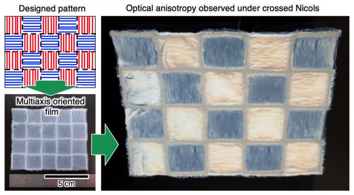
It’s not exactly three-dimensional chess but this nanocellulose film was produced by 3D printing methods,

Caption: Developed multiaxis nanocellulose-oriented film. Credit: Osaka University

A May 20, 2020 Osaka University press release (also on EurekAlert but dated May 19, 2020), which originated the news item, provides more detail,

Many existing optical devices, including liquid-crystal displays (LCDs) found in older flat-screen televisions, rely on long needle-shaped molecules aligned in the same direction. However, getting fibers to line up in multiple directions on the same device is much more difficult. Having a method that can reliably and cheaply produce optical fibers would accelerate the manufacture of low-cost displays or even “paper electronics”–computers that could be printed from biodegradable materials on demand.

Cellulose, the primary component of cotton and wood, is an abundant renewable resource made of long molecules. Nanocelluloses are nanofibers made of uniaxially aligned cellulose molecular chains that have different optical and heat conduction properties along one direction compared to the another.

In newly published research from the Institute of Scientific and Industrial Research at Osaka University, nanocellulose was harvested from sea pineapples, a kind of sea squirt. They then used liquid-phase 3D-pattering, which combined the wet spinning of nanofibers with the precision of 3D-printing. A custom-made triaxial robot dispensed a nanocellulose aqueous suspension into an acetone coagulation bath.

“We developed this liquid-phase three-dimensional patterning technique to allow for nanocellulose alignment along any preferred axis,” says first author Kojiro Uetani. The direction of the patterns could be programmed so that it formed an alternating checkerboard pattern of vertically- and horizontally-aligned fibers.

To demonstrate the method, a film was sandwiched between two orthogonal polarizing films. Under the proper viewing conditions, a birefringent checkerboard pattern appeared. They also measured the thermal transfer and optical retardation properties.

“Our findings could aid in the development of next-generation optical materials and paper electronics,” says senior author Masaya Nogi. “This could be the start of bottom-up techniques for building sophisticated and energy-efficient optical and thermal materials.”

Here’s a link to and a citation for the paper,

Checkered Films of Multiaxis Oriented Nanocelluloses by Liquid-Phase Three-Dimensional Patterning by Kojiro Uetani, Hirotaka Koga and Masaya Nogi. Nanomaterials 2020, 10(5), 958; DOI: https://doi.org/10.3390/nano10050958 Published: 18 May 2020

This is an open access paper.

Fourth Industrial Revolution and its impact on charity organizations

Andy Levy-Ajzenkopf’s February 21, 2020 article (Technology and innovation: How the Fourth Industrial Revolution is impacting the charitable sector) for Charity Village has an ebullient approach to adoption of new and emerging technologies in the charitable sector (Note: A link has been removed),

Almost daily, new technologies are being developed to help innovate the way people give or the way organizations offer opportunities to advance their causes. There is no going back.

The charitable sector – along with society at large – is now fully in the midst of what is being called the Fourth Industrial Revolution, a term first brought to prominence among CEOs, thought leaders and policy makers at the 2016 World Economic Forum. And if you haven’t heard the phrase yet, get ready to hear it tons more as economies around the world embrace it.

To be clear, the Fourth Industrial Revolution is the newest disruption in the way our world works. When you hear someone talk about it, what they’re describing is the massive technological shift in our business and personal ecosystems that now rely heavily on things like artificial intelligence, quantum computing, 3D printing and the general “Internet of things.”

Still, now more than ever, charitable business is getting done and being advanced by sector pioneers who aren’t afraid to make use of new technologies on offer to help civil society.

It seems like everywhere one turns, the topic of artificial intelligence (A.I.) is increasingly becoming subject of choice.

This is no different in the charitable sector, and particularly so for a new company called Fundraise Wisely (aka Wisely). Its co-founder and CEO, Artiom Komarov, explains a bit about what exactly his tech is doing for the sector.

“We help accelerate fundraising, with A.I. At a product level, we connect to your CRM (content relationship management system) and predict the next gift and next gift date for every donor. We then use that information to help you populate and prioritize donor portfolios,” Komarov states.

He notes that his company is seeing increased demand for innovative technologies from charities over the last while.

“What we’re hearing is that… A.I. tech is compelling because at the end of the day it’s meant to move the bottom line, helping nonprofits grow their revenue. We’ve also found that internally [at a charitable organization] there’s always a champion that sees the potential impact of technology; and that’s a great place to start with change,” Komarov says. “If it’s done right, tech can be an enabler of better work for organizations. From both research and experience, we know that tech adoption usually fails because of culture rather than the underlying technology. We’re here to work with the client closely to help that transition.”

I would like to have seen some numbers. For example, Komarov says that AI is having a positive impact on a charity’s bottom line. So, how much money did one of these charities raise? Was it more money than they would have made without AI? Assuming they did manage to raise greater funds, could another technology been more cost effective?

For another perspective (equally positive) on technology and charity, there’s a November 29, 2012 posting (Why technology and innovation are key to increasing charity donations) on the Guardian blogs by Henna Butt and Renita Shah (Note: Links have been removed),

At the beginning of this year the [UK] Cabinet Office and Nesta [formerly National Endowment for Science, Technology and the Arts {NESTA}] announced a £10m fund to invest in innovation in giving. The first tranche of this money has already been invested in promising initiatives such as Timto which allows you to create a gift list that includes a charity donation and Pennies, whose electronic money box allows customers to donate when paying for something in a shop using a credit card. Small and sizeable organisations alike are now using web and mobile technologies to make giving more convenient, more social and more compelling.

Butt’s and Shah’s focus was on mobile technologies and social networks. Like Levy-Ajzenkopf’s article, there’s no discussion of any possible downside to these technologies, e.g., privacy issues. As well, the inevitability of this move toward more technology for charity is explicitly stated by Levy-Ajzenkopf “There is no going back” and noted less starkly by Butt and Shah “… innovation is becoming increasingly important for the success of charities.” To rephrase my concern, are we utilizing technology in our work or are we serving the needs of our technology?

Finally, for anyone who’s curious about the Fourth Industrial Revolution, I have a December 3, 2015 posting about it.

Flexible graphene-rubber sensor for wearables

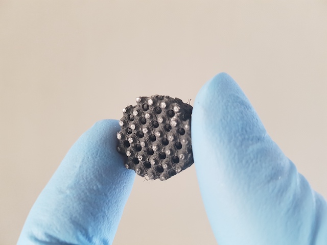
Courtesy: University of Waterloo

This waffled, greyish thing may not look like much but scientists are hopeful that it can be useful as a health sensor in athletic shoes and elsewhere. A March 6, 2020 news item on Nanowerk describes the work in more detail (Note: Links have been removed),

Researchers have utilized 3D printing and nanotechnology to create a durable, flexible sensor for wearable devices to monitor everything from vital signs to athletic performance (ACS Nano, “3D-Printed Ultra-Robust Surface-Doped Porous Silicone Sensors for Wearable Biomonitoring”).

The new technology, developed by engineers at the University of Waterloo [Ontario, Canada], combines silicone rubber with ultra-thin layers of graphene in a material ideal for making wristbands or insoles in running shoes.

A March 6, 2020 University of Waterloo news release, which originated the news item, delves further,

When that rubber material bends or moves, electrical signals are created by the highly conductive, nanoscale graphene embedded within its engineered honeycomb structure.

“Silicone gives us the flexibility and durability required for biomonitoring applications, and the added, embedded graphene makes it an effective sensor,” said Ehsan Toyserkani, research director at the Multi-Scale Additive Manufacturing (MSAM) Lab at Waterloo. “It’s all together in a single part.”

Fabricating a silicone rubber structure with such complex internal features is only possible using state-of-the-art 3D printing – also known as additive manufacturing – equipment and processes.

The rubber-graphene material is extremely flexible and durable in addition to highly conductive.

“It can be used in the harshest environments, in extreme temperatures and humidity,” said Elham Davoodi, an engineering PhD student at Waterloo who led the project. “It could even withstand being washed with your laundry.”

The material and the 3D printing process enable custom-made devices to precisely fit the body shapes of users, while also improving comfort compared to existing wearable devices and reducing manufacturing costs due to simplicity.

Toyserkani, a professor of mechanical and mechatronics engineering, said the rubber-graphene sensor can be paired with electronic components to make wearable devices that record heart and breathing rates, register the forces exerted when athletes run, allow doctors to remotely monitor patients and numerous other potential applications.

Researchers from the University of California, Los Angeles and the University of British Columbia collaborated on the project.

Here’s a link to and a citation for the paper,

3D-Printed Ultra-Robust Surface-Doped Porous Silicone Sensors for Wearable Biomonitoring by Elham Davoodi, Hossein Montazerian, Reihaneh Haghniaz, Armin Rashidi, Samad Ahadian, Amir Sheikhi, Jun Chen, Ali Khademhosseini, Abbas S. Milani, Mina Hoorfar, Ehsan Toyserkani. ACS Nano 2020, 14, 2, 1520-1532 DOI: https://doi.org/10.1021/acsnano.9b06283 Publication Date: January 6, 2020 Copyright © 2020 American Chemical Society

This paper is behind a paywall.

Nanocellulose sensors: 3D printed and biocompatible

I do like to keep up with nanocellulose doings, especially when there’s some Canadian involvement, and an October 8, 2019 news item on Nanowerk alerted me to a newish application for the product,

Physiological parameters in our blood can be determined without painful punctures. Empa researchers are currently working with a Canadian team to develop flexible, biocompatible nanocellulose sensors that can be attached to the skin. The 3D-printed analytic chips made of renewable raw materials will even be biodegradable in future.

The idea of measuring parameters that are relevant for our health via the skin has already taken hold in medical diagnostics. Diabetics, for example, can painlessly determine their blood sugar level with a sensor instead of having to prick their fingers.

An October 8, 2019 Empa (Swiss Federal Laboratories for Materials Science and Technology) press release, which originated the news item, provides more detail,

A transparent foil made of wood

Nanocellulose is an inexpensive, renewable raw material, which can be obtained in form of crystals and fibers, for example from wood. However, the original appearance of a tree no longer has anything to do with the gelatinous substance, which can consist of cellulose nanocrystals and cellulose nanofibers. Other sources of the material are bacteria, algae or residues from agricultural production. Thus, nanocellulose is not only relatively easy and sustainable to obtain. Its mechanical properties also make the “super pudding” an interesting product. For instance, new composite materials based on nanocellulose can be developed that could be used as surface coatings, transparent packaging films or even to produce everyday objects like beverage bottles.

Researchers at Empa’s Cellulose & Wood Materials lab and Woo Soo Kim from the Simon Fraser University [SFU] in Burnaby, Canada, are also focusing on another feature of nanocellulose: biocompatibility. Since the material is obtained from natural resources, it is particularly suitable for biomedical research.

With the aim of producing biocompatible sensors that can measure important metabolic values, the researchers used nanocellulose as an “ink” in 3D printing processes. To make the sensors electrically conductive, the ink was mixed with silver nanowires. The researchers determined the exact ratio of nanocellulose and silver threads so that a three-dimensional network could form.

Just like spaghetti – only a wee bit smaller

It turned out that cellulose nanofibers are better suited than cellulose nanocrystals to produce a cross-linked matrix with the tiny silver wires. “Cellulose nanofibers are flexible similar to cooked spaghetti, but with a diameter of only about 20 nanometers and a length of just a few micrometers,” explains Empa researcher Gilberto Siqueira.

The team finally succeeded in developing sensors that measure medically relevant metabolic parameters such as the concentration of calcium, potassium and ammonium ions. The electrochemical skin sensor sends its results wirelessly to a computer for further data processing. The tiny biochemistry lab on the skin is only half a millimeter thin.

While the tiny biochemistry lab on the skin – which is only half a millimeter thin – is capable of determining ion concentrations specifically and reliably, the researchers are already working on an updated version. “In the future, we want to replace the silver [nano] particles with another conductive material, for example on the basis of carbon compounds,” Siqueira explains. This would make the medical nanocellulose sensor not only biocompatible, but also completely biodegradable.

I like the images from Empa better than the ones from SFU,

Using a 3D printer, the nanocellulose “ink” is applied to a carrier plate. Silver particles provide the electrical conductivity of the material. Image: Empa
Empa researcher Gilberto Siqueira demonstrates the newly printed nanocellulose circuit. After a subsequent drying, the material can be further processed. Image: Empa

SFU produced a news release about this work back in February 2019. Again, I prefer what the Swiss have done because they’re explaining/communicating the science, as well as , communicating benefits. From a February 13, 2019 SFU news release (Note: Links have been removed),

Simon Fraser University and Swiss researchers are developing an eco-friendly, 3D printable solution for producing wireless Internet-of-Things (IoT) sensors that can be used and disposed of without contaminating the environment. Their research has been published as the cover story in the February issue of the journal Advanced Electronic Materials.

SFU professor Woo Soo Kim is leading the research team’s discovery, which uses a wood-derived cellulose material to replace the plastics and polymeric materials currently used in electronics.

Additionally, 3D printing can give flexibility to add or embed functions onto 3D shapes or textiles, creating greater functionality.

“Our eco-friendly, 3D-printed cellulose sensors can wirelessly transmit data during their life, and then can be disposed without concern of environmental contamination,” says Kim, a professor in the School of Mechatronic Systems Engineering. The SFU research is being carried out at PowerTech Labs in Surrey, which houses several state-of-the-art 3D printers used to advance the research.

“This development will help to advance green electronics. For example, the waste from printed circuit boards is a hazardous source of contamination to the environment. If we are able to change the plastics in PCB to cellulose composite materials, recycling of metal components on the board could be collected in a much easier way.”

Kim’s research program spans two international collaborative projects, including the latest focusing on the eco-friendly cellulose material-based chemical sensors with collaborators from the Swiss Federal Laboratories for Materials Science.

He is also collaborating with a team of South Korean researchers from the Daegu Gyeongbuk Institute of Science and Technology’s (DGIST)’s department of Robotics Engineering, and PROTEM Co Inc, a technology-based company, for the development of printable conductive ink materials.

In this second project, researchers have developed a new breakthrough in the embossing process technology, one that can freely imprint fine circuit patterns on flexible polymer substrate, a necessary component of electronic products.

Embossing technology is applied for the mass imprinting of precise patterns at a low unit cost. However, Kim says it can only imprint circuit patterns that are imprinted beforehand on the pattern stamp, and the entire, costly stamp must be changed to put in different patterns.

The team succeeded in developing a precise location control system that can imprint patterns directly resulting in a new process technology. The result will have widespread implications for use in semiconductor processes, wearable devices and the display industry.

This paper was made available online back in December 2018 and then published in print in February 2019. As to why there’d be such large gaps between the paper’s publication dates and the two institution’s news/press releases, it’s a mystery to me. In any event, here’s a link to and a citation for the paper,

3D Printed Disposable Wireless Ion Sensors with Biocompatible Cellulose Composites by Taeil Kim, Chao Bao, Michael Hausmann, Gilberto Siqueira, Tanja Zimmermann, Woo Soo Kim. Advanced Electronic Materials DOI: https://doi.org/10.1002/aelm.201970007 First published online December 19, 2018. First published in print: 08 February 2019 (Adv. Electron. Mater. 2/2109) Volume 5, Issue 2 February 2019 1970007

This paper is behind a paywall.

Nanocellulosic 3D-printed ears

It’s been a while since I’ve had a story abut cellulose nanocrystals (CNC) and this one comes from Switzerland’s Empa (Swiss Federal Laboratories for Materials Science and Technology) in a January 15, 2019 news item on Nanowerk (Note: A link has been removed),

Cellulose obtained from wood has amazing material properties. Empa researchers are now equipping the biodegradable material with additional functionalities to produce implants for cartilage diseases using 3D printing (ACS Nano, “Dynamics of Cellulose Nanocrystal Alignment during 3D Printing”).

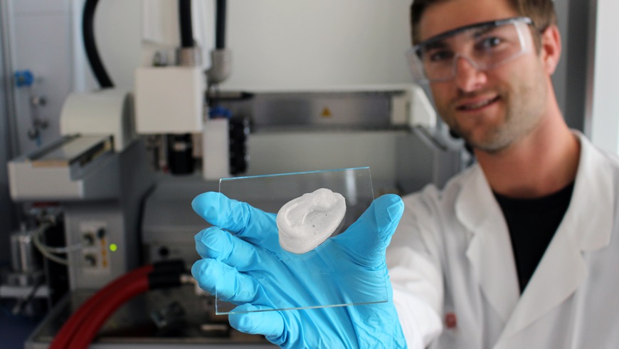
It all starts with an ear. Empa researcher Michael Hausmann removes the object shaped like a human ear from the 3D printer and explains: “In viscous state cellulose nanocrystals can easily be shaped together with nother biopolymers into complex 3-dimensional structures using a 3D printer, such as the Bioplotter.”

Once cross-linked, the structures remain stable despite their soft mechanical properties. Hausmann is currently investigating the characteristics of the nanocellulose composite hydrogels in order to further optimize their stability as well as the printing process. The researcher already used X-ray analysis to determine how cellulose is distributed and organized within the printed structures.

At this point in time the printed ear is entirely and solely made of cellulose nanocrystals and a biopolymer. However, the objective is to incorporate both human cells and therapeutics into the base structure in order to produce biomedical implants.

Here’s one of the researchers (Michael Hausmann) showing off their ‘ear’,

A 3D-printed ear: Empa researcher Michael Hausmann uses nanocellulose as the basis for novel implants (Image: Empa)

Doesn’t look like much does, eh? It’s scaffolding or, you could say, a kind of skeleton and a January 15, 2019 Empa press release, which originated the news item, describes it and explains how it will house new cells,

A new project is currently underway, looking into how chondrocytes (cartilage cells) can be integrated into the scaffold to yield artificial cartilage tissue. As soon as the colonization of the hydrogel with cells is established, nanocellulose based composites in the shape of an ear could serve as an implant for children with an inherited auricular malformation as for instance, in microtia, where the external ears are only incompletely developed. A reconstruction of the auricle can esthetically and medically correct the malformation; otherwise the hearing ability can be severely impaired. In the further course of the project, cellulose nanocrystals containing hydrogels will also be used for the replacement of articular cartilage (e.g. knee) in cases of joint wear due to, for example, chronic arthritis.

Once the artificial tissue has been implanted in the body, the biodegradable polymer material is expected to degrade over time. The cellulose itself is not degradable in the body, but biocompatible. However, it is not only its biocompatibility that makes nanocellulose the perfect material for implant scaffolds. “It is also the mechanical performance of cellulose nanocrystals that make them such promising candidates because the tiny but highly stable fibers can extremely well reinforce the produced implant,” said Hausmann.

Moreover, nanocellulose allows the incorporation of various functions by chemical modifications into the viscous hydrogel. Thus, the structure, the mechanical properties and the interactions of the nanocellulose with its environment can be specifically tailored to the desired end product. “For instance, we can incorporate active substances that promote the growth of chondrocytes or that sooth joint inflammation into the hydrogel,” says the Empa researcher.

And last but not least, as raw material cellulose is the most abundant natural polymer on earth. Therefore, the use of cellulose nanocrystals not only benefits from the mere elegance of the novel process but also from the availability of the raw material.

The white nanocellulose ear lies glossy on the glass carrier. Just out of the Bioplotter, it is already robust and dimensionally stable. Hausmann can give the go-ahead for the next steps. 

Here’s a link to and a citation for the paper,

Dynamics of Cellulose Nanocrystal Alignment during 3D Printing by Michael K. Hausmann, Patrick A. Rühs, Gilberto Siqueira, Jörg Läuger, Rafael Libanori, Tanja Zimmermann, and André R. Studart. ACS Nano, 2018, 12 (7), pp 6926–6937 DOI: 10.1021/acsnano.8b02366 Publication Date (Web): July 5, 2018

Copyright © 2018 American Chemical Society

This paper is behind a paywall.

Gene editing and personalized medicine: Canada

Back in the fall of 2018 I came across one of those overexcited pieces about personalized medicine and gene editing tha are out there. This one came from an unexpected source, an author who is a “PhD Scientist in Medical Science (Blood and Vasculature” (from Rick Gierczak’s LinkedIn profile).

It starts our promisingly enough although I’m beginning to dread the use of the word ‘precise’  where medicine is concerned, (from a September 17, 2018 posting on the Science Borealis blog by Rick Gierczak (Note: Links have been removed),

CRISPR-Cas9 technology was accidentally discovered in the 1980s when scientists were researching how bacteria defend themselves against viral infection. While studying bacterial DNA called clustered regularly interspaced short palindromic repeats (CRISPR), they identified additional CRISPR-associated (Cas) protein molecules. Together, CRISPR and one of those protein molecules, termed Cas9, can locate and cut precise regions of bacterial DNA. By 2012, researchers understood that the technology could be modified and used more generally to edit the DNA of any plant or animal. In 2015, the American Association for the Advancement of Science chose CRISPR-Cas9 as science’s “Breakthrough of the Year”.

Today, CRISPR-Cas9 is a powerful and precise gene-editing tool [emphasis mine] made of two molecules: a protein that cuts DNA (Cas9) and a custom-made length of RNA that works like a GPS for locating the exact spot that needs to be edited (CRISPR). Once inside the target cell nucleus, these two molecules begin editing the DNA. After the desired changes are made, they use a repair mechanism to stitch the new DNA into place. Cas9 never changes, but the CRISPR molecule must be tailored for each new target — a relatively easy process in the lab. However, it’s not perfect, and occasionally the wrong DNA is altered [emphasis mine].

Note that Gierczak makes a point of mentioning that CRISPR/Cas9 is “not perfect.” And then, he gets excited (Note: Links have been removed),

CRISPR-Cas9 has the potential to treat serious human diseases, many of which are caused by a single “letter” mutation in the genetic code (A, C, T, or G) that could be corrected by precise editing. [emphasis mine] Some companies are taking notice of the technology. A case in point is CRISPR Therapeutics, which recently developed a treatment for sickle cell disease, a blood disorder that causes a decrease in oxygen transport in the body. The therapy targets a special gene called fetal hemoglobin that’s switched off a few months after birth. Treatment involves removing stem cells from the patient’s bone marrow and editing the gene to turn it back on using CRISPR-Cas9. These new stem cells are returned to the patient ready to produce normal red blood cells. In this case, the risk of error is eliminated because the new cells are screened for the correct edit before use.

The breakthroughs shown by companies like CRISPR Therapeutics are evidence that personalized medicine has arrived. [emphasis mine] However, these discoveries will require government regulatory approval from the countries where the treatment is going to be used. In the US, the Food and Drug Administration (FDA) has developed new regulations allowing somatic (i.e., non-germ) cell editing and clinical trials to proceed. [emphasis mine]

The potential treatment for sickle cell disease is exciting but Gierczak offers no evidence that this treatment or any unnamed others constitute proof that “personalized medicine has arrived.” In fact, Goldman Sachs, a US-based investment bank, makes the case that it never will .

Cost/benefit analysis

Edward Abrahams, president of the Personalized Medicine Coalition (US-based), advocates for personalized medicine while noting in passing, market forces as represented by Goldman Sachs in his May 23, 2018 piece for statnews.com (Note: A link has been removed),

One of every four new drugs approved by the Food and Drug Administration over the last four years was designed to become a personalized (or “targeted”) therapy that zeros in on the subset of patients likely to respond positively to it. That’s a sea change from the way drugs were developed and marketed 10 years ago.

Some of these new treatments have extraordinarily high list prices. But focusing solely on the cost of these therapies rather than on the value they provide threatens the future of personalized medicine.

… most policymakers are not asking the right questions about the benefits of these treatments for patients and society. Influenced by cost concerns, they assume that prices for personalized tests and treatments cannot be justified even if they make the health system more efficient and effective by delivering superior, longer-lasting clinical outcomes and increasing the percentage of patients who benefit from prescribed treatments.

Goldman Sachs, for example, issued a report titled “The Genome Revolution.” It argues that while “genome medicine” offers “tremendous value for patients and society,” curing patients may not be “a sustainable business model.” [emphasis mine] The analysis underlines that the health system is not set up to reap the benefits of new scientific discoveries and technologies. Just as we are on the precipice of an era in which gene therapies, gene-editing, and immunotherapies promise to address the root causes of disease, Goldman Sachs says that these therapies have a “very different outlook with regard to recurring revenue versus chronic therapies.”

Let’s just chew on this one (contemplate)  for a minute”curing patients may not be ‘sustainable business model’!”

Coming down to earth: policy

While I find Gierczak to be over-enthused, he, like Abrahams, emphasizes the importance of new policy, in his case, the focus is Canadian policy. From Gierczak’s September 17, 2018 posting (Note: Links have been removed),

In Canada, companies need approval from Health Canada. But a 2004 law called the Assisted Human Reproduction Act (AHR Act) states that it’s a criminal offence “to alter the genome of a human cell, or in vitroembryo, that is capable of being transmitted to descendants”. The Actis so broadly written that Canadian scientists are prohibited from using the CRISPR-Cas9 technology on even somatic cells. Today, Canada is one of the few countries in the world where treating a disease with CRISPR-Cas9 is a crime.

On the other hand, some countries provide little regulatory oversight for editing either germ or somatic cells. In China, a company often only needs to satisfy the requirements of the local hospital where the treatment is being performed. And, if germ-cell editing goes wrong, there is little recourse for the future generations affected.

The AHR Act was introduced to regulate the use of reproductive technologies like in vitrofertilization and research related to cloning human embryos during the 1980s and 1990s. Today, we live in a time when medical science, and its role in Canadian society, is rapidly changing. CRISPR-Cas9 is a powerful tool, and there are aspects of the technology that aren’t well understood and could potentially put patients at risk if we move ahead too quickly. But the potential benefits are significant. Updated legislation that acknowledges both the risks and current realities of genomic engineering [emphasis mine] would relieve the current obstacles and support a path toward the introduction of safe new therapies.

Criminal ban on human gene-editing of inheritable cells (in Canada)

I had no idea there was a criminal ban on the practice until reading this January 2017 editorial by Bartha Maria Knoppers, Rosario Isasi, Timothy Caulfield, Erika Kleiderman, Patrick Bedford, Judy Illes, Ubaka Ogbogu, Vardit Ravitsky, & Michael Rudnicki for (Nature) npj Regenerative Medicine (Note: Links have been removed),

Driven by the rapid evolution of gene editing technologies, international policy is examining which regulatory models can address the ensuing scientific, socio-ethical and legal challenges for regenerative and personalised medicine.1 Emerging gene editing technologies, including the CRISPR/Cas9 2015 scientific breakthrough,2 are powerful, relatively inexpensive, accurate, and broadly accessible research tools.3 Moreover, they are being utilised throughout the world in a wide range of research initiatives with a clear eye on potential clinical applications. Considering the implications of human gene editing for selection, modification and enhancement, it is time to re-examine policy in Canada relevant to these important advances in the history of medicine and science, and the legislative and regulatory frameworks that govern them. Given the potential human reproductive applications of these technologies, careful consideration of these possibilities, as well as ethical and regulatory scrutiny must be a priority.4

With the advent of human embryonic stem cell research in 1978, the birth of Dolly (the cloned sheep) in 1996 and the Raelian cloning hoax in 2003, the environment surrounding the enactment of Canada’s 2004 Assisted Human Reproduction Act (AHRA) was the result of a decade of polarised debate,5 fuelled by dystopian and utopian visions for future applications. Rightly or not, this led to the AHRA prohibition on a wide range of activities, including the creation of embryos (s. 5(1)(b)) or chimeras (s. 5(1)(i)) for research and in vitro and in vivo germ line alterations (s. 5(1)(f)). Sanctions range from a fine (up to $500,000) to imprisonment (up to 10 years) (s. 60 AHRA).

In Canada, the criminal ban on gene editing appears clear, the Act states that “No person shall knowingly […] alter the genome of a cell of a human being or in vitro embryo such that the alteration is capable of being transmitted to descendants;” [emphases mine] (s. 5(1)(f) AHRA). This approach is not shared worldwide as other countries such as the United Kingdom, take a more regulatory approach to gene editing research.1 Indeed, as noted by the Law Reform Commission of Canada in 1982, criminal law should be ‘an instrument of last resort’ used solely for “conduct which is culpable, seriously harmful, and generally conceived of as deserving of punishment”.6 A criminal ban is a suboptimal policy tool for science as it is inflexible, stifles public debate, and hinders responsiveness to the evolving nature of science and societal attitudes.7 In contrast, a moratorium such as the self-imposed research moratorium on human germ line editing called for by scientists in December 20158 can at least allow for a time limited pause. But like bans, they may offer the illusion of finality and safety while halting research required to move forward and validate innovation.

On October 1st, 2016, Health Canada issued a Notice of Intent to develop regulations under the AHRA but this effort is limited to safety and payment issues (i.e. gamete donation). Today, there is a need for Canada to revisit the laws and policies that address the ethical, legal and social implications of human gene editing. The goal of such a critical move in Canada’s scientific and legal history would be a discussion of the right of Canadians to benefit from the advancement of science and its applications as promulgated in article 27 of the Universal Declaration of Human Rights9 and article 15(b) of the International Covenant on Economic, Social and Cultural Rights,10 which Canada has signed and ratified. Such an approach would further ensure the freedom of scientific endeavour both as a principle of a liberal democracy and as a social good, while allowing Canada to be engaged with the international scientific community.

Even though it’s a bit old, I still recommend reading the open access editorial in full, if you have the time.

One last thing abut the paper, the acknowledgements,

Sponsored by Canada’s Stem Cell Network, the Centre of Genomics and Policy of McGill University convened a ‘think tank’ on the future of human gene editing in Canada with legal and ethics experts as well as representatives and observers from government in Ottawa (August 31, 2016). The experts were Patrick Bedford, Janetta Bijl, Timothy Caulfield, Judy Illes, Rosario Isasi, Jonathan Kimmelman, Erika Kleiderman, Bartha Maria Knoppers, Eric Meslin, Cate Murray, Ubaka Ogbogu, Vardit Ravitsky, Michael Rudnicki, Stephen Strauss, Philip Welford, and Susan Zimmerman. The observers were Geneviève Dubois-Flynn, Danika Goosney, Peter Monette, Kyle Norrie, and Anthony Ridgway.

Competing interests

The authors declare no competing interests.

Both McGill and the Stem Cell Network pop up again. A November 8, 2017 article about the need for new Canadian gene-editing policies by Tom Blackwell for the National Post features some familiar names (Did someone have a budget for public relations and promotion?),

It’s one of the most exciting, and controversial, areas of health science today: new technology that can alter the genetic content of cells, potentially preventing inherited disease — or creating genetically enhanced humans.

But Canada is among the few countries in the world where working with the CRISPR gene-editing system on cells whose DNA can be passed down to future generations is a criminal offence, with penalties of up to 10 years in jail.

This week, one major science group announced it wants that changed, calling on the federal government to lift the prohibition and allow researchers to alter the genome of inheritable “germ” cells and embryos.

The potential of the technology is huge and the theoretical risks like eugenics or cloning are overplayed, argued a panel of the Stem Cell Network.

The step would be a “game-changer,” said Bartha Knoppers, a health-policy expert at McGill University, in a presentation to the annual Till & McCulloch Meetings of stem-cell and regenerative-medicine researchers [These meetings were originally known as the Stem Cell Network’s Annual General Meeting {AGM}]. [emphases mine]

“I’m completely against any modification of the human genome,” said the unidentified meeting attendee. “If you open this door, you won’t ever be able to close it again.”

If the ban is kept in place, however, Canadian scientists will fall further behind colleagues in other countries, say the experts behind the statement say; they argue possible abuses can be prevented with good ethical oversight.

“It’s a human-reproduction law, it was never meant to ban and slow down and restrict research,” said Vardit Ravitsky, a University of Montreal bioethicist who was part of the panel. “It’s a sort of historical accident … and now our hands are tied.”

There are fears, as well, that CRISPR could be used to create improved humans who are genetically programmed to have certain facial or other features, or that the editing could have harmful side effects. Regardless, none of it is happening in Canada, good or bad.

In fact, the Stem Cell Network panel is arguably skirting around the most contentious applications of the technology. It says it is asking the government merely to legalize research for its own sake on embryos and germ cells — those in eggs and sperm — not genetic editing of embryos used to actually get women pregnant.

The highlighted portions in the last two paragraphs of the excerpt were written one year prior to the claims by a Chinese scientist that he had run a clinical trial resulting in gene-edited twins, Lulu and Nana. (See my my November 28, 2018 posting for a comprehensive overview of the original furor). I have yet to publish a followup posting featuring the news that the CRISPR twins may have been ‘improved’ more extensively than originally realized. The initial reports about the twins focused on an illness-related reason (making them HIV ‘immune’) but made no mention of enhanced cognitive skills a side effect of eliminating the gene that would make them HIV ‘immune’. To date, the researcher has not made the bulk of his data available for an in-depth analysis to support his claim that he successfully gene-edited the twins. As well, there were apparently seven other pregnancies coming to term as part of the researcher’s clinical trial and there has been no news about those births.

Risk analysis innovation

Before moving onto the innovation of risk analysis, I want to focus a little more on at least one of the risks that gene-editing might present. Gierczak noted that CRISPR/Cas9 is “not perfect,” which acknowledges the truth but doesn’t convey all that much information.

While the terms ‘precision’ and ‘scissors’ are used frequently when describing the CRISPR technique, scientists actually mean that the technique is significantly ‘more precise’ than other techniques but they are not referencing an engineering level of precision. As for the ‘scissors’, it’s an analogy scientists like to use but in fact CRISPR is not as efficient and precise as a pair of scissors.

Michael Le Page in a July 16, 2018 article for New Scientist lays out some of the issues (Note: A link has been removed),

A study of CRIPSR suggests we shouldn’t rush into trying out CRISPR genome editing inside people’s bodies just yet. The technique can cause big deletions or rearrangements of DNA [emphasis mine], says Allan Bradley of the Wellcome Sanger Institute in the UK, meaning some therapies based on CRISPR may not be quite as safe as we thought.

The CRISPR genome editing technique is revolutionising biology, enabling us to create new varieties of plants and animals and develop treatments for a wide range of diseases.

The CRISPR Cas9 protein works by cutting the DNA of a cell in a specific place. When the cell repairs the damage, a few DNA letters get changed at this spot – an effect that can be exploited to disable genes.

At least, that’s how it is supposed to work. But in studies of mice and human cells, Bradley’s team has found that in around a fifth of cells, CRISPR causes deletions or rearrangements more than 100 DNA letters long. These surprising changes are sometimes thousands of letters long.

“I do believe the findings are robust,” says Gaetan Burgio of the Australian National University, an expert on CRISPR who has debunked previous studies questioning the method’s safety. “This is a well-performed study and fairly significant.”

I covered the Bradley paper and the concerns in a July 17, 2018 posting ‘The CRISPR ((clustered regularly interspaced short palindromic repeats)-CAS9 gene-editing technique may cause new genetic damage kerfuffle‘. (The ‘kerfufle’ was in reference to a report that the CRISPR market was affected by the publication of Bradley’s paper.)

Despite Health Canada not moving swiftly enough for some researchers, they have nonetheless managed to release an ‘outcome’ report about a consultation/analysis started in October 2016. Before getting to the consultation’s outcome, it’s interesting to look at how the consultation’s call for response was described (from Health Canada’s Toward a strengthened Assisted Human Reproduction Act ; A Consultation with Canadians on Key Policy Proposals webpage),

In October 2016, recognizing the need to strengthen the regulatory framework governing assisted human reproduction in Canada, Health Canada announced its intention to bring into force the dormant sections of the Assisted Human Reproduction Act  and to develop the necessary supporting regulations.

This consultation document provides an overview of the key policy proposals that will help inform the development of regulations to support bringing into force Section 10, Section 12 and Sections 45-58 of the Act. Specifically, the policy proposals describe the Department’s position on the following:

Section 10: Safety of Donor Sperm and Ova

  • Scope and application
  • Regulated parties and their regulatory obligations
  • Processing requirements, including donor suitability assessment
  • Record-keeping and traceability

Section 12: Reimbursement

  • Expenditures that may be reimbursed
  • Process for reimbursement
  • Creation and maintenance of records

Sections 45-58: Administration and Enforcement

  • Scope of the administration and enforcement framework
  • Role of inspectors designated under the Act

The purpose of the document is to provide Canadians with an opportunity to review the policy proposals and to provide feedback [emphasis mine] prior to the Department finalizing policy decisions and developing the regulations. In addition to requesting stakeholders’ general feedback on the policy proposals, the Department is also seeking input on specific questions, which are included throughout the document.

It took me a while to find the relevant section (in particular, take note of ‘Federal Regulatory Oversight’),

3.2. AHR in Canada Today

Today, an increasing number of Canadians are turning to AHR technologies to grow or build their families. A 2012 Canadian studyFootnote 1 found that infertility is on the rise in Canada, with roughly 16% of heterosexual couples experiencing infertility. In addition to rising infertility, the trend of delaying marriage and parenthood, scientific advances in cryopreserving ova, and the increasing use of AHR by LGBTQ2 couples and single parents to build a family are all contributing to an increase in the use of AHR technologies.

The growing use of reproductive technologies by Canadians to help build their families underscores the need to strengthen the AHR Act. While the approach to regulating AHR varies from country to country, Health Canada has considered international best practices and the need for regulatory alignment when developing the proposed policies set out in this document. …

3.2.1 Federal Regulatory Oversight

Although the scope of the AHR Act was significantly reduced in 2012 and some of the remaining sections have not yet been brought into force, there are many important sections of the Act that are currently administered and enforced by Health Canada, as summarized generally below:

Section 5: Prohibited Scientific and Research Procedures
Section 5 prohibits certain types of scientific research and clinical procedures that are deemed unacceptable, including: human cloning, the creation of an embryo for non-reproductive purposes, maintaining an embryo outside the human body beyond the fourteenth day, sex selection for non-medical reasons, altering the genome in a way that could be transmitted to descendants, and creating a chimera or a hybrid. [emphasis mine]

….

It almost seems as if the they were hiding the section that broached the human gene-editing question. It doesn’t seem to have worked as it appears, there are some very motivated parties determined to reframe the discussion. Health Canada’s ‘outocme’ report, published March 2019, What we heard: A summary of scanning and consultations on what’s next for health product regulation reflects the success of those efforts,

1.0 Introduction and Context

Scientific and technological advances are accelerating the pace of innovation. These advances are increasingly leading to the development of health products that are better able to predict, define, treat, and even cure human diseases. Globally, many factors are driving regulators to think about how to enable health innovation. To this end, Health Canada has been expanding beyond existing partnerships and engaging both domestically and internationally. This expanding landscape of products and services comes with a range of new challenges and opportunities.

In keeping up to date with emerging technologies and working collaboratively through strategic partnerships, Health Canada seeks to position itself as a regulator at the forefront of health innovation. Following the targeted sectoral review of the Health and Biosciences Sector Regulatory Review consultation by the Treasury Board Secretariat, Health Canada held a number of targeted meetings with a broad range of stakeholders.

This report outlines the methodologies used to look ahead at the emerging health technology environment, [emphasis mine] the potential areas of focus that resulted, and the key findings from consultations.

… the Department identified the following key drivers that are expected to shape the future of health innovation:

  1. The use of “big data” to inform decision-making: Health systems are generating more data, and becoming reliant on this data. The increasing accuracy, types, and volume of data available in real time enable automation and machine learning that can forecast activity, behaviour, or trends to support decision-making.
  2. Greater demand for citizen agency: Canadians increasingly want and have access to more information, resources, options, and platforms to manage their own health (e.g., mobile apps, direct-to-consumer services, decentralization of care).
  3. Increased precision and personalization in health care delivery: Diagnostic tools and therapies are increasingly able to target individual patients with customized therapies (e.g., individual gene therapy).
  4. Increased product complexity: Increasingly complex products do not fit well within conventional product classifications and standards (e.g., 3D printing).
  5. Evolving methods for production and distribution: In some cases, manufacturers and supply chains are becoming more distributed, challenging the current framework governing production and distribution of health products.
  6. The ways in which evidence is collected and used are changing: The processes around new drug innovation, research and development, and designing clinical trials are evolving in ways that are more flexible and adaptive.

With these key drivers in mind, the Department selected the following six emerging technologies for further investigation to better understand how the health product space is evolving:

  1. Artificial intelligence, including activities such as machine learning, neural networks, natural language processing, and robotics.
  2. Advanced cell therapies, such as individualized cell therapies tailor-made to address specific patient needs.
  3. Big data, from sources such as sensors, genetic information, and social media that are increasingly used to inform patient and health care practitioner decisions.
  4. 3D printing of health products (e.g., implants, prosthetics, cells, tissues).
  5. New ways of delivering drugs that bring together different product lines and methods (e.g., nano-carriers, implantable devices).
  6. Gene editing, including individualized gene therapies that can assist in preventing and treating certain diseases.

Next, to test the drivers identified and further investigate emerging technologies, the Department consulted key organizations and thought leaders across the country with expertise in health innovation. To this end, Health Canada held seven workshops with over 140 representatives from industry associations, small-to-medium sized enterprises and start-ups, larger multinational companies, investors, researchers, and clinicians in Ottawa, Toronto, Montreal, and Vancouver. [emphases mine]

The ‘outocme’ report, ‘What we heard …’, is well worth reading in its entirety; it’s about 9 pp.

I have one comment, ‘stakeholders’ don’t seem to include anyone who isn’t “from industry associations, small-to-medium sized enterprises and start-ups, larger multinational companies, investors, researchers, and clinician” or from “Ottawa, Toronto, Montreal, and Vancouver.” Aren’t the rest of us stakeholders?

Innovating risk analysis

This line in the report caught my eye (from Health Canada’s Toward a strengthened Assisted Human Reproduction Act ; A Consultation with Canadians on Key Policy Proposals webpage),

There is increasing need to enable innovation in a flexible, risk-based way, with appropriate oversight to ensure safety, quality, and efficacy. [emphases mine]

It reminded me of the 2019 federal budget (from my March 22, 2019 posting). One comment before proceeding, regulation and risk are tightly linked and, so, by innovating regulation they are by exttension alos innovating risk analysis,

… Budget 2019 introduces the first three “Regulatory Roadmaps” to specifically address stakeholder issues and irritants in these sectors, informed by over 140 responses [emphasis mine] from businesses and Canadians across the country, as well as recommendations from the Economic Strategy Tables.

Introducing Regulatory Roadmaps

These Roadmaps lay out the Government’s plans to modernize regulatory frameworks, without compromising our strong health, safety, and environmental protections. They contain proposals for legislative and regulatory amendments as well as novel regulatory approaches to accommodate emerging technologies, including the use of regulatory sandboxes and pilot projects—better aligning our regulatory frameworks with industry realities.

Budget 2019 proposes the necessary funding and legislative revisions so that regulatory departments and agencies can move forward on the Roadmaps, including providing the Canadian Food Inspection Agency, Health Canada and Transport Canada with up to $219.1 million over five years, starting in 2019–20, (with $0.5 million in remaining amortization), and $3.1 million per year on an ongoing basis.

In the coming weeks, the Government will be releasing the full Regulatory Roadmaps for each of the reviews, as well as timelines for enacting specific initiatives, which can be grouped in the following three main areas:

What Is a Regulatory Sandbox? Regulatory sandboxes are controlled “safe spaces” in which innovative products, services, business models and delivery mechanisms can be tested without immediately being subject to all of the regulatory requirements.
– European Banking Authority, 2017

Establishing a regulatory sandbox for new and innovative medical products
The regulatory approval system has not kept up with new medical technologies and processes. Health Canada proposes to modernize regulations to put in place a regulatory sandbox for new and innovative products, such as tissues developed through 3D printing, artificial intelligence, and gene therapies targeted to specific individuals. [emphasis mine]

Modernizing the regulation of clinical trials
Industry and academics have expressed concerns that regulations related to clinical trials are overly prescriptive and inconsistent. Health Canada proposes to implement a risk-based approach [emphasis mine] to clinical trials to reduce costs to industry and academics by removing unnecessary requirements for low-risk drugs and trials. The regulations will also provide the agri-food industry with the ability to carry out clinical trials within Canada on products such as food for special dietary use and novel foods.

Does the government always get 140 responses from a consultation process? Moving on, I agree with finding new approaches to regulatory processes and oversight and, by extension, new approaches to risk analysis.

Earlier in this post, I asked if someone had a budget for public relations/promotion. I wasn’t joking. My March 22, 2019 posting also included these line items in the proposed 2019 budget,

Budget 2019 proposes to make additional investments in support of the following organizations:
Stem Cell Network: Stem cell research—pioneered by two Canadians in the 1960s [James Till and Ernest McCulloch]—holds great promise for new therapies and medical treatments for respiratory and heart diseases, spinal cord injury, cancer, and many other diseases and disorders. The Stem Cell Network is a national not-for-profit organization that helps translate stem cell research into clinical applications and commercial products. To support this important work and foster Canada’s leadership in stem cell research, Budget 2019 proposes to provide the Stem Cell Network with renewed funding of $18 million over three years, starting in 2019–20.

Genome Canada: The insights derived from genomics—the study of the entire genetic information of living things encoded in their DNA and related molecules and proteins—hold the potential for breakthroughs that can improve the lives of Canadians and drive innovation and economic growth. Genome Canada is a not-for-profit organization dedicated to advancing genomics science and technology in order to create economic and social benefits for Canadians. To support Genome Canada’s operations, Budget 2019 proposes to provide Genome Canada with $100.5 million over five years, starting in 2020–21. This investment will also enable Genome Canada to launch new large-scale research competitions and projects, in collaboration with external partners, ensuring that Canada’s research community continues to have access to the resources needed to make transformative scientific breakthroughs and translate these discoveries into real-world applications.

Years ago, I managed to find a webpage with all of the proposals various organizations were submitting to a government budget committee. It was eye-opening. You can tell which organizations were able to hire someone who knew the current government buzzwords and the things that a government bureaucrat would want to hear and the organizations that didn’t.

Of course, if the government of the day is adamantly against or uninterested, no amount of persusasion will work to get your organization more money in the budget.

Finally

Reluctantly, I am inclined to explore the topic of emerging technologies such as gene-editing not only in the field of agriculture (for gene-editing of plants, fish, and animals see my November 28, 2018 posting) but also with humans. At the very least, it needs to be discussed whether we choose to participate or not.

If you are interested in the arguments against changing Canada’s prohibition against gene-editing of humans, there’s an Ocotber 2, 2017 posting on Impact Ethics by Françoise Baylis, Professor and Canada Research Chair in Bioethics and Philosophy at Dalhousie University, and Alana Cattapan, Johnson Shoyama Graduate School of Public Policy at the University of Saskatchewan, which makes some compelling arguments. Of course, it was written before the CRISPR twins (my November 28, 2018 posting).

Recaliing CRISPR Therapeutics (mentioned by Gierczak), the company received permission to run clinical trials in the US in October 2018 after the FDA (US Food and Drug Administration) lifted an earlier ban on their trials according to an Oct. 10, 2018 article by Frank Vinhuan for exome,

The partners also noted that their therapy is making progress outside of the U.S. They announced that they have received regulatory clearance in “multiple countries” to begin tests of the experimental treatment in both sickle cell disease and beta thalassemia, …

It seems to me that the quotes around “multiple countries” are meant to suggest doubt of some kind. Generally speaking, company representatives make those kinds of generalizations when they’re trying to pump up their copy. E.g., 50% increase in attendance  but no whole numbers to tell you what that means. It could mean two people attended the first year and then brought a friend the next year or 100 people attended and the next year there were 150.

Despite attempts to declare personalized medicine as having arrived, I think everything is still in flux with no preordained outcome. The future has yet to be determined but it will be and I , for one, would like to have some say in the matter.
Structural colo(u)r from transparent 3D printed nanostructures

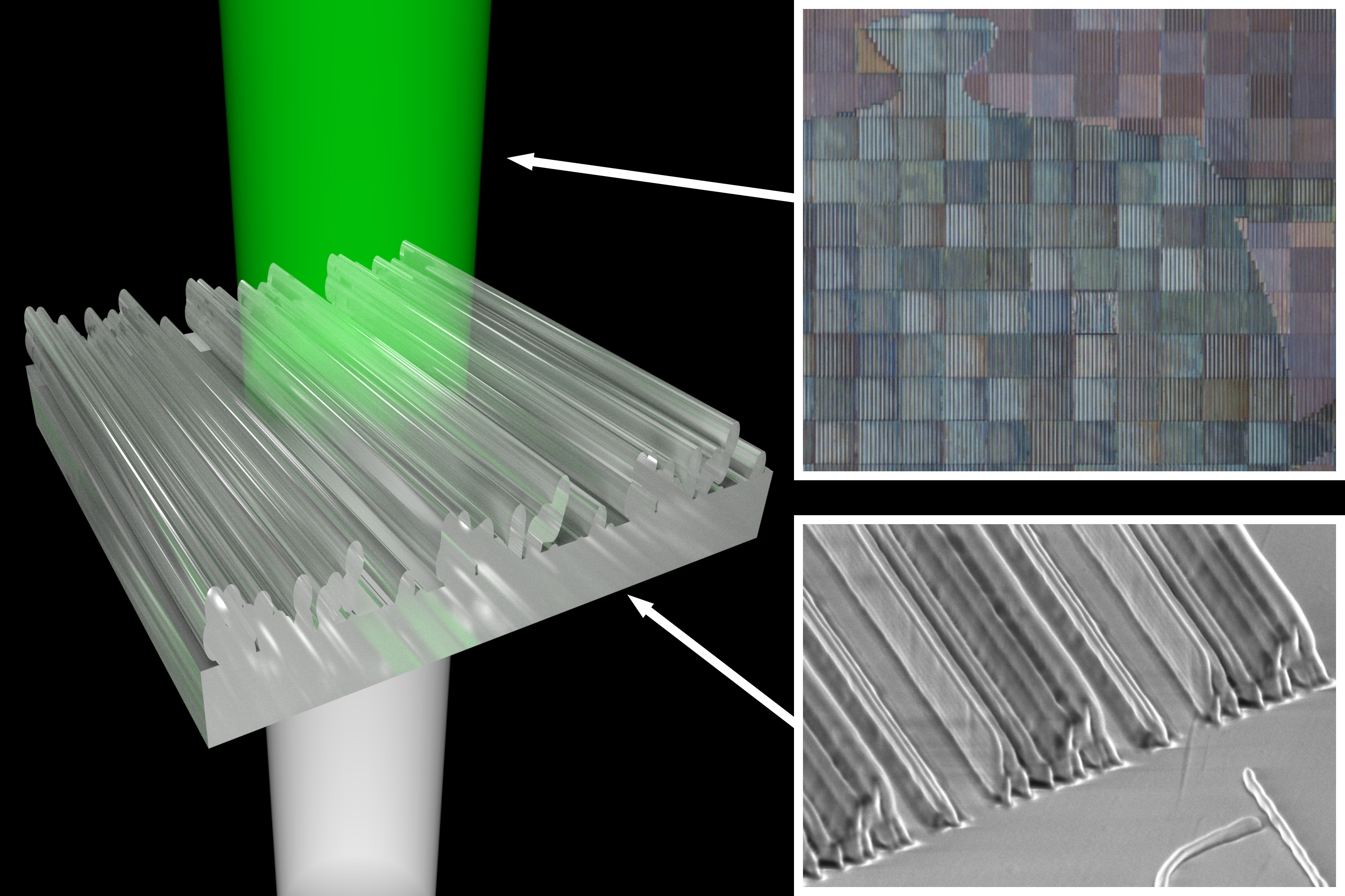
Caption: Light hits the 3-D printed nanostructures from below. After it is transmitted through, the viewer sees only green light — the remaining colors are redirected. Credit: Thomas Auzinger [downloaded from http://visualcomputing.ist.ac.at/publications/2018/StructCol/]

An August 17, 2018 news item on ScienceDaily announces the work illustrated by the image above,

Most of the objects we see are colored by pigments, but using pigments has disadvantages: such colors can fade, industrial pigments are often toxic, and certain color effects are impossible to achieve. The natural world, however, also exhibits structural coloration, where the microstructure of an object causes various colors to appear. Peacock feathers, for instance, are pigmented brown, but — because of long hollows within the feathers — reflect the gorgeous, iridescent blues and greens we see and admire. Recent advances in technology have made it practical to fabricate the kind of nanostructures that result in structural coloration, and computer scientists from the Institute of Science and Technology Austria (IST Austria) and the King Abdullah University of Science and Technology (KAUST) have now created a computational tool that automatically creates 3D-print templates for nanostructures that correspond to user-defined colors. Their work demonstrates the great potential for structural coloring in industry, and opens up possibilities for non-experts to create their own designs. This project will be presented at this year’s top computer graphics conference, SIGGRAPH 2018, by first author and IST Austria postdoc Thomas Auzinger. This is one of five IST Austria presentations at the conference this year.

SIGGRAPH 2018, now ended, was mentioned in my Aug. 9, 2018 posting.but since this presentation is accompanied by a paper, it rates its own posting. For one more excuse, there’s my fascination with structural colour.

An August 17, 2018 Institute of Science and Technology Austria press release (also on EurekAlert), which originated the news item, delves into the work,

The changing colors of a chameleon and the iridescent blues and greens of the morpho butterfly, among many others in nature, are the result of structural coloration, where nanostructures cause interference effects in light, resulting in a variety of colors when viewed macroscopically. Structural coloration has certain advantages over coloring with pigments (where particular wavelengths are absorbed), but until recently, the limits of technology meant fabricating such nanostructures required highly specialized methods. New “direct laser writing” set-ups, however, cost about as much as a high-quality industrial 3D printer, and allow for printing at the scale of hundreds of nanometers (hundred to thousand time thinner than a human hair), opening up possibilities for scientists to experiment with structural coloration.

So far, scientists have primarily experimented with nanostructures that they had observed in nature, or with simple, regular nanostructural designs (e.g. row after row of pillars). Thomas Auzinger and Bernd Bickel of IST Austria, together with Wolfgang Heidrich of KAUST, however, took an innovative new approach that differs in several key ways. First, they solve the inverse design task: the user enters the color they want to replicate, and then the computer creates a nanostructure pattern that gives that color, rather than attempting to reproduce structures found in nature. Moreover, “our design tool is completely automatic,” says Thomas Auzinger. “No extra effort is required on the part of the user.”

Second, the nanostructures in the template do not follow a particular pattern or have a regular structure; they appear to be randomly composed—a radical break from previous methods, but one with many advantages. “When looking at the template produced by the computer I cannot tell by the structure alone, if I see a pattern for blue or red or green,” explains Auzinger. “But that means the computer is finding solutions that we, as humans, could not. This free-form structure is extremely powerful: it allows for greater flexibility and opens up possibilities for additional coloring effects.” For instance, their design tool can be used to print a square that appears red from one angle, and blue from another (known as directional coloring).

Finally, previous efforts have also stumbled when it came to actual fabrication: the designs were often impossible to print. The new design tool, however, guarantees that the user will end up with a printable template, which makes it extremely useful for the future development of structural coloration in industry. “The design tool can be used to prototype new colors and other tools, as well as to find interesting structures that could be produced industrially,” adds Auzinger. Initial tests of the design tool have already yielded successful results. “It’s amazing to see something composed entirely of clear materials appear colored, simply because of structures invisible to the human eye,” says Bernd Bickel, professor at IST Austria, “we’re eager to experiment with additional materials, to expand the range of effects we can achieve.”

“It’s particularly exciting to witness the growing role of computational tools in fabrication,” concludes Auzinger, “and even more exciting to see the expansion of ‘computer graphics’ to encompass physical as well as virtual images.”

Here’s a link to and a citation for the paper,

Computational Design of Nanostructural Color for Additive Manufacturing by Thomas Auzinger, Wolfgang Heidrich, and Bernd Bickel. ACM Trans. Graph. 37, 4, Article 159 (August 2018). 16 pages. doi.org/10.1145/3197517.3201376

This appears to be open access.

There is also a project page bearing the same title as the paper, Computational Design of Nanostructural Color for Additive Manufacturing.

3D printed all liquid electronics

Even after watching the video, I still don’t quite believe it. A March 28, 2018 news item on ScienceDaily announces the work,

Scientists from the Department of Energy’s Lawrence Berkeley National Laboratory (Berkeley Lab [or LBNL]) have developed a way to print 3-D structures composed entirely of liquids. Using a modified 3-D printer, they injected threads of water into silicone oil — sculpting tubes made of one liquid within another liquid.

They envision their all-liquid material could be used to construct liquid electronics that power flexible, stretchable devices. The scientists also foresee chemically tuning the tubes and flowing molecules through them, leading to new ways to separate molecules or precisely deliver nanoscale building blocks to under-construction compounds.

A March 28, 2018 Berkeley Lab March 26, 2018 news release (also on EurekAlert), which originated the news item, describe the work in more detail,

The researchers have printed threads of water between 10 microns and 1 millimeter in diameter, and in a variety of spiraling and branching shapes up to several meters in length. What’s more, the material can conform to its surroundings and repeatedly change shape.

“It’s a new class of material that can reconfigure itself, and it has the potential to be customized into liquid reaction vessels for many uses, from chemical synthesis to ion transport to catalysis,” said Tom Russell, a visiting faculty scientist in Berkeley Lab’s Materials Sciences Division. He developed the material with Joe Forth, a postdoctoral researcher in the Materials Sciences Division, as well as other scientists from Berkeley Lab and several other institutions. They report their research March 24 [2018] in the journal Advanced Materials.

The material owes its origins to two advances: learning how to create liquid tubes inside another liquid, and then automating the process.

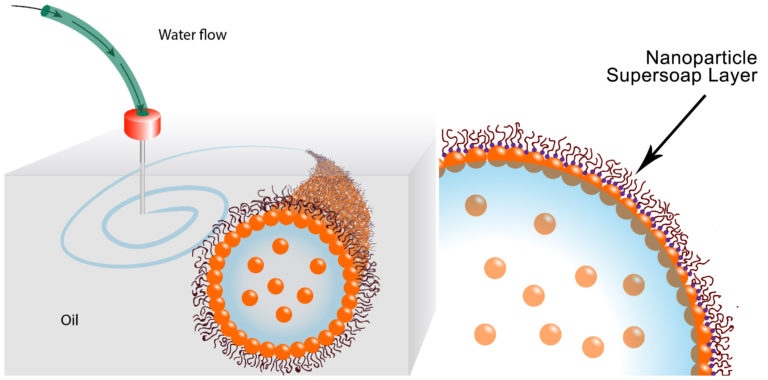
For the first step, the scientists developed a way to sheathe tubes of water in a special nanoparticle-derived surfactant that locks the water in place. The surfactant, essentially soap, prevents the tubes from breaking up into droplets. Their surfactant is so good at its job, the scientists call it a nanoparticle supersoap.

The supersoap was achieved by dispersing gold nanoparticles into water and polymer ligands into oil. The gold nanoparticles and polymer ligands want to attach to each other, but they also want to remain in their respective water and oil mediums. The ligands were developed with help from Brett Helms at the Molecular Foundry, a DOE Office of Science User Facility located at Berkeley Lab.

In practice, soon after the water is injected into the oil, dozens of ligands in the oil attach to individual nanoparticles in the water, forming a nanoparticle supersoap. These supersoaps jam together and vitrify, like glass, which stabilizes the interface between oil and water and locks the liquid structures in position.

This stability means we can stretch water into a tube, and it remains a tube. Or we can shape water into an ellipsoid, and it remains an ellipsoid,” said Russell. “We’ve used these nanoparticle supersoaps to print tubes of water that last for several months.”

Next came automation. Forth modified an off-the-shelf 3-D printer by removing the components designed to print plastic and replacing them with a syringe pump and needle that extrudes liquid. He then programmed the printer to insert the needle into the oil substrate and inject water in a predetermined pattern.

“We can squeeze liquid from a needle, and place threads of water anywhere we want in three dimensions,” said Forth. “We can also ping the material with an external force, which momentarily breaks the supersoap’s stability and changes the shape of the water threads. The structures are endlessly reconfigurable.”

This image illustrates how the water is printed,

These schematics show the printing of water in oil using a nanoparticle supersoap. Gold nanoparticles in the water combine with polymer ligands in the oil to form an elastic film (nanoparticle supersoap) at the interface, locking the structure in place. (Credit: Berkeley Lab)

Here’s a link to and a citation for the paper,

Reconfigurable Printed Liquids by Joe Forth, Xubo Liu, Jaffar Hasnain, Anju Toor, Karol Miszta, Shaowei Shi, Phillip L. Geissler, Todd Emrick, Brett A. Helms, Thomas P. Russell. Advanced Materials https://doi.org/10.1002/adma.201707603 First published: 24 March 2018

This paper is behind a paywall.

A 3D printed eye cornea and a 3D printed copy of your brain (also: a Brad Pitt connection)

Sometimes it’s hard to keep up with 3D tissue printing news. I have two news bits, one concerning eyes and another concerning brains.

3D printed human corneas

A May 29, 2018 news item on ScienceDaily trumpets the news,

The first human corneas have been 3D printed by scientists at Newcastle University, UK.

It means the technique could be used in the future to ensure an unlimited supply of corneas.

As the outermost layer of the human eye, the cornea has an important role in focusing vision.

Yet there is a significant shortage of corneas available to transplant, with 10 million people worldwide requiring surgery to prevent corneal blindness as a result of diseases such as trachoma, an infectious eye disorder.

In addition, almost 5 million people suffer total blindness due to corneal scarring caused by burns, lacerations, abrasion or disease.

The proof-of-concept research, published today [May 29, 2018] in Experimental Eye Research, reports how stem cells (human corneal stromal cells) from a healthy donor cornea were mixed together with alginate and collagen to create a solution that could be printed, a ‘bio-ink’.

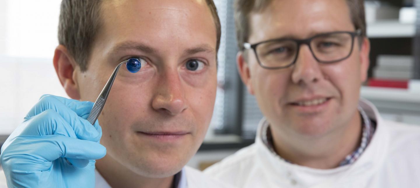
Here are the proud researchers with their cornea,

Caption: Dr. Steve Swioklo and Professor Che Connon with a dyed cornea. Credit: Newcastle University, UK

A May 30,2018 Newcastle University press release (also on EurekAlert but published on May 29, 2018), which originated the news item, adds more details,

Using a simple low-cost 3D bio-printer, the bio-ink was successfully extruded in concentric circles to form the shape of a human cornea. It took less than 10 minutes to print.

The stem cells were then shown to culture – or grow.

Che Connon, Professor of Tissue Engineering at Newcastle University, who led the work, said: “Many teams across the world have been chasing the ideal bio-ink to make this process feasible.

“Our unique gel – a combination of alginate and collagen – keeps the stem cells alive whilst producing a material which is stiff enough to hold its shape but soft enough to be squeezed out the nozzle of a 3D printer.

“This builds upon our previous work in which we kept cells alive for weeks at room temperature within a similar hydrogel. Now we have a ready to use bio-ink containing stem cells allowing users to start printing tissues without having to worry about growing the cells separately.”

The scientists, including first author and PhD student Ms Abigail Isaacson from the Institute of Genetic Medicine, Newcastle University, also demonstrated that they could build a cornea to match a patient’s unique specifications.

The dimensions of the printed tissue were originally taken from an actual cornea. By scanning a patient’s eye, they could use the data to rapidly print a cornea which matched the size and shape.

Professor Connon added: “Our 3D printed corneas will now have to undergo further testing and it will be several years before we could be in the position where we are using them for transplants.

“However, what we have shown is that it is feasible to print corneas using coordinates taken from a patient eye and that this approach has potential to combat the world-wide shortage.”

Here’s a link to and a citation for the paper,

3D bioprinting of a corneal stroma equivalent by Abigail Isaacson, Stephen Swioklo, Che J. Connon. Experimental Eye Research Volume 173, August 2018, Pages 188–193 and 2018 May 14 pii: S0014-4835(18)30212-4. doi: 10.1016/j.exer.2018.05.010. [Epub ahead of print]

This paper is behind a paywall.

A 3D printed copy of your brain

I love the title for this May 30, 2018 Wyss Institute for Biologically Inspired Engineering news release: Creating piece of mind by Lindsay Brownell (also on EurekAlert),

What if you could hold a physical model of your own brain in your hands, accurate down to its every unique fold? That’s just a normal part of life for Steven Keating, Ph.D., who had a baseball-sized tumor removed from his brain at age 26 while he was a graduate student in the MIT Media Lab’s Mediated Matter group. Curious to see what his brain actually looked like before the tumor was removed, and with the goal of better understanding his diagnosis and treatment options, Keating collected his medical data and began 3D printing his MRI [magnetic resonance imaging] and CT [computed tomography] scans, but was frustrated that existing methods were prohibitively time-intensive, cumbersome, and failed to accurately reveal important features of interest. Keating reached out to some of his group’s collaborators, including members of the Wyss Institute at Harvard University, who were exploring a new method for 3D printing biological samples.

“It never occurred to us to use this approach for human anatomy until Steve came to us and said, ‘Guys, here’s my data, what can we do?” says Ahmed Hosny, who was a Research Fellow with at the Wyss Institute at the time and is now a machine learning engineer at the Dana-Farber Cancer Institute. The result of that impromptu collaboration – which grew to involve James Weaver, Ph.D., Senior Research Scientist at the Wyss Institute; Neri Oxman, [emphasis mine] Ph.D., Director of the MIT Media Lab’s Mediated Matter group and Associate Professor of Media Arts and Sciences; and a team of researchers and physicians at several other academic and medical centers in the US and Germany – is a new technique that allows images from MRI, CT, and other medical scans to be easily and quickly converted into physical models with unprecedented detail. The research is reported in 3D Printing and Additive Manufacturing.

“I nearly jumped out of my chair when I saw what this technology is able to do,” says Beth Ripley, M.D. Ph.D., an Assistant Professor of Radiology at the University of Washington and clinical radiologist at the Seattle VA, and co-author of the paper. “It creates exquisitely detailed 3D-printed medical models with a fraction of the manual labor currently required, making 3D printing more accessible to the medical field as a tool for research and diagnosis.”

Imaging technologies like MRI and CT scans produce high-resolution images as a series of “slices” that reveal the details of structures inside the human body, making them an invaluable resource for evaluating and diagnosing medical conditions. Most 3D printers build physical models in a layer-by-layer process, so feeding them layers of medical images to create a solid structure is an obvious synergy between the two technologies.

However, there is a problem: MRI and CT scans produce images with so much detail that the object(s) of interest need to be isolated from surrounding tissue and converted into surface meshes in order to be printed. This is achieved via either a very time-intensive process called “segmentation” where a radiologist manually traces the desired object on every single image slice (sometimes hundreds of images for a single sample), or an automatic “thresholding” process in which a computer program quickly converts areas that contain grayscale pixels into either solid black or solid white pixels, based on a shade of gray that is chosen to be the threshold between black and white. However, medical imaging data sets often contain objects that are irregularly shaped and lack clear, well-defined borders; as a result, auto-thresholding (or even manual segmentation) often over- or under-exaggerates the size of a feature of interest and washes out critical detail.

The new method described by the paper’s authors gives medical professionals the best of both worlds, offering a fast and highly accurate method for converting complex images into a format that can be easily 3D printed. The key lies in printing with dithered bitmaps, a digital file format in which each pixel of a grayscale image is converted into a series of black and white pixels, and the density of the black pixels is what defines the different shades of gray rather than the pixels themselves varying in color.

Similar to the way images in black-and-white newsprint use varying sizes of black ink dots to convey shading, the more black pixels that are present in a given area, the darker it appears. By simplifying all pixels from various shades of gray into a mixture of black or white pixels, dithered bitmaps allow a 3D printer to print complex medical images using two different materials that preserve all the subtle variations of the original data with much greater accuracy and speed.

The team of researchers used bitmap-based 3D printing to create models of Keating’s brain and tumor that faithfully preserved all of the gradations of detail present in the raw MRI data down to a resolution that is on par with what the human eye can distinguish from about 9-10 inches away. Using this same approach, they were also able to print a variable stiffness model of a human heart valve using different materials for the valve tissue versus the mineral plaques that had formed within the valve, resulting in a model that exhibited mechanical property gradients and provided new insights into the actual effects of the plaques on valve function.

“Our approach not only allows for high levels of detail to be preserved and printed into medical models, but it also saves a tremendous amount of time and money,” says Weaver, who is the corresponding author of the paper. “Manually segmenting a CT scan of a healthy human foot, with all its internal bone structure, bone marrow, tendons, muscles, soft tissue, and skin, for example, can take more than 30 hours, even by a trained professional – we were able to do it in less than an hour.”

The researchers hope that their method will help make 3D printing a more viable tool for routine exams and diagnoses, patient education, and understanding the human body. “Right now, it’s just too expensive for hospitals to employ a team of specialists to go in and hand-segment image data sets for 3D printing, except in extremely high-risk or high-profile cases. We’re hoping to change that,” says Hosny.

In order for that to happen, some entrenched elements of the medical field need to change as well. Most patients’ data are compressed to save space on hospital servers, so it’s often difficult to get the raw MRI or CT scan files needed for high-resolution 3D printing. Additionally, the team’s research was facilitated through a joint collaboration with leading 3D printer manufacturer Stratasys, which allowed access to their 3D printer’s intrinsic bitmap printing capabilities. New software packages also still need to be developed to better leverage these capabilities and make them more accessible to medical professionals.

Despite these hurdles, the researchers are confident that their achievements present a significant value to the medical community. “I imagine that sometime within the next 5 years, the day could come when any patient that goes into a doctor’s office for a routine or non-routine CT or MRI scan will be able to get a 3D-printed model of their patient-specific data within a few days,” says Weaver.

Keating, who has become a passionate advocate of efforts to enable patients to access their own medical data, still 3D prints his MRI scans to see how his skull is healing post-surgery and check on his brain to make sure his tumor isn’t coming back. “The ability to understand what’s happening inside of you, to actually hold it in your hands and see the effects of treatment, is incredibly empowering,” he says.

“Curiosity is one of the biggest drivers of innovation and change for the greater good, especially when it involves exploring questions across disciplines and institutions. The Wyss Institute is proud to be a space where this kind of cross-field innovation can flourish,” says Wyss Institute Founding Director Donald Ingber, M.D., Ph.D., who is also the Judah Folkman Professor of Vascular Biology at Harvard Medical School (HMS) and the Vascular Biology Program at Boston Children’s Hospital, as well as Professor of Bioengineering at Harvard’s John A. Paulson School of Engineering and Applied Sciences (SEAS).

Here’s an image illustrating the work,

Caption: This 3D-printed model of Steven Keating’s skull and brain clearly shows his brain tumor and other fine details thanks to the new data processing method pioneered by the study’s authors. Credit: Wyss Institute at Harvard University

Here’s a link to and a citation for the paper,

From Improved Diagnostics to Presurgical Planning: High-Resolution Functionally Graded Multimaterial 3D Printing of Biomedical Tomographic Data Sets by Ahmed Hosny , Steven J. Keating, Joshua D. Dilley, Beth Ripley, Tatiana Kelil, Steve Pieper, Dominik Kolb, Christoph Bader, Anne-Marie Pobloth, Molly Griffin, Reza Nezafat, Georg Duda, Ennio A. Chiocca, James R.. Stone, James S. Michaelson, Mason N. Dean, Neri Oxman, and James C. Weaver. 3D Printing and Additive Manufacturing http://doi.org/10.1089/3dp.2017.0140 Online Ahead of Print:May 29, 2018

This paper appears to be open access.

A tangential Brad Pitt connection

It’s a bit of Hollywood gossip. There was some speculation in April 2018 that Brad Pitt was dating Dr. Neri Oxman highlighted in the Wyss Institute news release. Here’s a sample of an April 13, 2018 posting on Laineygossip (Note: A link has been removed),

It took him a long time to date, but he is now,” the insider tells PEOPLE. “He likes women who challenge him in every way, especially in the intellect department. Brad has seen how happy and different Amal has made his friend (George Clooney). It has given him something to think about.”

While a Pitt source has maintained he and Oxman are “just friends,” they’ve met up a few times since the fall and the insider notes Pitt has been flying frequently to the East Coast. He dropped by one of Oxman’s classes last fall and was spotted at MIT again a few weeks ago.

Pitt and Oxman got to know each other through an architecture project at MIT, where she works as a professor of media arts and sciences at the school’s Media Lab. Pitt has always been interested in architecture and founded the Make It Right Foundation, which builds affordable and environmentally friendly homes in New Orleans for people in need.

“One of the things Brad has said all along is that he wants to do more architecture and design work,” another source says. “He loves this, has found the furniture design and New Orleans developing work fulfilling, and knows he has a talent for it.”

It’s only been a week since Page Six first broke the news that Brad and Dr Oxman have been spending time together.

I’m fascinated by Oxman’s (and her colleagues’) furniture. Rose Brook writes about one particular Oxman piece in her March 27, 2014 posting for TCT magazine (Note: Links have been removed),

MIT Professor and 3D printing forerunner Neri Oxman has unveiled her striking acoustic chaise longue, which was made using Stratasys 3D printing technology.

Oxman collaborated with Professor W Craig Carter and Composer and fellow MIT Professor Tod Machover to explore material properties and their spatial arrangement to form the acoustic piece.

Christened Gemini, the two-part chaise was produced using a Stratasys Objet500 Connex3 multi-colour, multi-material 3D printer as well as traditional furniture-making techniques and it will be on display at the Vocal Vibrations exhibition at Le Laboratoire in Paris from March 28th 2014.

An Architect, Designer and Professor of Media, Arts and Science at MIT, Oxman’s creation aims to convey the relationship of twins in the womb through material properties and their arrangement. It was made using both subtractive and additive manufacturing and is part of Oxman’s ongoing exploration of what Stratasys’ ground-breaking multi-colour, multi-material 3D printer can do.

Brook goes on to explain how the chaise was made and the inspiration that led to it. Finally, it’s interesting to note that Oxman was working with Stratasys in 2014 and that this 2018 brain project is being developed in a joint collaboration with Statasys.

That’s it for 3D printing today.