There’s another Park Systems webinar coming up on July 9, 2015 (the last one concerning Nanostructured Polymers and Nanomaterials for Oil & Gas was mentioned in my June 9, 2015 posting).
This latest webinar series is focused on graphene, from a June 29, 2015 Park Systems news release,
Park Systems, world-leader in atomic force microscopy (AFM) is hosting a webinar to provide advanced scientific research into new classes of Nanoscale Graphene-based materials poised to revolutionize industries such as semiconductor, material science, bio science and energy. Touted as ‘the wonder material of the 21st Century’ by the researchers who were awarded the 2010 Nobel Prize in physics for their graphene research, this carbon-based lightweight material is 200 times stronger than steel and one of the most promising and versatile materials ever discovered.
The Park Systems Webinar titled Graphene Based Nanomaterials and Films will be given by Professor Rigoberto Advincula of Case Western Reserve University on July 9, 2015 at 9am PST. Prof. Advincula is an eminent professor, researcher and expert in the area of polymers, smart coatings, nanomaterials, surface analytical methods for a variety of applications.
“The discovery of graphene is but a continuing evolution on how we analyze, treat, synthesize carbon based nanomaterials which includes the fullerenes, nanotubes, and now C polymorph platelets called graphene,” explains Dr. Advincula. “Graphene is used in many areas of research and potential applications for electronics, solid-state devices, biosensors, coatings and much more for numerous industries where there are opportunities to make quantum improvements in methods and materials.”
Graphene is part of the C polymorph family of nanomaterials and because of the platy nature of the basal plane, it’s reactivity on the edges, and various redox forms, it is an excellent thin film additive and component that can be grown by vapor deposition methods as well as exfoliation. Current research into dispersion, preparations, and patterning of graphene using Park Systems AFM to identify nanoscale characteristics and surface properties as well as conductivity indicates that numerous breakthroughs in materials and chemicals are on the horizon.
“Park AFM is the natural tool to investigate Graphene’s adsorbed state on a flat substrate as well as characterize its surface properties and conductivity because of the reliability and accuracy of the equipment,” adds Dr. Advincula who will give the Webinar on July 9. “AFM is useful in understanding the surface properties of these products but is equally valuable in failure analysis because of the capability to do in-situ or real time measurements of failure with a temperature stage or a magnetic field.”
Graphene-based Nanomaterials offer many innovations in industries such as electronics, semiconductor, life science, material science and bio science. Some potential advancements already being researched include flexible electronics, anti bacterial paper, actuators, electrochoromic devices and transistors.
“Park Systems is presenting this webinar as part of Park Nano Academy, which will offer valuable education and shared knowledge across many Nano Science Disciplines and Industries as a way to further enable NanoScale advancements,” comments Keibock Lee, Park Systems President. “We invite all curious Nano Researchers to join our webinars and educational forums to launch innovative ideas that propel us into future Nano Scientific Technologies.”
The webinar will highlight how the research into is conducted and present some of the findings by Professor Rigoberto Advincula of Case Western Reserve University.
This webinar is available at no cost and is part of Park Systems Nano Academy.
To register go to: http://www.parkafm.com/index.php/medias/nano-academy/webinars/115-webinars/486-nanomaterials-webinar-july-9-2015
Enjoy!
![February 2014 / Gems [downloaded from http://kids.nationalgeographic.com/kids/guinness-world-record-smallest-magazine/]](http://www.frogheart.ca/wp-content/uploads/2014/03/NtnlGeoKidsContestCover.jpg)
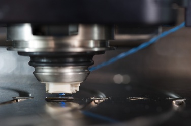
![Nanofriction at the tip of the microscope. Courtesy SISSA [Scuola Internazionale Superiore di Studi Avanzat], Italy](http://www.frogheart.ca/wp-content/uploads/2013/12/Nanofriction.jpg)