Tag Archives: AgNPs

Mechanism behind interaction of silver nanoparticles with the cells of the immune system

Scientists have come to a better understanding of the mechanism affecting silver nanoparticle toxicity according to an Aug. 30, 2016 news item on Nanowerk (Note: A link has been removed),

A senior fellow at the Faculty of Chemistry, MSU (Lomonosov Moscow State University), Vladimir Bochenkov together with his colleagues from Denmark succeeded in deciphering the mechanism of interaction of silver nanoparticles with the cells of the immune system. The study is published in the journal Nature Communications (“Dynamic protein coronas revealed as a modulator of silver nanoparticle sulphidation in vitro”).

‘Currently, a large number of products are containing silver nanoparticles: antibacterial drugs, toothpaste, polishes, paints, filters, packaging, medical and textile items. The functioning of these products lies in the capacity of silver to dissolve under oxidation and form ions Ag+ with germicidal properties. At the same time there are research data in vitro, showing the silver nanoparticles toxicity for various organs, including the liver, brain and lungs. In this regard, it is essential to study the processes occurring with silver nanoparticles in biological environments, and the factors affecting their toxicity,’ says Vladimir Bochenkov.
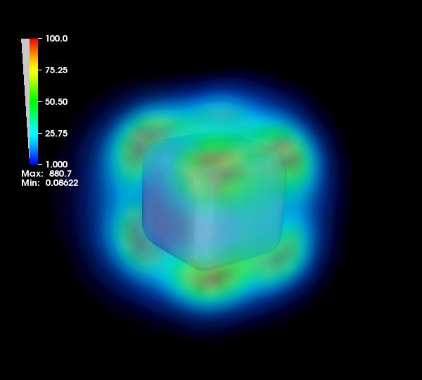
Caption: Increased intensity of the electric field near the silver nanoparticle surface in the excitation of plasmon resonance. Credit: Vladimir Bochenkov

Caption: Increased intensity of the electric field near the silver nanoparticle surface in the excitation of plasmon resonance. Credit: Vladimir Bochenkov

An Aug. 30, 2016 MSU press release on EurekAlert, which originated the news item, provides more information about the research,

The study is devoted to the protein corona — a layer of adsorbed protein molecules, which is formed on the surface of the silver nanoparticles during their contact with the biological environment, for example in blood. Protein crown masks nanoparticles and largely determines their fate: the speed of the elimination from the body, the ability to penetrate to a particular cell type, the distribution between the organs, etc.

According to the latest research, the protein corona consists of two layers: a rigid hard corona — protein molecules tightly bound with silver nanoparticles, and soft corona, consisting of weakly bound protein molecules in a dynamic equilibrium with the solution. Hitherto soft corona has been studied very little because of the experimental difficulties: the weakly bound nanoparticles separated from the protein solution easily desorbed (leave a particle remaining in the solution), leaving only the rigid corona on the nanoparticle surface.

The size of the studied silver nanoparticles was of 50-88 nm, and the diameter of the proteins that made up the crown — 3-7 nm. Scientists managed to study the silver nanoparticles with the protein corona in situ, not removing them from the biological environment. Due to the localized surface plasmon resonance used for probing the environment near the surface of the silver nanoparticles, the functions of the soft corona have been primarily investigated.

‘In the work we showed that the corona may affect the ability of the nanoparticles to dissolve to silver cations Ag+, which determine the toxic effect. In the absence of a soft corona (quickly sharing the medium protein layer with the environment) silver cations are associated with the sulfur-containing amino acids in serum medium, particularly cysteine and methionine, and precipitate as nanocrystals Ag2S in the hard corona,’ says Vladimir Bochenkov.

Ag2S (silver sulfide) famously easily forms on the silver surface even on the air in the presence of the hydrogen sulfide traces. Sulfur is also part of many biomolecules contained in the body, provoking the silver to react and be converted into sulfide. Forming of the nano-crystals Ag2S due to low solubility reduces the bioavailability of the Ag+ ions, reducing the toxicity of silver nanoparticles to null. With a sufficient amount of amino acid sulfur sources available for reaction, all the potentially toxic silver is converted into the nontoxic insoluble sulfide. Scientists have shown that what happens in the absence of a soft corona.

In the presence of a soft corona, the Ag2S silver sulfide nanocrystals are formed in smaller quantities or not formed at all. Scientists attribute this to the fact that the weakly bound protein molecules transfer the Ag+ ions from nanoparticles into the solution, thereby leaving the sulfide not crystallized. Thus, the soft corona proteins are ‘vehicles’ for the silver ions.

This effect, scientists believe, be taken into account when analyzing the stability of silver nanoparticles in a protein environment, and in interpreting the results of the toxicity studies. Studies of the cells viability of the immune system (J774 murine line macrophages) confirmed the reduction in cell toxicity of silver nanoparticles at the sulfidation (in the absence of a soft corona).

Vladimir Bochenkov’s challenge was to simulate the plasmon resonance spectra of the studied systems and to create the theoretical model that allowed quantitative determination of silver sulfide content in situ around nanoparticles, following the change in the absorption bands in the experimental spectra. Since the frequency of the plasmon resonance is sensitive to a change in dielectric constant near the nanoparticle surface, changes in the absorption spectra contain information about the amount of silver sulfide formed.

Knowledge of the mechanisms of formation and dynamics of the behavior of the protein corona, information about its composition and structure are extremely important for understanding the toxicity and hazards of nanoparticles for the human body. In prospect the protein corona formation can be used to deliver drugs in the body, including the treatment of cancer. For this purpose it will be enough to pick such a content of the protein corona, which enables silver nanoparticles penetrate only in the cancer cell and kill it.

Here’s a link to and a citation for the paper describing this fascinating work,

Dynamic protein coronas revealed as a modulator of silver nanoparticle sulphidation in vitro by Teodora Miclăuş, Christiane Beer, Jacques Chevallier, Carsten Scavenius, Vladimir E. Bochenkov, Jan J. Enghild, & Duncan S. Sutherland. Nature Communications 7,
Article number: 11770 doi:10.1038/ncomms11770 Published  09 June 2016

This paper is open access.

Using mung bean extract to synthesize silver nanoparticles

Not everyone is quite as enthusiastic as whoever wrote this press release about silver nanoparticles (due to potential environmental issues as more manufactured silver nanoparticles enter the ecosystem). Still, it’s good news that there may be a greener way to synthesize them. A July 20, 2016 news item on Azonano ‘spills the beans’,

… researchers from the Guru Nanak National College, the Institute of Microbial Technology, Mata Gujri College and Punjab University, have found a simple, non-toxic and environmentally friendly way to synthesize AgNPs [silver nanoparticles] using seed extract of Vigna radiata, commonly known as the mung bean or green gram.

Manoj Kumar Choudhary and colleagues used aqueous seed extract of mung beans to break up aqueous silver nitrate solution into NPs, as well as to reduce and stabilize the particles. The NPs were characterized by UV–visible spectroscopy, Fourier transform infrared spectroscopy, transmission electron microscopy, atomic absorption spectroscopy and X-ray diffraction. They were then tested for antimicrobial effectiveness.

As reported in Applied Nanoscience, the researchers found that phytochemicals present in the seed extract were effective at reducing and stabilizing the Ag metal ions. They found they could synthesize crystalline, spherically-shaped NPs, with a size range of 5 to 30 nm. The particles remained highly stable for months at room temperature, even after five months.

Antibacterial activity was assayed by the standard well-diffusion method, which showed that the biogenic silver NPs had broad-spectrum antibacterial activity against the Gram-negative bacteria Escherichia coli and the Gram-positive bacteria Staphylococcus aureus.

“In the present paper, we report a simple, eco-friendly and cost-effective synthesis method of AgNPs at ambient conditions using seed extract of Vigna radiata as a reducing and stabilizing agent,” say Choudhary and team. “The AgNPs synthesized by this method have efficient antimicrobial activity against pathogenic bacteria.”

The researchers say the next step would be further investigation of the potential applications of the synthesized AgNPs, as the outcome of the study could be useful for applications in nanotechnology-based applications in pharmacology and medicine.

Fig. 1: A Vigna radiata seeds. b Reddish brown solution of silver nanoparticles formed after 3 h due to reduction of silver ions [downloaded from http://link.springer.com/article/10.1007/s13204-015-0418-6]

Fig. 1: A Vigna radiata seeds. b Reddish brown solution of silver nanoparticles formed after 3 h due to reduction of silver ions [downloaded from http://link.springer.com/article/10.1007/s13204-015-0418-6]

Here’s a link to and a citation for the paper,

A facile biomimetic preparation of highly stabilized silver nanoparticles derived from seed extract of Vigna radiata and evaluation of their antibacterial activity by Manoj Kumar Choudhary, Jyoti Kataria, Swaranjit Singh Cameotra, Jagdish Singh. Appl Nanosci (2016) 6: 105. doi:10.1007/s13204-015-0418-6 First Online: 19 February 2015

This paper is open access.

Toxicity, nanoparticles, soil, and Europe’s NANO-ECOTOXICITY Project

I have featured pieces on nanoparticles, toxicity, and soil in the past (this Aug. 15, 2011 posting about Duke University’s mesocosm project is probably the most relevant) but this study is the first one I’ve seen focusing on earthworms. From the Sept. 23, 2013 news item on Nanowerk (Note: A link has been removed),

From the clothes and make-up we wear to the electronic devices we use every day, nanotechnology is becoming ubiquitous. But while industry has mastered the production of such materials, little is known about their fate once their service life comes to an end. The NANO-ECOTOXICITY project looked into their impact on soil organisms.

The Sept. 23, 2013 CORDIS (European Commission Community Research and Development Information Service) news release, which originated the new item, offers a Q&A (Question and Answer) with the project research leader,

Dr Maria Diez-Ortiz, research leader of the NANO-ECOTOXICITY project, tells us about her research findings and how she expects them to help increase knowledge and shape tools allowing for standard environmental hazard and risk-assessment methodologies.

What is the background of the NANO-ECOTOXICITY project?

Nanotechnology is based on the idea that, by engineering the size and shape of materials at the scale of atoms, i.e. nanometres (nm), distinct optical, electronic, or magnetic properties can be tuned to produce novel properties of commercial value. However, there is an obvious concern that such novel properties may also lead to novel behaviour when interacting with biological organisms, and thus to potentially novel toxic effects.

Since nanoparticles (NPs) are similar in size to viruses, their uptake by and transport through tissues are based on mechanisms distinct from those of molecular uptake and transport. Therefore, there is concern that standard toxicological tests may not be applicable or reliable in relation to NPs, hence compromising current risk-assessment procedures.

The majority of research on nano-safety in the environment has so far focused on the aquatic environment. Current research on environmental fate, however, indicates that soils will become the biggest environmental sink for nanoparticles. Following their entry into liquid waste streams, nanoparticles will pass through wastewater-treatment. processes, ending up in waste sludge which may accumulate in the agricultural land where this sludge is often applied.

What are the main objectives of the project?

This project deals with the toxicokinetics – that is, the rate at which a chemical enters a body and affects it – of metal nanoparticles coming into contact with soil-dwelling organisms. The aim is to determine NPs’ fate and effects in terrestrial ecosystems by means of case studies with zinc oxide and silver NPs, which represent different fate kinetics.

The project’s main objectives are to assess the toxicity of metal nanoparticles in soils in the short and long term; the main route of exposure for earthworms and whether it differs from those of ionic metals; and, finally, the influence of the exposure media on metal nanoparticle toxicity.

What is new or innovative about the project and the way it is addressing these issues?

We have been running a long-term study where soils with AgNP [silver nanoparticles] were stored and left to age for up to a year; their toxicity was tested at the start and after three, seven and 12 months of ageing. The results showed that silver toxicity increased over time, meaning that short-term standard toxicity tests may underestimate the environmental risk of silver nanoparticles.

In parallel, we found that organisms exposed to silver nanoparticles in short-term studies accumulated higher silver concentrations than organisms that were exposed to the same mass concentration of ionic silver. However, these NP exposed organisms actually suffered lower toxic effects. This observation contradicts the prevailing assumption in toxicology that the internalised concentration is directly related to chemical concentration at the target site and hence to its toxicity. This observation creates a new paradigm for nano-ecotoxicology.

What is not yet known is whether the accumulated NP metal may in the longer-term ultimately become toxic (e.g. through dissolution and ion release) in cells and tissues where AgNPs may be stored. Should this occur, the high concentrations accumulated may ultimately result in greater long-term toxicity for NPs than for ionic forms. This may reveal these accumulated NPs as internalised ‘time bombs’ relevant to long-term effects and toxicity.

However, it has to be borne in mind that the redicted environmental concentrations resulting from current use of nanoparticles (e.g. results from EU projects like NANOFATE2) are many times smaller than those used in these studies, meaning that such accumulations of nanoparticle-related silver are unlikely to occur in the environment or, ultimately, in humans.

What difficulties did you encounter and how did you solve them?

The main problems encountered relate to the tracking of nanoparticles inside the tissues and soils, as both are complex matrices. The analysis of the particles is a challenge in itself, even when in water, but to get information about their state in these matrices often requires unrealistic exposure concentrations (due to low detection limits of the highly specialised techniques used for analysis) or extraction of the particles from the matrices, which could potentially change the state of the particles.

In this project, I travelled to University of Kentucky to work with Jason Unrine and used gentle water-based extractions of soil samples immediately before analysing them using ‘Field-flow fractionation’ and ‘Inductively coupled plasma mass spectrometry’ to identify the state of nanoparticles in my aged soils.

To look at what form (speciation) of silver and zinc from the nanoparticle exposures could be found inside worms I collaborated with NANOFATE researchers at Cardiff University who fixed and thinly sectioned the worm tissues. I was lucky to be given the time to use specialist facilities like the UK’s Diamond Light Source synchrotron to investigate where and in what form the metals and potential nanoparticles could be found in these tissues.

The main challenge is that as soon as you take nanoparticles out of the manufacturers’ bottle they start changing, particularly when put into environments likes natural soils and waters, or even organisms. Therefore a lot of characterisation is needed during exposure to establish the state of the nanoparticles the organisms have been exposed to and how fast they are changing from pristine particles to dissolved ions, or particles with completely different surfaces.

Technical solutions to characterisation have been found during this short project, but this will remain a logistical challenge for many years to come as the analysis equipment is still very specialised and expensive and therefore not generally available.

What are the concrete results from the research so far?

The project has helped us draw various conclusions regarding the impact of NPs on the environment and how to assess them. First, we now know that soil acidity, or pH, influences the dissolution and toxicity of ZnO nanoparticles [zinc oxide].

Then, we found that toxicity of silver nanoparticles’ increases over time and that the particles’ coating affects their toxicity to soil invertebrates.

As previously mentioned, earthworms exposed to silver nanoparticles for 28 days accumulated higher silver concentrations than earthworms exposed to silver ions, without the excess silver from the nanoparticles having a toxic effect. [emphasis mine] Moreover, soil ingestion was identified as the main route of exposure to AgNP and ZnONP in earthworms.

How can industry and decision-makers ensure that nanomaterials do not impact our environment?

We hope that this project, and the larger EU project NANOFATE to which it is linked, will provide knowledge and tools enabling standard environmental-hazard and risk-assessment methodologies to be applied to engineered nanoparticles (ENPs) with just a few judicious modifications. The current systems and protocols for chemical risk assessment have been developed over decades, and where no novel toxic mechanisms exist, our results tend to say that nano fits in as long as we measure the right things and characterise realistic exposures properly.

Our research aims to determine the minimum methodological tweaks needed. So far everything indicates that the potential benefits from nanotechnology can be realised and managed safely alongside other chemicals. While we are fairly confident at this stage that ENPs impose no greater acute effects on important biological parameters – like reproduction – than their ionic forms, the NANO-ECOTOXICITY results demonstrate that we have some way to go before we can state loud and clear that we do not believe there is any novel low-level or long-term effect.

As for all chemicals, proving such a negative is impossible using short-term tests. We think the final conclusions by industry and regulators on safe use of nanoparticles should and will have to be made according to a ‘weight of evidence’ approach – proving there is a gap between predicted likely exposure levels and those levels seen to cause any effects or accumulations within ecosystem species.

What are the next topics for your research?

This project has finished but the next step for any other funding opportunity would be to address increasingly environmentally relevant exposure scenarios by analysing how nanoparticles modify in the environment and interact with living tissues and organisms at different trophic levels. I would like to investigate nanoparticle transformation and interactions in living tissues. To date, the studies that have identified this ‘excess’ accumulation of non-toxic metal loads in nanoparticleexposed organisms have only been short term.

Apart from the obviously increased food-chain transfer potential, is also not known whether, over the longer term, the accumulated NP-derived metal ultimately becomes toxic when present in tissues and cells. Such transformation and release of metal ions within tissues may ultimately result in greater longterm toxicity for NPs than for ionic forms.

Furthermore, I want to test exposures in a functioning model ecosystem including interspecific interactions and trophic transfer. Since interactions between biota and nanoparticles are relevant in natural soil systems, caution is needed when attempting to predict the ecological consequences of nanoparticles based on laboratory assays conducted with only a single species. In the presence of the full complement of biological components of soil systems, complex NPs may follow a range of pathways in which coatings may be removed and replaced with exudate materials. Studies to quantify the nature of these interactions are therefore needed to identify the fate, bioavailability and toxicity of realistic ‘non-pristine’ forms of NPs present in real soil environments.

New to me was the material about ageing silver nanoparticles and their increased toxicity over time. While this is an interesting piece of information it’s not necessarily all that useful. It seems even with their increased uptake compared to silver ions, silver nanoparticles (Diez-Ortiz doesn’t indicate whether or not * they tested variously aged silver nanoparticles) did not have toxic effects on the earthworms tested.

The NANO-ECOTOXICITY website doesn’t appear to exist anymore but you can find the NANOFATE (Nanoparticle Fate Assessment and Toxicity in the Environment) website here.

* ‘not’ removed to clarify meaning, Oct. 9, 2013. (Note: I had on Oct. 8, 2013 removed ‘not’ in a second place from the sentence in an attempt t o clarify the meaning and ended up not making any sense at all.) Please read Maria Diez-Ortiz in the Comments, as she clarifies matters in a way I could never hope to.