Tag Archives: American Friends of Tel Aviv University

First 3D heart printed using patient’s biological materials

This is very exciting news and it’s likely be at least 10 years before this technology could be made available to the public.
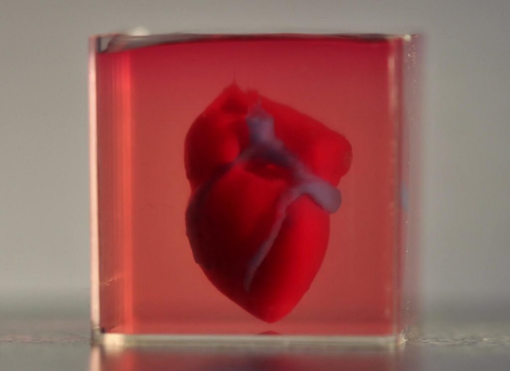
Caption: A 3D-printed, small-scaled human heart engineered from the patient’s own materials and cells. Credit: Advanced Science. © 2019 The Authors.

An April 15, 2019 news item on ScienceDaily makes a remarkable announcement,

In a major medical breakthrough, Tel Aviv University researchers have “printed” the world’s first 3D vascularised engineered heart using a patient’s own cells and biological materials. Their findings were published on April 15 [2019] in a study in Advanced Science.

Until now, scientists in regenerative medicine — a field positioned at the crossroads of biology and technology — have been successful in printing only simple tissues without blood vessels.

“This is the first time anyone anywhere has successfully engineered and printed an entire heart replete with cells, blood vessels, ventricles and chambers,” says Prof. Tal Dvir of TAU’s School of Molecular Cell Biology and Biotechnology, Department of Materials Science and Engineering, Center for Nanoscience and Nanotechnology and Sagol Center for Regenerative Biotechnology, who led the research for the study.

An April 15, 2019 Amricna Friends of Tel Aviv University (TAU) news release (also on EurekAlert), which originated the news item, provides more detail,

Heart disease is the leading cause of death among both men and women in the United States. Heart transplantation is currently the only treatment available to patients with end-stage heart failure. Given the dire shortage of heart donors, the need to develop new approaches to regenerate the diseased heart is urgent.

“This heart is made from human cells and patient-specific biological materials. In our process these materials serve as the bioinks, substances made of sugars and proteins that can be used for 3D printing of complex tissue models,” Prof. Dvir says. “People have managed to 3D-print the structure of a heart in the past, but not with cells or with blood vessels. Our results demonstrate the potential of our approach for engineering personalized tissue and organ replacement in the future.

Research for the study was conducted jointly by Prof. Dvir, Dr. Assaf Shapira of TAU’s Faculty of Life Sciences and Nadav Moor, a doctoral student in Prof. Dvir’s lab.

“At this stage, our 3D heart is small, the size of a rabbit’s heart, [emphasis mine] ” explains Prof. Dvir. “But larger human hearts require the same technology.”

For the research, a biopsy of fatty tissue was taken from patients. The cellular and a-cellular materials of the tissue were then separated. While the cells were reprogrammed to become pluripotent stem cells, the extracellular matrix (ECM), a three-dimensional network of extracellular macromolecules such as collagen and glycoproteins, were processed into a personalized hydrogel that served as the printing “ink.”

After being mixed with the hydrogel, the cells were efficiently differentiated to cardiac or endothelial cells to create patient-specific, immune-compatible cardiac patches with blood vessels and, subsequently, an entire heart.

According to Prof. Dvir, the use of “native” patient-specific materials is crucial to successfully engineering tissues and organs.

“The biocompatibility of engineered materials is crucial to eliminating the risk of implant rejection, which jeopardizes the success of such treatments,” Prof. Dvir says. “Ideally, the biomaterial should possess the same biochemical, mechanical and topographical properties of the patient’s own tissues. Here, we can report a simple approach to 3D-printed thick, vascularized and perfusable cardiac tissues that completely match the immunological, cellular, biochemical and anatomical properties of the patient.”

The researchers are now planning on culturing the printed hearts in the lab and “teaching them to behave” like hearts, Prof. Dvir says. They then plan to transplant the 3D-printed heart in animal models.

“We need to develop the printed heart further,” he concludes. “The cells need to form a pumping ability; they can currently contract, but we need them to work together. Our hope is that we will succeed and prove our method’s efficacy and usefulness.

“Maybe, in ten years, there will be organ printers in the finest hospitals around the world, and these procedures will be conducted routinely.”

Growing the heart to human size and getting the cells to work together so the heart will pump makes it seem like the 10 years Dvir imagines as the future date when there will be organ printers in hospitals routinely printing up hearts seems a bit optimistic. Regardless, I hope he’s right. Bravo to these Israeli researchers!

Here’s a link to and a citation for the paper,

3D Printing of Personalized Thick and Perfusable Cardiac Patches and Hearts by Nadav Noor, Assaf Shapira, Reuven Edri, Idan Gal, Lior Wertheim, Tal Dvir. Advanced Science DOI: https://doi.org/10.1002/advs.201900344 First published: 15 April 2019

This paper is open access.

Nanotechnology-enabled electronic tattoo from Tel Aviv University (Israel)

This is the first stick-on, nanotechnology-enabled tattoo I’ve seen that’s designed for the face. From a July 11, 2016 news item on ScienceDaily,

A new temporary “electronic tattoo” developed by Tel Aviv University [TAU] that can measure the activity of muscle and nerve cells researchers is poised to revolutionize medicine, rehabilitation, and even business and marketing research.

A July 11, 2016 American Friends of Tel Aviv University news release (also on EurekAlert), which originated the news item, provides more detail (Note: Some formatting has been changed),

The tattoo consists of a carbon electrode, an adhesive surface that attaches to the skin, and a nanotechnology-based conductive polymer coating that enhances the electrode’s performance. It records a strong, steady signal for hours on end without irritating the skin.

The electrode, developed by Prof. Yael Hanein, head of TAU’s Center for Nanoscience and Nanotechnology, may improve the therapeutic restoration of damaged nerves and tissue — and may even lead to new insights into our emotional life.

Prof. Hanein’s research was published last month in Scientific Reports and presented at an international nanomedicine program held at TAU.

“Stick it on and forget about it”

One major application of the new electrode is the mapping of emotion by monitoring facial expressions through electric signals received from facial muscles. “The ability to identify and map people’s emotions has many potential uses,” said Prof. Hanein. “Advertisers, pollsters, media professionals, and others — all want to test people’s reactions to various products and situations. Today, with no accurate scientific tools available, they rely mostly on inevitably subjective questionnaires.

“Researchers worldwide are trying to develop methods for mapping emotions by analyzing facial expressions, mostly via photos and smart software,” Prof. Hanein continued. “But our skin electrode provides a more direct and convenient solution.”

The device was first developed as an alternative to electromyography, a test that assesses the health of muscles and nerve cells. It’s an uncomfortable and unpleasant medical procedure that requires patients to lie sedentary in the lab for hours on end. Often a needle is stuck into muscle tissue to record its electrical activity, or patients are swabbed with a cold, sticky gel and attached to unwieldy surface electrodes.

“Our tattoo permits patients to carry on with their daily routines, while the electrode monitors their muscle and nerve activity,” said Prof. Hanein. “The idea is: stick it on and forget about it.”

Applications for rehabilitation and more

According to Prof. Hanein, the new skin electrode has other important therapeutic applications. The tattoo will be used to monitor the muscle activity of patients with neurodegenerative diseases in a study at Tel Aviv Medical Center.

“But that’s not all,” said Prof. Hanein. “The physiological data measured in specific muscles may be used in the future to indicate the alertness of drivers on the road; patients in rehabilitation following stroke or brain injury may utilize the ‘tattoo’ to improve muscle control; and amputees may employ it to move artificial limbs with remaining muscles.”

As it often is, the funding sources prove to be interesting (from the news release),

The electrode is the product of a European Research Council (ERC) project and received support from the BSMT Consortium of Israel’s Ministry of Economy.

The involvement of the European Research Council underlines the very close relationship Israel has to the European Union even though it is not an official member.

Here’s a link to and a citation for the paper,

Temporary-tattoo for long-term high fidelity biopotential recordings by Lilach Bareket, Lilah Inzelberg, David Rand, Moshe David-Pur, David Rabinovich, Barak Brandes & Yael Hanein. Scientific Reports 6, Article number: 25727 (2016)  doi:10.1038/srep25727 Published online: 12 May 2016

This paper is open access.

Tel Aviv University and the quest for super-slim, bendable displays

It’s beginning to seem like the quest for the Holy Grail. That is, the search for an object more myth than fact, but researchers at Tel Aviv University (TAU) believe they are on the right track to develop a slim, flexible screen according to a March 30, 2015 news item on Nanowerk (Note: A link has been removed),

From smartphones and tablets to computer monitors and interactive TV screens, electronic displays are everywhere. As the demand for instant, constant communication grows, so too does the urgency for more convenient portable devices — especially devices, like computer displays, that can be easily rolled up and put away, rather than requiring a flat surface for storage and transportation.

A new Tel Aviv University study, published recently in Nature Nanotechnology (“Light-emitting self-assembled peptide nucleic acids exhibit both stacking interactions and Watson–Crick base pairing”), suggests that a novel DNA-peptide structure can be used to produce thin, transparent, and flexible screens. The research, conducted by Prof. Ehud Gazit and doctoral student Or Berger of the Department of Molecular Microbiology and Biotechnology at TAU’s Faculty of Life Sciences, in collaboration with Dr. Yuval Ebenstein and Prof. Fernando Patolsky of the School of Chemistry at TAU’s Faculty of Exact Sciences, harnesses bionanotechnology to emit a full range of colors in one pliable pixel layer — as opposed to the several rigid layers that constitute today’s screens.

A March 30, 2015 American Friends of Tel Aviv University news release, which originated the news item, describes the material’s advantages and how the researchers developed it,

“Our material is light, organic, and environmentally friendly,” said Prof. Gazit. “It is flexible, and a single layer emits the same range of light that requires several layers today. By using only one layer, you can minimize production costs dramatically, which will lead to lower prices for consumers as well.”

For the purpose of the study, a part of Berger’s Ph.D. thesis, the researchers tested different combinations of peptides: short protein fragments, embedded with DNA elements which facilitate the self-assembly of a unique molecular architecture.

Peptides and DNA are two of the most basic building blocks of life. Each cell of every life form is composed of such building blocks. In the field of bionanotechnology, scientists utilize these building blocks to develop novel technologies with properties not available for inorganic materials such as plastic and metal.

“Our lab has been working on peptide nanotechnology for over a decade, but DNA nanotechnology is a distinct and fascinating field as well. When I started my doctoral studies, I wanted to try and converge the two approaches,” said Berger. “In this study, we focused on PNA — peptide nucleic acid, a synthetic hybrid molecule of peptides and DNA. We designed and synthesized different PNA sequences, and tried to build nano-metric architectures with them.”

Using methods such as electron microscopy and X-ray crystallography, the researchers discovered that three of the molecules they synthesized could self-assemble, in a few minutes, into ordered structures. The structures resembled the natural double-helix form of DNA, but also exhibited peptide characteristics. This resulted in a very unique molecular arrangement that reflects the duality of the new material.

“Once we discovered the DNA-like organization, we tested the ability of the structures to bind to DNA-specific fluorescent dyes,” said Berger. “To our surprise, the control sample, with no added dye, emitted the same fluorescence as the variable. This proved that the organic structure is itself naturally fluorescent.”

The structures were found to emit light in every color, as opposed to other fluorescent materials that shine only in one specific color. Moreover, light emission was observed also in response to electric voltage — which make it a perfect candidate for opto-electronic devices like display screens.

Here’s a link to and a citation for the paper,

Light-emitting self-assembled peptide nucleic acids exhibit both stacking interactions and Watson–Crick base pairing by Or Berger, Lihi Adler-Abramovich, Michal Levy-Sakin, Assaf Grunwald, Yael Liebes-Peer, Mor Bachar, Ludmila Buzhansky, Estelle Mossou, V. Trevor Forsyth, Tal Schwartz, Yuval Ebenstein, Felix Frolow, Linda J. W. Shimon, Fernando Patolsky, & Ehud Gazit. Nature Nanotechnology (2015) doi:10.1038/nnano.2015.27 Published online 16 March 2015

This paper is behind a paywall but a free preview is available via ReadCube Access.

Tackling ‘untreatable’ brain tumours

Isreal’s Tel Aviv University (TAU) has announced research that combines a nanoparticle-platform with RNA (ribonucleic acid) interference (RNAi) therapy for a difficult to treat brain cancer. From a Feb. 24, 2015 news item on Nanowerk,

There are no effective available treatments for sufferers of Glioblastoma multiforme (GBM), the most aggressive and devastating form of brain tumor. The disease, always fatal, has a survival rate of only 6-18 months.

Now a new Tel Aviv University study may offer hope to the tens of thousands diagnosed with gliomas every year. A pioneer of cancer-busting nanoscale therapeutics, Prof. Dan Peer of TAU’s Department of Department of Cell Research and Immunology and Scientific Director of TAU’s Center for NanoMedicine has adapted an earlier treatment modality — one engineered to tackle ovarian cancer tumors — to target gliomas, with promising results.

A Feb. 24, 2015 American Friends of Tel Aviv University news release (also on EurekAlert), which originated the news item, describes how the two lead researchers came to collaborate on this project,

“I was approached by a neurosurgeon insistent on finding a solution, any solution, to a desperate situation,” said Prof. Peer. “Their patients were dying on them, fast, and they had virtually no weapons in their arsenal. Prof. Zvi Cohen heard about my earlier nanoscale research and suggested using it as a basis for a novel mechanism with which to treat gliomas.”

Dr. Cohen had acted as the primary investigator in several glioma clinical trials over the last decade, in which new treatments were delivered surgically into gliomas or into the surrounding tissues following tumor removal. “Unfortunately, gene therapy, bacterial toxin therapy, and high-intensity focused ultrasound therapy had all failed as approaches to treat malignant brain tumors,” said Dr. Cohen. “I realized that we must think differently. When I heard about Dan’s work in the field of nanomedicine and cancer, I knew I found an innovative approach combining nanotechnology and molecular biology to tackle brain cancer.”

The news release then describes the research in more detail,

Dr. Peer’s new research is based on a nanoparticle platform, which transports drugs to target sites while minimizing adverse effects on the rest of the body. Prof. Peer devised a localized strategy to deliver RNA genetic interference (RNAi) directly to the tumor site using lipid-based nanoparticles coated with the polysugar hyaluronan (HA) that binds to a receptor expressed specifically on glioma cells. Prof. Peer and his team of researchers tested the therapy in mouse models affected with gliomas and control groups treated with standard forms of chemotherapy. The results were, according to the researchers, astonishing.

“We used a human glioma implanted in mice as our preclinical model,” said Prof. Peer. “Then we injected our designed particle with fluorescent dye to monitor its success entering the tumor cells. We were pleased and astonished to find that, a mere three hours later, the particles were situated within the tumor cells.”

Rather than chemotherapy, Prof. Peer’s nanoparticles contain nucleic acid with small interference RNAs, which silence the functioning of a key protein involved in cell proliferation. “Cancer cells, always dividing, are regulated by a specific protein,” said Prof. Peer. “We thought if we could silence this gene, they would die off. It is a basic, elegant mechanism and much less toxic than chemotherapy. This protein is not expressed in normal cells, so it only works where cells are highly proliferated.”

100 days following the treatment of four injections over 30 days, 60 percent of the afflicted mice were still alive. This represents a robust survival rate for mice, whose average life expectancy is only two years. The control mice died 30-34.5 days into treatment.

“This is a proof of concept study which can be translated into a novel clinical modality,” said Prof. Peer. “While it is in early stages, the data is so promising — it would be a crime not to pursue it.”

Here’s a link to and a citation for the paper,

Localized RNAi Therapeutics of Chemoresistant Grade IV Glioma Using Hyaluronan-Grafted Lipid-Based Nanoparticles by Zvi R. Cohen, Srinivas Ramishetti, Naama Peshes-Yaloz, Meir Goldsmith, Anton Wohl, Zion Zibly, and Dan Peer. ACS Nano, 2015, 9 (2), pp 1581–1591 DOI: 10.1021/nn506248s Publication Date (Web): January 5, 2015
Copyright © 2015 American Chemical Society

This study is behind a paywall.

Israeli scientists help us “sniff out” bombs

A July 23, 2014 news item on ScienceDaily describes the situation regarding bombs and other explosive devices and the Israelie research,

Security forces worldwide rely on sophisticated equipment, trained personnel, and detection dogs to safeguard airports and other public areas against terrorist attacks. A revolutionary new electronic chip with nano-sized chemical sensors is about to make their job much easier.

The groundbreaking nanotechnology-inspired sensor, devised by Prof. Fernando Patolsky of Tel Aviv University’s School of Chemistry and Center for Nanoscience and Nanotechnology, and developed by the Herzliya company Tracense, picks up the scent of explosives molecules better than a detection dog’s nose. Research on the sensor was recently published in the journal Nature Communications.

Existing explosives sensors are expensive, bulky and require expert interpretation of the findings. In contrast, the new sensor is mobile, inexpensive, and identifies in real time — and with great accuracy — explosives in the air at concentrations as low as a few molecules per 1,000 trillion.

A July 23, 2014 American Friends of Tel Aviv University news release (also on EurekAlert), which originated the news item, gives more detail about the research and potential product,

“Using a single tiny chip that consists of hundreds of supersensitive sensors, we can detect ultra low traces of extremely volatile explosives in air samples, and clearly fingerprint and differentiate them from other non-hazardous materials,” said Prof. Patolsky, a top researcher in the field of nanotechnology. “In real time, it detects small molecular species in air down to concentrations of parts-per-quadrillion, which is four to five orders of magnitude more sensitive than any existing technological method, and two to three orders of magnitude more sensitive than a dog’s nose.

“This chip can also detect improvised explosives, such as TATP (triacetone triperoxide), used in suicide bombing attacks in Israel and abroad,” Prof. Patolsky added.

The clusters of nano-sized transistors used in the prototype are extremely sensitive to chemicals, which cause changes in the electrical conductance of the sensors upon surface contact. When just a single molecule of an explosive comes into contact with the sensors, it binds with them, triggering a rapid and accurate mathematical analysis of the material.

“Animals are influenced by mood, weather, state of health and working hours, the oversaturation of olfactory system, and much more,” said Prof. Patolsky. “They also cannot tell us what they smell. Automatic sensing systems are superior candidates to dogs, working at least as well or better than nature. This is not an easy task, but was achieved through the development of novel technologies such as our sensor.”

The trace detector, still in prototype, identifies several different types of explosives several meters from the source in real time. It has been tested on the explosives TNT, RDX, and HMX, used in commercial blasting and military applications, as well as peroxide-based explosives like TATP and HMTD. The latter are commonly used in homemade bombs and are very difficult to detect using existing technology.

“Our breakthrough has the potential to change the way hazardous materials are detected, and of course should provide populations with more security,” said Prof. Patolsky. “The faster, more sensitive detection of tiny amounts of explosives in the air will provide for a better and safer world.”

Tracense has invested over $10M in research and development of the device since 2007, and expects to go to market next year [2015]. Prof.Patolsky and his team of researchers are currently performing multiple and extensive field tests of prototype devices of the sensor.

Here’s a link to and a citation for a recent paper by Professor Patolsky and his team,

Supersensitive fingerprinting of explosives by chemically modified nanosensors arrays by Amir Lichtenstein, Ehud Havivi, Ronen Shacham, Ehud Hahamy, Ronit Leibovich, Alexander Pevzner, Vadim Krivitsky, Guy Davivi, Igor Presman, Roey Elnathan, Yoni Engel, Eli Flaxer, & Fernando Patolsky. Nature Communications 5, Article number: 4195 doi:10.1038/ncomms5195 Published 24 June 2014 Updated online 09 July 2014

This paper is behind a paywall but a free preview is available via ReadCube Access.

Blood-, milk-, and mucus-powered electronics

Researchers at Tel Aviv University ([TAU] Israel) have already begun to develop biodegradable display screens in their quest to create electronic devices powered by blood, milk, and mucus proteins found in our bodies. From the March 7, 2012 news item on Nanowerk,

… a team including Ph.D. students Elad Mentovich and Netta Hendler of TAU’s Department of Chemistry and The Center for Nanoscience and Nanotechnology, with supervisor Dr. Shachar Richter and in collaboration with Prof. Michael Gozin and his Ph.D. student Bogdan Belgorodsky, has brought together cutting-edge techniques from multiple fields of science to create protein-based transistors — semi-conductors used to power electronic devices — from organic materials found in the human body. They could become the basis of a new generation of nano-sized technologies that are both flexible and biodegradable.

The March 7, 2012 news release on the American Friend of TAU website notes some of the issues with silicon-based electronics,

One of the challenges of using silicon as a semi-conductor is that a transistor must be created with a “top down” approach. Manufacturers start with a sheet of silicon and carve it into the shape that is needed, like carving a sculpture out of a rock. This method limits the capabilities of transistors when it comes to factors such as size and flexibility.

The TAU researchers turned to biology and chemistry for a different approach to building the ideal transistor. When they applied various combinations of blood, milk, and mucus proteins to any base material, the molecules self-assembled to create a semi-conducting film on a nano-scale. In the case of blood protein, for example, the film is approximately four nanometers high. The current technology in use now is 18 nanometers, says Mentovich.

Together, the three different kinds of proteins create a complete circuit with electronic and optical capabilities, each bringing something unique to the table. Blood protein has the ability to absorb oxygen, Mentovich says, which permits the “doping” of semi-conductors with specific chemicals in order to create specific technological properties. Milk proteins, known for their strength in difficult environments, form the fibers which are the building blocks of the transistors, while the mucosal proteins have the ability to keep red, green and, blue fluorescent dyes separate, together creating the white light emission that is necessary for advanced optics.

Overall, the natural abilities of each protein give the researchers “unique control” over the resulting organic transistor, allowing adjustments for conductivity, memory storage, and fluorescence among other characteristics.

I have previously featured work on vampire (blood-powered) fuel cells and batteries  in my July 18, 2012 posting and my April 3, 2009 posting so the notion of using blood (and presumably other bodily fluids) as a source for electrical power is generating (pun intended, weak though it is) interest in many research labs.

While the researchers don’t speculate about integrating these new carbon-based devices, which are smaller and more flexible than current devices, in bodies (from the American Friends of TAU news release),

Technology is now shifting from a silicon era to a carbon era, notes Mentovich, and this new type of transistor could play a big role. Transistors built from these proteins will be ideal for smaller, flexible devices that are made out of plastic rather than silicon, which exists in wafer form that would shatter like glass if bent. The breakthrough could lead to a new range of flexible technologies, such as screens, cell phones and tablets, biosensors, and microprocessor chips.

Just as significant, because the researchers are using natural proteins to build their transistor, the products they create will be biodegradable. It’s a far more environmentally friendly technology that addresses the growing problem of electronic waste, which is overflowing landfills worldwide.

The biodegradability of these proposed devices may be a problem if they are integrated into our bodies but it is certain that this will be attempted as we continue to explore machine/flesh possibilities.