Tag Archives: aptamers

Tapping into wound healing by harnessing the natural healing process

If you’re imagining an enhanced chakra balancing experience or more efficient digestion of your vitamin supplements, you will be a little disappointed in this latest news from the Imperial College of London (ICL). On the other hand, if you have damaged tissue, this discovery could make your recovery much easier. From a January 7, 2019 news item on phys.org,

Materials are widely used to help heal wounds: Collagen sponges help treat burns and pressure sores, and scaffold-like implants are used to repair bones. However, the process of tissue repair changes over time, so scientists are developing biomaterials that interact with tissues as healing takes place

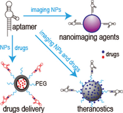
Now, Dr. Ben Almquist and his team at Imperial College London have created a new molecule that could change the way traditional materials work with the body. Known as traction force-activated payloads (TrAPs), their method lets materials talk to the body’s natural repair systems to drive healing.

The researchers say incorporating TrAPs into existing medical materials could revolutionise the way injuries are treated. Dr. Almquist, from Imperial’s Department of Bioengineering, said: “Our technology could help launch a new generation of materials that actively work with tissues to drive healing.”

A January 7, 2019 ICL press release (also on EurekAlert) by Caroline Brogan, which originated the news item, expands on the theme,

After an injury, cells ‘crawl’ through the collagen ‘scaffolds’ found in wounds, like spiders navigating webs. As they move, they pull on the scaffold, which activates hidden healing proteins that begin to repair injured tissue.

The researchers in the study designed TrAPs as a way to recreate this natural healing method. They folded the DNA segments into three-dimensional shapes known as aptamers that cling tightly to proteins. Then, they attached a customisable ‘handle’ that cells can grab onto on one end, before attaching the opposite end to a scaffold such as collagen.

During laboratory testing of their technique, they found that cells pulled on the TrAPs as they crawled through the collagen scaffolds. The pulling made the TrAPs unravel like shoelaces to reveal and activate the healing proteins. These proteins instruct the healing cells to grow and multiply

The researchers also found that by changing the cellular ‘handle’, they can change which type of cell can grab hold and pull, letting them tailor TrAPs to release specific therapeutic proteins based on which cells are present at a given point in time. In doing so, the TrAPs produce materials that can smartly interact with the correct type of cell at the correct time during wound repair.

This is the first time scientists have activated healing proteins using differing cell types in man-made materials. The technique mimics healing methods found in nature. Dr Almquist said: “Creatures from sea sponges to humans use cell movement to activate healing. Our approach mimics this by using the different cell varieties in wounds to drive healing.””

From lab to humans

This approach is adaptable to different cell types, so could be used in a variety of injuries such as fractured bones, scar tissue after heart attacks, and damaged nerves. New techniques are also desperately needed for patients whose wounds won’t heal despite current interventions, like diabetic foot ulcers, which are the leading cause of non-traumatic lower leg amputations.

TrAPs are relatively straightforward to create and are fully man-made, meaning they are easily recreated in different labs and can be scaled up to industrial quantities. Their adaptability also means they could help scientists create new methods for laboratory studies of diseases, stem cells, and tissue development.

Aptamers are currently used as drugs, meaning they are already proven safe and optimised for clinical use. Because TrAPs take advantage of aptamers that are safe for humans, they may be able to take a shorter path to the clinic than methods that start from ground zero.

Dr Almquist said: “TrAPs provide a flexible method of actively communicating with wounds, as well as key instructions when and where they are needed. This intelligent healing is useful during every phase of the healing process, has the potential to increase the body’s chance to recover, and has far-reaching uses on many different types of wounds. This technology could serve as a conductor of wound repair, orchestrating different cells over time to work together to heal damaged tissues.”

The researchers have made available an image and a video abstract illustrating their work,

TrAPs could harness the body’s natural healing powers to repair bone. Courtesy: Imperial College of London

By the way, the video was produced by www.animateyour.science (based in Adelaide, Australia) and they have a very interesting About page,

Our story

Your research is brilliant and novel. I’m sure of it. You might even be a pioneer in your field. But ask yourself honestly, is it enough? Is it truly enough to make a difference in the world?
 
My name is Tullio Rossi, and I founded Animate Your Science on my quest to make a positive impact on society through science.
 
During my Ph.D., I found that my peer-reviewed paper alone wasn’t cutting it. If I wanted to reach my peers, let alone the general public, I needed to communicate my findings in a fun and imaginative way.
 
This realization changed everything and inspired me to create “Lost at Sea,” an award-winning video that reached the hearts and minds of thousands of people.
 
The success of this first video blew my mind. And I got to thinking, maybe other scientists are lost at sea, so to speak. Maybe others want to reach the masses with their research, but just don’t know where to start.
 
This was the day Animate Your Science was born.

Why we do it

What we really want to do is bring science into society. That’s the true value of this company and the reason we believe in it.

We love science but we believe that, if not communicated properly, science is of limited use to society.​
 
As scientists, it’s our privilege and duty to unearth these revelations and package them in a way that appeals to our peers as well as the general public.

Getting back to TrAPS, here’s a link to and a citation for the paper,

Biologically Inspired, Cell‐Selective Release of Aptamer‐Trapped Growth Factors by Traction Forces by Anna Stejskalová, Nuria Oliva, Frances J. England, Benjamin D. Almquist. Advanced Materials DOI: https://doi.org/10.1002/adma.201806380 First published: 07 January 2019

This paper is open access.

Aptamers and theranostics (theragnostics)

A popular concept in some circles, theranostics (sometimes called theragnostics) is a conflation of the words ‘therapeutics’ and ‘diagnostics’. A Feb. 17, 2015 news item on Nanowerk features the use of aptamers as theranostic agents,

Aptamers are composed of short RNA or single-stranded DNA sequences that, when folded into their unique 3D conformation, can bind to their targets with high specifi city and affinity. Although functionally similar to protein antibodies, oligonucleotide aptamers offer several advantages over protein antibodies in biomedical and clinical applications.

Through the enhanced permeability and retention effect, nanomedicines can improve the therapeutic index of a treatment and reduce side effects by enhancing accumulation at the disease site. However, this targets tumors passively and, thus, may not be ideal for targeted therapy.

To construct ligand-directed “active targeting” nanobased delivery systems, aptamer-equipped nanomedicines have been tested for in vitro diagnosis, in vivo imaging, targeted cancer therapy, theranostic approaches, sub-cellular molecule detection, food safety, and environmental monitoring.

Here’s a link to and a citation for the paper,

Aptamers and Their Applications in Nanomedicine by Hongguang Sun and Youli Zu. Small DOI: 10.1002/smll.201403073 Article first published online: 11 FEB 2015

© 2015 WILEY-VCH Verlag GmbH & Co. KGaA, Weinheim

This paper is behind a paywall.

Here’s an illustration of the theranostic concept,

© Wiley

© Wiley

I have a bit more about aptamers in an Oct. 25, 2011 post featuring an interview with professor Maria DeRosa at the University of Ottawa.

Luminous bacteria sense pharmaceuticals and metals in wastewater

Scientists at the Helmholtz Association of German Research Centres have conceptualized a technique using luminescent bacterial proteins for sensing pharmaceuticals and metals in waste water. From the Helmholtz Association of German Research Centres June 12, 2013 press release,

While residual medications don’t belong in the water, trace metals from industrial process waters handled by the recycling industry are, in contrast, valuable resources. Scientists at the Helmholtz-Zentrum Dresden-Rossendorf (HZDR) have developed a simple color sensor principle which facilitates the easy detection of both materials as well as many other substances. This is the concept: If the analyzed sample shines red, then the water is ‘clean;’ if its color turns green, however, then it contains the substances the scientists wish to detect. The researchers recently published their concept in the scientific journal Sensors and Actuators B: Chemical (DOI: 10.1016/j.snb.2013.05.051).

Here’s the concept, from the press release,

The sensor principle is based on a red and a green fluorescent dye. If a substance to be detected is present in a water sample, then the sensor shines green; a red color, however, indicates that the substance is not present. What is the reason for the color difference? “The color molecules are located on a nanostructured surface consisting of bacterial proteins. The dyes are so close to one another that energy is transferred from the green to the red dye if these dyes are irradiated with light at a specific wavelength, for example, the light emitted by a laser. Then the sample shines red. This energy transfer, though, only occurs if the water sample is ‘clean.’ If, however, any foreign substances such as, for example, the pharmaceuticals or pollutants to be detected accumulate between the color molecules at specific binding sites, then the transfer is interrupted and only the green dyes shine,” explains Ulrike Weinert. Her doctoral dissertation revolves around the binding of color molecules on nano surfaces.

The network project (“AptaSens”) was subsidized by the Federal Ministry of Education and Research (BMBF). Nanostructured surfaces are an important part of the project. They are extracted from the envelope proteins of bacteria which are cultivated by the researchers in a lab. “The proteins form regular lattice structures at the nano level. They are ideally suited to evenly arrange functional groups and other molecules,” notes Weinert.

Another essential component of the sensor principle are the binding sites on the nano surface of the substances which are to be detected. That’s why so-called aptamers are used. These aptamers are short, single-stranded DNA oligonucleotides; the DNA segments can be designed in such a way that they are capable of specifically binding the most diverse substances such as the pharmaceuticals or the pollutants mentioned above. Dr. Beate Strehlitz from the Leipzig-based Helmholtz Centre for Environmental Research (UFZ) has specialized in this field. Within the scope of the AptaSens project, her team developed such a receptor for the antibiotic kanamycin which is used, for example, for the treatment of such bacterial infections of the eye as conjunctivitis, or in veterinary medicine.

The next step will be testing, from the press release,

What remains to be done now is to combine the kanamycin receptor with the dyes to test the color sensor principle with a sample substance. “From there, it’s just a small step to the development of a complete color sensor,” notes Katrin Pollmann [Dr. Katrin Pollmann, Team Leader Biotechnology at the HZDR {Holtzman Centres}]. For this, the researchers need to integrate the individual components – which include bacterial proteins, dyes, and aptamers – into a sensor chip. They have actually conducted a number of experiments with suitable substrates such as, for example, glass or silicon dioxide. “The sensor chip could be as small as a thumbnail. It could be wetted on site with the water sample to be analyzed. This would also include a laser light source which activates the chip as well as a detector that measures the change in color,” adds Pollmann. The scientists are now applying for a follow-up project.

I’d love to get a little more information about which metals (gold nanoparticles? silver nanoparticles? zinc oxide nanoparticles? etc.) could be detected in the water. If the information is in the research team’s published paper, that is available only behind a paywall.  H/T to Nanowerk (June 12, 2013 news item) for alerting me to this research work.

Here’s a citation (the link was provided earlier in this post),

U. Weinert, K. Pollmann, J. Raff. “Fluorescence Resonance Energy Transfer by S-layer coupled fluorescence dyes”, in Sensors and Actuators B: Chemical (2013), DOI: 10.1016/j.snb.2013.05.051

For anyone who’s interested in more information about aptamers, there’s my Oct. 25, 2011 posting which featured an interview with Dr. Maria DeRosa about her work with them.

Nanopore instruments, femtomolar concentrations, Ireland, and New Zealand

It was the word femtomolar that did it for me. While I have somehow managed to conceptualize the nanoscale, the other scales (pico, femto, atto, zetto, and yocto) continue to  elude me. If my experience with the ‘nanoscale ‘ is any guide, the only solution will be to find as much information as I can on these other ones and immerse myself in them. With that said, here’s more from the July 19, 2012 Izon press release,

Researchers at the Lee Bionanosciences Laboratory at UCD [University College Dublin] School of Chemistry and Chemical Biology in Dublin have demonstrated the detection and measurement of biological analytes down to femtomolar concentration levels using an off the shelf qNano instrument. This ultra low level biodetection capability has implications for biomedical research and clinical development as trace amounts of a biological substance in a sample can now be detected amd quantfied using standard commercially available equipment.

Platt [Dr Mark Platt] and colleagues’ [Professor Gil Lee and Dr Geoff Willmott] method for femtomolar-level detection uses magnetic particle systems and can be applied to any biological particle or protein for which specific aptamers or antibodies exist. Resistive pulse sensing, the underlying technology of the qNano [Izon product], was used to monitor individual and aggregated rod-shaped nanoparticles as they move through tunable pores in elastomeric membranes.

Dr Platt says, “The strength of using the qNano is the ability to interrogate individual particles through a nanopore. This allowed us to establish a very sensitive measurement of concentration because we could detect the interactions occurring down to individual particle level.

”The unique and technically innovative approach of the authors was to detect a molecule’s presence by a process that results in end on end or side by side aggregation of rod shaped nickel-gold particles. The rods were designed so that the aptamers could be attached to one end only.

“By comparing particles of similar dimensions we demonstrated that the resistive pulse signal is fundamentally different for rod and sphere-shaped particles, and for rod shaped particles of different lengths. We could exploit these differences in a new agglutina¬tion assay to achieve these low detection levels,” says Dr Platt.

In the agglutination assay particles with a particular aspect ratio can be distinguished by two measurements: the measured drop in current as particles traverse the pore (∆ip), which reveals the particle’s size; and the full width at half maximum (FWHM) duration of the resistive pulse, which relates to the particle’s speed and length. Limits of detection down to femtomolar levels were thus able to be demonstrated.

I’m a little unclear as to what qNano actually is. I did find this description on the qNano product page,

qNano uses unique nanopore-based detection to enable the physical properties of a wide range of particle types to be measured with unsurpassed accuracy.

Detailed Multi-Parameter Analysis.

Particle-by-particle measurement through qNano enables detailed determination of:

Nanopore-based detection allows thousands of particles to be measured individually, providing far greater detail and accuracy than light-based techniques.

Applications & Particle Types

A wide range of biological and synthetic particle types, spanning 50 nm – 10 μm, can be measured, across a broad range of research fields.

qNano Package

qNano is sold as a full system ready for use including the base instrument, variable pressure module, fluid cell and a starter kit of nanopores, buffer solution and standard particle sets.

Here’s what the product looks like,

qNano (from the Izon website)

As for what this all might mean to those of us who exist at the macroscale (from the Izon press release),

Izon Science will continue to work with Dr Platt at Loughborough, and with University College Dublin and various customers to develop a series of diagnostic kits that can be used with the qNano to identify and measure biomolecules, viruses, and microvesicles.“This is a real milestone for Izon’s technology as being able to measure biomolecules down to these extremely low levels opens up new bio-analysis options for researchers. 10 femtomolar was achieved, which is the equivalent dilution to 1 gram in 3.3 billion litres, or 1 gram in 1300 Olympic sized swimming pools,” says Hans van der Voorn, Executive Chairman of Izon Science.

For those interested in finding out about nanopores, these were mentioned in my July 18, 2012 posting while aptamers were discussed in my interview (Oct. 25, 2011 posting) with Dr. Maria DeRosa who researches them in her Carleton University laboratory (Ottawa, Canada).

Aptamers and Maria DeRosa

Today’s (Oct. 25, 2011) next interview is with Maria DeRosa of the DeRosa Lab at Carleton University (Ottawa, Canada) where she and her colleagues work on bionanotechnology projects. (The Highlighting the 2011 Dance Your Ph.D. contest posting featured a Ph.D student from her lab who is one of this year’s contest finalists.)

Before proceeding to the interview, here’s a little bit about the DeRosa Lab (from the website homepage),

The first step in the rational design of novel bionanotechnology is to find the right molecular components for the task. Our group seeks to investigate the use of chemically-modified nucleic acid aptamers, single stranded DNA or RNA sequences that specifically bind to a diverse variety of targets, in biosensing and catalysis.

Here’s some information about Dr. DeRosa,

Dr. Maria DeRosa’s research examines a type of nucleic acid called ‘aptamers’ that can fold into 3D nanoscale shapes capable of binding tightly to a specific molecular target.  Her group is focused on developing a better understanding of how these systems and using this information to design useful nanotechnology, like biosensors or “smart” delivery devices.  Dr. DeRosa received her Ph.D in Chemistry from Carleton University in 2003 and was presented with a University Senate Medal. She was awarded an NSERC Postdoctoral Fellowship to do research at the California Institute of Technology from 2004-2005 with Prof. Jackie Barton, a world-leader in DNA sensor research. In 2005, she returned to Carleton as a faculty member in the Chemistry Department. Her research group has received funding from the Natural Sciences and Engineering Research Council (NSERC), the Ontario Ministry of Agriculture, Food and Rural Affairs (OMAFRA), the Canada Foundation for Innovation (CFI) and Alberta Innovates Biosolutions.  DeRosa was a recipient of the John Charles Polanyi Research Award for new researchers in 2006 and an Ontario Early Researcher Award in 2010.

Here’s the interview,

*   Are you one of those people who always wanted to be a scientist or was this something you discovered later?

I was never one of those people who knew what they wanted to do from an early age.  I thought about being a doctor, pharmacist, plumber, engineer, bank teller…  In high school, I had many great math and science teachers that inspired me to go into science when I started at Carleton University.  Then, in my third year I got a summer job working in Dr. Bob Crutchley’s research lab.  He was a great mentor and it was then that I started seriously thinking about a career as a scientist.  I loved the idea of research, that I was working on a problem and no one knew what the answer would be.  I wanted the answers!

*   How did you get interested in aptamers (and could you briefly describe what they are)?

Aptamers are synthetic pieces of DNA that can recognize and stick to a molecular target.  The targets can vary from things that are very small, like a drug molecule to something much larger, like bacteria or viruses.  Because they can recognize and stick to other molecules, people are interested in using them as receptors for sensors.  I had never even heard of them until about 2005.

After my Ph.D., I went to Caltech to do something called a postdoctoral fellowship.  It was a research position in the lab of Dr. Jackie Barton, one of the world’s top DNA researchers (she just won a National Medal of Science a couple days ago).  She wasn’t working with aptamers but she opened me up to the idea of using DNA in an “unnatural” way.  Most of us, when we are thinking of DNA, we think of our genes and that it is the blueprint for life.  But from a chemistry point of view, DNA is just another material that has certain chemical properties that can be useful for other applications.  In Jackie’s lab, I learned how to make synthetic DNA and I started reading about aptamers.  I found the whole field fascinating and I knew that I wanted to be a part of it.

*   What applications are there for your work? (I noticed that you discussed fertilizers in your TEDxCarleton talk. Is agriculture an area of particular interest?)

Applications for aptamers mostly stem from their ability to bind tightly and selectively to other molecules.  So, they are typically used in technology such as biosensors where they can serve to detect low levels of something, like a toxin or a virus for example, in another matrix.  We’re developing aptamers for the detection of mycotoxins (toxins that come from moulds) in crops and food.  We’re also working on aptamers for norovirus (the virus that causes Norwalk, that awful stomach bug) so that we can catch it if it is in meat and other foods before they get sent off to stores.

We are also trying to use aptamers for triggered delivery of drugs and/or nutrients.  In many cases with drugs, we want them to act on certain cells or tissues and not on others.  So, we need to be able to control where the drug is released in the body.  There is a similar problem in agriculture.  We want to give crops certain nutrients from fertilizers but if we deliver them at the wrong time, they will be washed away and not taken up by the crop.  This leads to major economic losses for the farmer and problems for the environment.  With our work, the idea is that we use the aptamer to control the release of whatever we are delivering.  We incorporate the aptamer into a coating that covers the drug or nutrient.  The aptamer is there to recognize a stimulus that we want to use to release the contents.  For drug delivery, that stimulus might be a cancer cell or a disease biomarker.  For fertilizers, that stimulus might a be a plant signal that corresponds to the plant’s need for nutrients.  (We are working with Dr.Carlos Monreal from Agriculture and Agrifood Canada on the fertilizer project, and he is an expert in these plant signals and ‘smart fertilizers’.)  In the absence of that signal, the coating does not allow the release of the drug or nutrient.  But, once the aptamer recognizes that key signal, the aptamer distorts or destroys the coating and it allows the nutrient to be released.

*   According to the information on your lab website, you are the recipient of Canada Foundation for Innovation (CFI) Leaders Opportunity Fund (LOF) monies. Are these funds being applied to a particular project in your lab or are they used to support your general area of research?

CFI funds helped us to build our facility called the LADDER (Laboratory for Aptamer Discovery and Development of Emerging Research applications).  That funding allowed us to get the state-of-the-art equipment we need to support all of our research projects.  Without CFI funding, our work would not be possible!

*   Given your TEDxCarleton talk and your involvement in the 2011 Canadian Science Writers conference (researchers’ speed dating [I couldn’t confirm it but I’m pretty sure I saw your name listed for this event]), I gather you’re quite interested in public outreach. Why do you think it’s important?

Yes, I was at that ‘speed dating’ event and I am very committed to science outreach.  The public helps to support my research through funding like NSERC and CFI, so I think it is critical that I can explain to them what it is that I do, why it is important, and why their money is well-spent.  The general public may not know what an aptamer is, but they all realize the importance of keeping our food free of toxins or the need to make drugs that are better able to target disease.

*   I noticed that one of your students is a finalist in the Dance your Ph.D 2011 contest. And it’s not the first time. Do you find a lot of scientists with ‘dance’ tendencies are attracted to your lab? Are you one of those scientists?

My students won the competition last year and then they were finalists again this year!  I’m not sure if dancers are attracted to my lab or if my students are just as committed to outreach as I am!  My students are very excited to talk about their research with anyone who will listen.  This contest is a fun way to explain their work to everyday people.  Friends and family, after watching these dances online, have told me that they finally understand what is going on in my lab.  Maybe I should dance more!  (I’m not a dancer and you will not find me in either video…I support them from the sidelines!)

*   Is there anything you would like to add?

Thanks for profiling me and it has been fun!

Maria, thank you for this intriguing peek into your research, the field of DNA nanotechnology, and your (and shared by your students) commitment to public science outreach. I’m very happy you managed to cram the time to answer these questions into your schedule.

Highlighting the 2011 Dance Your Ph.D. contest

Science magazine (published by the American Association for the Advancement of Science [AAAS]) has been holding a Dance Your PhD contest since 2008* (as best I can determine from a Sept. 17, 2010 posting by Katherine for SciFri). In any case, this year they received a record number of entries (from an Oct. 14, 2011 posting by John Bohannon on Science Now),

Have you ever wondered what nanotube chemistry might look like as a dance? Or fruit fly sex? Or protein x-ray crystallography? Look no further. As part of the 2011 Dance Your Ph.D. contest, scientists who study those phenomena and more have converted their research into dance videos for your enjoyment and edification. And today the 16 finalists of this annual contest are revealed below.

A record 55 dances were created for this year’s contest, submitted by scientists around the globe, from the United States and Canada to Europe, India, and Australia. As the contest rules state, each dance must be based on the scientist’s own Ph.D. research thesis, and that scientist must participate in the dance. For many of the graduate students who danced, the research they depicted is still ongoing. For some of the older contestants, the project is a distant, perhaps harrowing memory from their early days in science. The dances are divided into four categories based on subject: physics, chemistry, biology, and social science. (The criteria for those categories are explained here.)

One of this year’s finalists is from the DeRosa lab at Carleton University in Ottawa, Canada. Titled, “DNA Aptamers as a Tool for Studying Mental Health Disease.” Erin McConnell and her troop are featured in the video below, *ETA February 27, 2024: The video is no longer here, please follow the link to Vimeo: DNA Aptamers as a Tool for Studying Mental Health Disease from Erin McConnell on Vimeo.*

I haven’t had time to review the other finalists but given this one, I can hardly wait.

The DeRosa lab also had a finalist in last year’s Dance Your PhD contest. It’s not the only reason I contacted the lab’s leader, Maria DeRosa but it did add a piquant flavour to my interview with her, which I will be posting tomorrow (Oct. 25, 2011).

*ETA Oct 24, 2011 1500 hours: There is an Oct. 18, 2011 article by Bob Weber for the Globe and Mail newspaper about the Canadian finalists in the 2011 Dance Your PhD contest. The contest was informally created in 2007 according to its originator John Bohannon.