Tag Archives: artificial organs

A cheaper way to make artificial organs

In the quest to develop artificial organs, the University of British Columbia (UBC) is the not the first research institution that comes to my mind. It seems I may need to reevaluate now that UBC (Okanagan) has announced some work on bio-inks and artificial organs in a Sept. 12, 2017 news  release (also on EurekAlert) by Patty Wellborn,,

A new bio-ink that may support a more efficient and inexpensive fabrication of human tissues and organs has been created by researchers at UBC’s Okanagan campus.

Keekyoung Kim, an assistant professor at UBC Okanagan’s School of Engineering, says this development can accelerate advances in regenerative medicine.

Using techniques like 3D printing, scientists are creating bio-material products that function alongside living cells. These products are made using a number of biomaterials including gelatin methacrylate (GelMA), a hydrogel that can serve as a building block in bio-printing. This type of bio-material—called bio-ink—are made of living cells, but can be printed and molded into specific organ or tissue shapes.

The UBC team analyzed the physical and biological properties of three different GelMA hydrogels—porcine skin, cold-water fish skin and cold-soluble gelatin. They found that hydrogel made from cold-soluble gelatin (gelatin which dissolves without heat) was by far the best performer and a strong candidate for future 3D organ printing.

“A big drawback of conventional hydrogel is its thermal instability. Even small changes in temperature cause significant changes in its viscosity or thickness,” says Kim. “This makes it problematic for many room temperature bio-fabrication systems, which are compatible with only a narrow range of hydrogel viscosities and which must generate products that are as uniform as possible if they are to function properly.”

Kim’s team created two new hydrogels—one from fish skin, and one from cold-soluble gelatin—and compared their properties to those of porcine skin GelMA. Although fish skin GelMA had some benefits, cold-soluble GelMA was the top overall performer. Not only could it form healthy tissue scaffolds, allowing cells to successfully grow and adhere to it, but it was also thermally stable at room temperature.

The UBC team also demonstrated that cold-soluble GelMA produces consistently uniform droplets at temperatures, thus making it an excellent choice for use in 3D bio-printing.

“We hope this new bio-ink will help researchers create improved artificial organs and lead to the development of better drugs, tissue engineering and regenerative therapies,” Kim says. “The next step is to investigate whether or not cold-soluble GelMA-based tissue scaffolds are can be used long-term both in the laboratory and in real-world transplants.”

Three times cheaper than porcine skin gelatin, cold-soluble gelatin is used primarily in culinary applications.

Here’s a link to and a citation for the paper,

Comparative study of gelatin methacrylate hydrogels from different sources for biofabrication applications by Zongjie Wang, Zhenlin Tian, Fredric Menard, and Keekyoung Kim. Biofabrication, Volume 9, Number 4 Special issue on Bioinks https://doi.org/10.1088/1758-5090/aa83cf Published 21 August 2017

© 2017 IOP Publishing Ltd

This paper is behind a paywall.
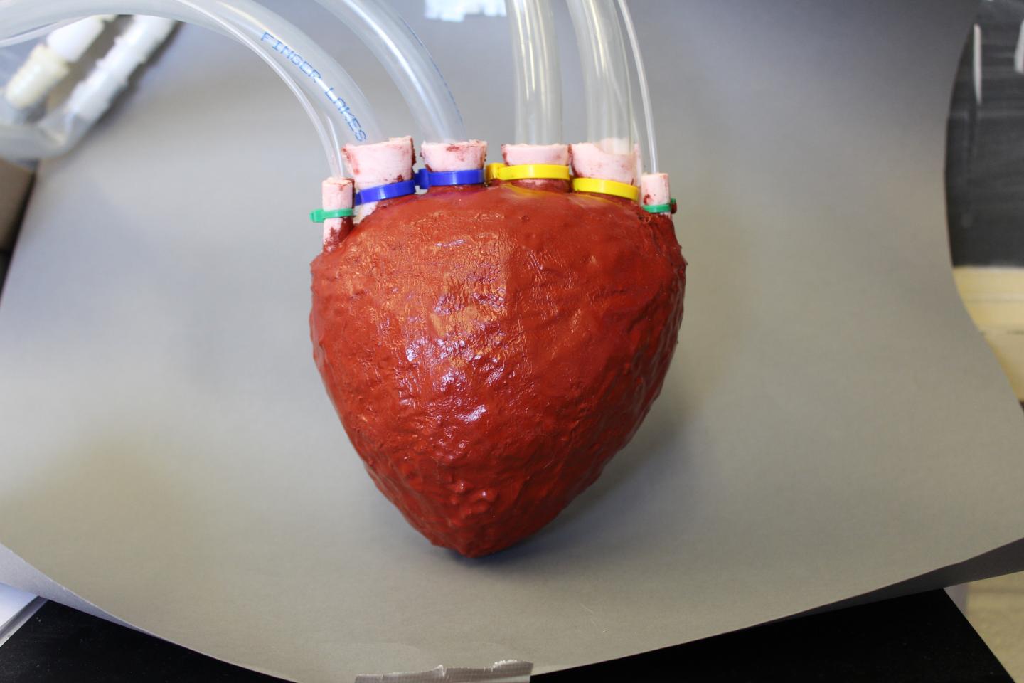
A soft heart from Cornell University (US)

Caption: This is an artificial foam heart created by Rob Shepherd and his engineering team at Cornell University. Credit: Cornell University

Caption: This is an artificial foam heart created by Rob Shepherd and his engineering team at Cornell University.
Credit: Cornell University

It’s not exactly what I imagined on seeing the words “foam heart” but this is what researchers at Cornell University have produced as a ‘working concept’. From an Oct. 14, 2015 Cornell University news release (also on EurekAlert but dated Oct. 15, 2015) describes the research in more detail,

Cornell University researchers have developed a new lightweight and stretchable material with the consistency of memory foam that has potential for use in prosthetic body parts, artificial organs and soft robotics. The foam is unique because it can be formed and has connected pores that allow fluids to be pumped through it.

The polymer foam starts as a liquid that can be poured into a mold to create shapes, and because of the pathways for fluids, when air or liquid is pumped through it, the material moves and can change its length by 300 percent.

While applications for use inside the body require federal approval and testing, Cornell researchers are close to making prosthetic body parts with the so-called “elastomer foam.”

“We are currently pretty far along for making a prosthetic hand this way,” said Rob Shepherd, assistant professor of mechanical and aerospace engineering, and senior author of a paper appearing online and in an upcoming issue of the journal Advanced Materials. Benjamin Mac Murray, a graduate student in Shepherd’s lab, is the paper’s first author.

In the paper, the researchers demonstrated a pump they made into a heart, mimicking both shape and function.

The researchers used carbon fiber and silicone on the outside to fashion a structure that expands at different rates on the surface – to make a spherical shape into an egg shape, for example, that would hold its form when inflated.

“This paper was about exploring the effect of porosity on the actuator, but now we would like to make the foam actuators faster and with higher strength, so we can apply more force. We are also focusing on biocompatibility,” Shepherd said.

Cornell has made a video of researcher Rob Shepherd describing the work,

Here’s a link to and a citation for the paper,

Poroelastic Foams for Simple Fabrication of Complex Soft Robots by Benjamin C. Mac Murray, Xintong An, Sanlin S. Robinson, Ilse M. van Meerbeek, Kevin W. O’Brien, Huichan Zhao, andRobert F. Shepherd. Advanced Materials DOI: 10.1002/adma.201503464 Article first published online: 19 SEP 2015

© 2015 WILEY-VCH Verlag GmbH & Co. KGaA, Weinheim

This paper is behind a paywall.

Brushing your way to nanofibres

The scientists are using what looks like a hairbrush to create nanofibres ,

Figure 2: Brush-spinning of nanofibers. (Reprinted with permission by Wiley-VCH Verlag)) [downloaded from http://www.nanowerk.com/spotlight/spotid=41398.php]

Figure 2: Brush-spinning of nanofibers. (Reprinted with permission by Wiley-VCH Verlag)) [downloaded from http://www.nanowerk.com/spotlight/spotid=41398.php]

A Sept. 23, 2015 Nanowerk Spotlight article by Michael Berger provides an in depth look at this technique (developed by a joint research team of scientists from the University of Georgia, Princeton University, and Oxford University) which could make producing nanofibers for use in scaffolds (tissue engineering and other applications) more easily and cheaply,

Polymer nanofibers are used in a wide range of applications such as the design of new composite materials, the fabrication of nanostructured biomimetic scaffolds for artificial bones and organs, biosensors, fuel cells or water purification systems.

“The simplest method of nanofiber fabrication is direct drawing from a polymer solution using a glass micropipette,” Alexander Tokarev, Ph.D., a Research Associate in the Nanostructured Materials Laboratory at the University of Georgia, tells Nanowerk. “This method however does not scale up and thus did not find practical applications. In our new work, we introduce a scalable method of nanofiber spinning named touch-spinning.”

James Cook in a Sept. 23, 2015 article for Materials Views provides a description of the technology,

A glass rod is glued to a rotating stage, whose diameter can be chosen over a wide range of a few centimeters to more than 1 m. A polymer solution is supplied, for example, from a needle of a syringe pump that faces the glass rod. The distance between the droplet of polymer solution and the tip of the glass rod is adjusted so that the glass rod contacts the polymer droplet as it rotates.

Following the initial “touch”, the polymer droplet forms a liquid bridge. As the stage rotates the bridge stretches and fiber length increases, with the diameter decreasing due to mass conservation. It was shown that the diameter of the fiber can be precisely controlled down to 40 nm by the speed of the stage rotation.

The method can be easily scaled-up by using a round hairbrush composed of 600 filaments.

When the rotating brush touches the surface of a polymer solution, the brush filaments draw many fibers simultaneously producing hundred kilometers of fibers in minutes.

The drawn fibers are uniform since the fiber diameter depends on only two parameters: polymer concentration and speed of drawing.

Returning to Berger’s Spotlight article, there is an important benefit with this technique,

As the team points out, one important aspect of the method is the drawing of single filament fibers.

These single filament fibers can be easily wound onto spools of different shapes and dimensions so that well aligned one-directional, orthogonal or randomly oriented fiber meshes with a well-controlled average mesh size can be fabricated using this very simple method.

“Owing to simplicity of the method, our set-up could be used in any biomedical lab and facility,” notes Tokarev. “For example, a customized scaffold by size, dimensions and othermorphologic characteristics can be fabricated using donor biomaterials.”

Berger’s and Cook’s articles offer more illustrations and details.

Here’s a link to and a citation for the paper,

Touch- and Brush-Spinning of Nanofibers by Alexander Tokarev, Darya Asheghal, Ian M. Griffiths, Oleksandr Trotsenko, Alexey Gruzd, Xin Lin, Howard A. Stone, and Sergiy Minko. Advanced Materials DOI: 10.1002/adma.201502768ViewFirst published: 23 September 2015

This paper is behind a paywall.