Tag Archives: biomimetic

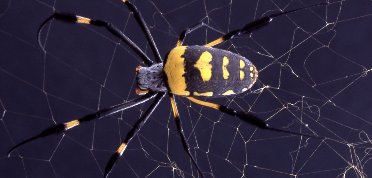
Spider webs inspire liquid wire

Courtesy University of Oxford

Courtesy University of Oxford

Usually, when science talk runs to spider webs the focus is on strength but this research from the UK and France is all about resilience. From a May 16, 2016 news item on phys.org,

Why doesn’t a spider’s web sag in the wind or catapult flies back out like a trampoline? The answer, according to new research by an international team of scientists, lies in the physics behind a ‘hybrid’ material produced by spiders for their webs.

Pulling on a sticky thread in a garden spider’s orb web and letting it snap back reveals that the thread never sags but always stays taut—even when stretched to many times its original length. This is because any loose thread is immediately spooled inside the tiny droplets of watery glue that coat and surround the core gossamer fibres of the web’s capture spiral.

This phenomenon is described in the journal PNAS by scientists from the University of Oxford, UK and the Université Pierre et Marie Curie, Paris, France.

The researchers studied the details of this ‘liquid wire’ technique in spiders’ webs and used it to create composite fibres in the laboratory which, just like the spider’s capture silk, extend like a solid and compress like a liquid. These novel insights may lead to new bio-inspired technology.

A May 16, 2016 University of Oxford press release (also on EurekAlert), which originated the news item, provides more detail,

Professor Fritz Vollrath of the Oxford Silk Group in the Department of Zoology at Oxford University said: ‘The thousands of tiny droplets of glue that cover the capture spiral of the spider’s orb web do much more than make the silk sticky and catch the fly. Surprisingly, each drop packs enough punch in its watery skins to reel in loose bits of thread. And this winching behaviour is used to excellent effect to keep the threads tight at all times, as we can all observe and test in the webs in our gardens.’

The novel properties observed and analysed by the scientists rely on a subtle balance between fibre elasticity and droplet surface tension. Importantly, the team was also able to recreate this technique in the laboratory using oil droplets on a plastic filament. And this artificial system behaved just like the spider’s natural winch silk, with spools of filament reeling and unreeling inside the oil droplets as the thread extended and contracted.

Dr Hervé Elettro, the first author and a doctoral researcher at Institut Jean Le Rond D’Alembert, Université Pierre et Marie Curie, Paris, said: ‘Spider silk has been known to be an extraordinary material for around 40 years, but it continues to amaze us. While the web is simply a high-tech trap from the spider’s point of view, its properties have a huge amount to offer the worlds of materials, engineering and medicine.

‘Our bio-inspired hybrid threads could be manufactured from virtually any components. These new insights could lead to a wide range of applications, such as microfabrication of complex structures, reversible micro-motors, or self-tensioned stretchable systems.’

Here’s a link to and a citation for the paper,

In-drop capillary spooling of spider capture thread inspires hybrid fibers with mixed solid–liquid mechanical properties by Hervé Elettro, Sébastien Neukirch, Fritz Vollrath, and Arnaud Antkowiak. PNAS doi: 10.1073/pnas.1602451113

This paper appears to be open access.
Mimicking the sea urchin’s mouth and teeth for space exploration

Researchers at the University of California at San Diego (UCSD) have designed a new device for use in space exploration that is based on the structure and mechanics of a sea urchin’s mouth and teeth. From a May 2, 2016 news item on ScienceDaily,

The sea urchin’s intricate mouth and teeth are the model for a claw-like device developed by a team of engineers and marine biologists at the University of California, San Diego to sample sediments on other planets, such as Mars. The researchers detail their work in a recent issue of the Journal of Visualized Experiments.

A May 2, 2016 UCSD press release (also on EurekAlert), which originated the news item, expands on the theme by hearkening back to Aristotle (a Greek philosopher),

The urchin’s mouthpiece was first described in detail by the Greek philosopher Aristotle, earning it the nickname “Aristotle’s lantern.” It is comprised of an intricate framework of muscles and five curved teeth with triangle-shaped tips that can scrape, cut, chew and bore holes into the toughest rocks—a colony of sea urchins can destroy an entire kelp forest by churning through rock and uprooting seaweed.  The teeth are arranged in a dome-like formation that opens outwards and closes inwards in a smooth motion, similar to a claw in an arcade prize-grabbing machine.

The news release goes on to describe the methodology,

Bio-inspiration for the study came from pink sea urchins (Strongylocentrotus fragilis), which live off the West Coast of North America, at depths ranging from 100 to 1000 meters in the Pacific Ocean. The urchins were collected for scientific research by the Scripps Institution of Oceanography at UC San Diego.

Researchers extracted the urchins’ mouthpieces, scanned them with microCT, essentially a 3D microscopy technique, and analyzed the structures at the National Center for Microscopy and Imaging Research at the School of Medicine at UC San Diego. This allowed engineers to build a highly accurate model of the mouthpiece’s geometry.

Researchers also used finite element analysis to investigate the structure of the teeth, a method that allowed them to determine the importance of the keel to the teeth’s performance.

Engineers then turned the microCT data into a user-friendly file that a team of undergraduate engineering students at UC San Diego used to start iterating prototypes of the claw-like device, under the supervision of Ph.D. students in McKittrick’s lab.

The first iteration was very close to the mouthpiece’s natural structure, but didn’t do a very good job at grasping sand.  In the second iteration, students flattened the pointed end of the teeth so the device would scoop up sand better. But the device wasn’t opening quite right. Finally, on the third iteration, they connected the teeth differently to the rest of the device, which allowed it to open much easier. The students were able to quickly modify each prototype by using 3D printers in the UC San Diego Design Studio.

The device was then attached to a remote-controlled small rover. The researchers first tested the claw on beach sand, where it performed well. They then used the claw on sand that simulates Martian soil in density and humidity (or lack thereof). The device was able to scoop up sand efficiently. Researchers envision a fleet of mini rovers equipped with the claw that could be deployed to collect samples and bring them back to a main rover. Frank hopes that this design will be of interest to NASA [US National Aeronautics and Space Administraton] and SpaceX [a private enterprise for designing, manufacturing, and launching craft bound for space].

Here’s a link to and a citation for the paper,

A Protocol for Bioinspired Design: A Ground Sampler Based on Sea Urchin Jaws by Michael B. Frank, Steven E. Naleway, Taylor S. Wirth, Jae-Young Jung, Charlene L. Cheung, Faviola B. Loera, Sandra Medina, Kirk N. Sato, Jennifer R. A. Taylor, Joanna McKittrick. Journal of Visualized Experiments, 2016; (110) DOI: 10.3791/53554 Date Published: 4/24/2016

This paper and its video are behind a paywall. For those unfamiliar with the Journal of Visualized Experiments (JOVE), it is focused largely on videos which demonstrate the various techniques and protocols being described in the accompanying papers.

The researchers have made an introductory video available courtesy of UCSD,

Nanotechnology-enabled flame retardant coating

This is a pretty remarkable demonstration made more so when you find out the flame retardant is naturally derived and nontoxic. From an Oct. 5, 2015 news item on Nanowerk,

Inspired by a naturally occurring material found in marine mussels, researchers at The University of Texas at Austin have created a new flame retardant to replace commercial additives that are often toxic and can accumulate over time in the environment and living animals, including humans.

An Oct. 5, 2015 University of Texas news release, which originated the news item, describes the situation with regard to standard flame retardants and what makes this new flame retardant technology so compelling,

Flame retardants are added to foams found in mattresses, sofas, car upholstery and many other consumer products. Once incorporated into foam, these chemicals can migrate out of the products over time, releasing toxic substances into the air and environment. Throughout the United States, there is pressure on state legislatures to ban flame retardants, especially those containing brominated compounds (BRFs), a mix of human-made chemicals thought to pose a risk to public health.

A team led by Cockrell School of Engineering associate professor Christopher Ellison found that a synthetic coating of polydopamine — derived from the natural compound dopamine — can be used as a highly effective, water-applied flame retardant for polyurethane foam. Dopamine is a chemical compound found in humans and animals that helps in the transmission of signals in the brain and other vital areas. The researchers believe their dopamine-based nanocoating could be used in lieu of conventional flame retardants.

“Since polydopamine is natural and already present in animals, this question of toxicity immediately goes away,” Ellison said. “We believe polydopamine could cheaply and easily replace the flame retardants found in many of the products that we use every day, making these products safer for both children and adults.”

Using far less polydopamine by weight than typical of conventional flame retardant additives, the UT Austin team found that the polydopamine coating on foams leads to a 67 percent reduction in peak heat release rate, a measure of fire intensity and imminent danger to building occupants or firefighters. The polydopamine flame retardant’s ability to reduce the fire’s intensity is about 20 percent better than existing flame retardants commonly used today.

Researchers have studied the use of synthetic polydopamaine for a number of health-related applications, including cancer drug delivery and implantable biomedical devices. However, the UT Austin team is thought to be one of the first to pursue the use of polydopamine as a flame retardant. To the research team’s surprise, they did not have to change the structure of the polydopamine from its natural form to use it as a flame retardant. The polydopamine was coated onto the interior and exterior surfaces of the polyurethane foam by simply dipping it into a water solution of dopamine for several days.

Ellison said he and his team were drawn to polydopamine because of its ability to adhere to surfaces as demonstrated by marine mussels who use the compound to stick to virtually any surface, including Teflon, the material used in nonstick cookware. Polydopamine also contains a dihydroxy-ring structure linked with an amine group that can be used to scavenge or remove free radicals. Free radicals are produced during the fire cycle as a polymer degrades, and their removal is critical to stopping the fire from continuing to spread. Polydopamine also produces a protective coating called char, which blocks fire’s access to its fuel source — the polymer. The synergistic combination of both these processes makes polydopamine an attractive and powerful flame retardant.

Ellison and his team are now testing to see whether they can shorten the nanocoating treatment process or develop a more convenient application process.

“We believe this alternative to flame retardants can prove very useful to removing potential hazards from products that children and adults use every day,” Ellison said. “We weren’t expecting to find a flame retardant in nature, but it was a serendipitous discovery.”

Here’s a link to and a citation for the paper,

Bioinspired Catecholic Flame Retardant Nanocoating for Flexible Polyurethane Foams by Joon Hee Cho, Vivek Vasagar, Kadhiravan Shanmuganathan, Amanda R. Jones, Sergei Nazarenko, and Christopher J. Ellison. Chem. Mater., 2015, 27 (19), pp 6784–6790 DOI: 10.1021/acs.chemmater.5b03013
Publication Date (Web): September 9, 2015
Copyright © 2015 American Chemical Society

This paper is behind a paywall. It should be noted that researchers from the University of Southern Mississippi and the Council of Scientific & Industrial Research (CSIR)-National Chemical Laboratory in Pune, India were also involved in this work.

Building nanocastles in the sand

Scientists have taken inspiration from sandcastles to build robots made of nanoparticles. From an Aug. 5, 2015 news item on ScienceDaily,

If you want to form very flexible chains of nanoparticles in liquid in order to build tiny robots with flexible joints or make magnetically self-healing gels, you need to revert to childhood and think about sandcastles.

In a paper published this week in Nature Materials, researchers from North Carolina State University and the University of North Carolina-Chapel Hill show that magnetic nanoparticles encased in oily liquid shells can bind together in water, much like sand particles mixed with the right amount of water can form sandcastles.

An Aug. 5, 2015 North Carolina State University (NCSU) news release (also on EurekAlert) by Mick Kulikowski, which originated the news item, expands on the theme,

“Because oil and water don’t mix, the oil wets the particles and creates capillary bridges between them so that the particles stick together on contact,” said Orlin Velev, INVISTA Professor of Chemical and Biomolecular Engineering at NC State and the corresponding author of the paper.

“We then add a magnetic field to arrange the nanoparticle chains and provide directionality,” said Bhuvnesh Bharti, research assistant professor of chemical and biomolecular engineering at NC State and first author of the paper.

Chilling the oil is like drying the sandcastle. Reducing the temperature from 45 degrees Celsius to 15 degrees Celsius freezes the oil and makes the bridges fragile, leading to breaking and fragmentation of the nanoparticle chains. Yet the broken nanoparticles chains will re-form if the temperature is raised, the oil liquefies and an external magnetic field is applied to the particles.

“In other words, this material is temperature responsive, and these soft and flexible structures can be pulled apart and rearranged,” Velev said. “And there are no other chemicals necessary.”

The paper is also co-authored by Anne-Laure Fameau, a visiting researcher from INRA [French National Institute for Agricultural Research or Institut National de la Recherche Agronomique], France. …

Here’s a link to and a citation for the paper,

Nanocapillarity-mediated magnetic assembly of nanoparticles into ultraflexible filaments and reconfigurable networks by Bhuvnesh Bharti, Anne-Laure Fameau, Michael Rubinstein, & Orlin D. Velev. Nature Materials (2015) doi:10.1038/nmat4364 Published online 03 August 2015

This paper is behind a paywall.

Micro-supercapacitor, leaves, and Korea’s Institute for Basic Sciences

South Korea’s research on creating micro-supercapacitors (MSC) was first published online in February 2015 but it seems the researchers decided to promote the work after its print publication in May 2015.

A July 2, 2015 news item on Nanotechnology Now makes the announcement,

There was a time during the early development of portable electronics when the biggest hurdle to overcome was making the device small enough to be considered portable.  After the invention of the microprocessor in the early 1970s, miniature, portable electronics have become commonplace and ever since the next challenge has been finding an equally small and reliable power source.  Chemical batteries store a lot of energy but require a long period of time for that energy to charge and discharge plus have a limited lifespan.  Capacitors charge quickly but cannot store enough charge to work for long enough to be practical.  One possible solution is something called a solid-state micro-supercapacitor (MSC).  Supercapacitors are armed with the power of a battery and can also sustain that power for a prolonged period time.  Researchers have attempted to create MSCs in the past using various hybrids of metals and polymers but none were suitable for practical use.  In more recent trials using graphene and carbon nanotubes to make MSCs, the results were similarly lackluster.

An international team of researchers led by Young Hee Lee, including scientists from the Center for Integrated Nanostructure Physics at the Institute for Basic Science (IBS) and Department of Energy Science at Sungkyunkwan University in South Korea, has devised a new technique for creating an MSC that doesn’t have the shortcomings of previous attempts but instead delivers high electrochemical performance.

A June 29, 2015 South Korea Institute for Basic Science (IBS) press release by Daniel Kopperud, which originated the news item, reveals this research is bioinspired,

When designing something new and complex, sometimes the best inspiration is one already found in nature.  The team modeled their MSC film structure on natural vein-textured leaves in order to take advantage of the natural transport pathways which enable efficient ion diffusion parallel to the graphene planes found within them.

To create this final, efficient shape, the team layered a graphene-hybrid film with copper hydroxide nanowires.  After many alternating layers they achieved the desired thickness, and added an acid solution to dissolve the nanowires so that a thin film with nano-impressions was all that remained.

To fabricate the MSCs the film was applied to a plastic layer with thin, ~5μm long parallel gold strips placed on top.  Everything not covered by the gold strips was chemically etched away so that only the gold strips on top of a layer of film were left.  Gold contact pads perpendicular to the gold strips were added and a conductive gel filled in the remaining spaces and was allowed to solidify.  Once peeled from the plastic layer, the finished MSCs resemble clear tape with gold electrical leads on opposite sides.

The team produced stunning test results. In addition to its superior energy density, the film is highly flexible and actually increases capacitance after initial use.  The volumetric energy density was 10 times higher than currently available commercial supercapacitors and also far superior to any other recent research.  The MSCs are displaying electrical properties about five orders of magnitude higher than similar lithium batteries and are comparable to existing, larger supercapacitors.  According to Lee, “To our knowledge, the volumetric energy density and the maximum volumetric power density in our work are the highest values among all carbon-based solid-state MSCs reported to date.”

In the future, consumers will likely power their devices with MSCs instead of batteries.  Applications for light, reliable energy storage combined with a long lifespan and fast charge/discharge time.  The team’s MSCs could be embedded into an electronic circuit chip as power sources for practical applications such as implantable medical devices, active radio frequency identification tags, and micro robots.  If engineers utilize the material’s incredible flexibility, these MSCs could be utilized in portable, stretchable, and even wearable electronic devices.

Here’s a link to and a citation for the paper,

Leaf Vein-Inspired Nanochanneled Graphene Film for Highly Efficient Micro-Supercapacitors by Jian Chang, Subash Adhikari, Tae Hoon Lee, Bing Li, Fei Yao, Duy Tho Pham, Viet Thong Le, and Young Hee Lee. Advanced Energy Materials, Volume 5, Issue 9, May 6, 2015 First published online Feb. 20, 2015 DOI: 10.1002/aenm.201500003

This paper is behind a paywall.

Reducing friction with snakeskin-inspired surface

A June 30, 2015 Institute of Physics press release (also on EurekAlert) explains how snakeskin may inspire a whole new generation of robots bound for outer space along with other more earth-bound applications,

Snakeskin-inspired surfaces smash records, providing an astonishing 40% friction reduction in tests of high performance materials.

These new surfaces could improve the reliability of mechanical components in machines such as high performance cars and add grist to the mill of engineers designing a new generation of space exploration robots.

The skin of many snakes and lizards has been studied by biologists and has long been known to provide friction reduction to the animal as it moves. It is also resistant to wear, particularly in environments that are dry and dusty or sandy.

Dr Greiner and his team used a laser to etch the surface of a steel pin so that it closely resembled the texture of snakeskin. They then tested the friction created when the pin moved against another surface.

In dry conditions, i.e. with no oil or other lubricant, the scale-like surface created far less friction – 40% less – than its smooth counterpart.

Lead researcher Dr Christian Greiner said: “If we’d managed just a 1% reduction in friction, our engineering colleagues would have been delighted; 40% really is a leap forward and everyone is very excited.”

Applications are likely to be in mechanical devices that are made to a micro or nano scale. Familiar examples include the sensors in car anti-lock braking systems, computer hard disk drives, and accelerometers in mobile phones, which enable the device to determine for example whether it’s in portrait or landscape mode.

“Our new surface texture will mainly come into its own when engineers are really looking to push the envelope,” Dr Greiner said.

The snakeskin surface could be used in very high-end automotive engineering, such as Formula 1 racing cars. It could also be used in highly sensitive scientific equipment, including sensors installed in synchrotrons such as the Diamond Light Source in the UK or the Large Hadron Collider in Switzerland, and anywhere the engineering challenge is to further miniaturise moving parts.

There is interest in snakeskin-inspired materials from the robotics sector, too, which is designing robots inspired by snakes, which could aid exploration of very dusty environments, including those in space. This raises a new challenge for Dr Greiner’s team: to make a material that decreases friction in only one direction.

Anyone who has felt snakeskin will know that the scales all lie in the same direction and are articulated to aid the snake in its forward motion, while resisting backwards motion. The steel pins tested in this research mimic only the overall surface texture of snakeskin and reduce friction in at least two directions. Dr Greiner has made some progress with polymers that even more closely mimic snakeskin to reduce friction in only one direction. It is, he says, early days and this later work is not yet scheduled for publication.

The only caution is that this new surface doesn’t work well in an environment where oil or another lubricant is present. In fact, the snakeskin effect created three times more friction with lubricant than an equivalent smooth surface.

“This wasn’t a huge surprise,” Dr Greiner explained, “since we were looking to nature for inspiration and the species we mimicked – the royal python and a lizard called a sandfish skink – live in very dry environments and don’t secrete oils or other liquids onto their skin.”

Here’s a link to and citation for the paper,

Bio-inspired scale-like surface textures and their tribological properties by Christian Greiner and Michael Schäfer. Bioinspir. Biomim. 10 044001 doi:10.1088/1748-3190/10/4/044001 Published 30 June 2015

This paper is open access.

Researchers at Karolinska Institute (Sweden) build an artificial neuron

Unlike my post earlier today (June 26, 2015) about BrainChip, this is not about neuromorphic engineering (artificial brain), although I imagine this new research from the Karolinska Institute (Institutet) will be of some interest to that community. This research was done in the interest of developing* therapeutic interventions for brain diseases. One aspect of this news item/press release I find particularly interesting is the insistence that “no living parts” were used to create the artificial neuron,

A June 24, 2015 news item on ScienceDaily describes what the artificial neuron can do,

Scientists have managed to build a fully functional neuron by using organic bioelectronics. This artificial neuron contain [sic] no ‘living’ parts, but is capable of mimicking the function of a human nerve cell and communicate in the same way as our own neurons do. [emphasis mine]

A June 24, 2015 Karolinska Institute press release (also on EurekAlert), which originated the news item, describes how neurons communicate in the brain, standard techniques for stimulating neuronal cells, and the scientists’ work on a technique to improve stimulation,

Neurons are isolated from each other and communicate with the help of chemical signals, commonly called neurotransmitters or signal substances. Inside a neuron, these chemical signals are converted to an electrical action potential, which travels along the axon of the neuron until it reaches the end. Here at the synapse, the electrical signal is converted to the release of chemical signals, which via diffusion can relay the signal to the next nerve cell.

To date, the primary technique for neuronal stimulation in human cells is based on electrical stimulation. However, scientists at the Swedish Medical Nanoscience Centre (SMNC) at Karolinska Institutet in collaboration with collegues at Linköping University, have now created an organic bioelectronic device that is capable of receiving chemical signals, which it can then relay to human cells.

“Our artificial neuron is made of conductive polymers and it functions like a human neuron,” says lead investigator Agneta Richter-Dahlfors, professor of cellular microbiology. “The sensing component of the artificial neuron senses a change in chemical signals in one dish, and translates this into an electrical signal. This electrical signal is next translated into the release of the neurotransmitter acetylcholine in a second dish, whose effect on living human cells can be monitored.”

The research team hope that their innovation, presented in the journal Biosensors & Bioelectronics, will improve treatments for neurologial disorders which currently rely on traditional electrical stimulation. The new technique makes it possible to stimulate neurons based on specific chemical signals received from different parts of the body. In the future, this may help physicians to bypass damaged nerve cells and restore neural function.

“Next, we would like to miniaturize this device to enable implantation into the human body,” says Agneta Richer-Dahlfors. “We foresee that in the future, by adding the concept of wireless communication, the biosensor could be placed in one part of the body, and trigger release of neurotransmitters at distant locations. Using such auto-regulated sensing and delivery, or possibly a remote control, new and exciting opportunities for future research and treatment of neurological disorders can be envisaged.”

Here’s a link to and a citation for the paper,

An organic electronic biomimetic neuron enables auto-regulated neuromodulation by Daniel T. Simon, Karin C. Larsson, David Nilsson, Gustav Burström, b, Dagmar Galter, Magnus Berggren, and Agneta Richter-Dahlfors. Biosensors and Bioelectronics Volume 71, 15 September 2015, Pages 359–364         doi:10.1016/j.bios.2015.04.058

This paper is behind a paywall.

As to anyone (other than myself) who may be curious about exactly what they used (other than “living parts”) to create an artificial neuron, there’s the paper’s abstract,

Current therapies for neurological disorders are based on traditional medication and electric stimulation. Here, we present an organic electronic biomimetic neuron, with the capacity to precisely intervene with the underlying malfunctioning signalling pathway using endogenous substances. The fundamental function of neurons, defined as chemical-to-electrical-to-chemical signal transduction, is achieved by connecting enzyme-based amperometric biosensors and organic electronic ion pumps. Selective biosensors transduce chemical signals into an electric current, which regulates electrophoretic delivery of chemical substances without necessitating liquid flow. Biosensors detected neurotransmitters in physiologically relevant ranges of 5–80 µM, showing linear response above 20 µm with approx. 0.1 nA/µM slope. When exceeding defined threshold concentrations, biosensor output signals, connected via custom hardware/software, activated local or distant neurotransmitter delivery from the organic electronic ion pump. Changes of 20 µM glutamate or acetylcholine triggered diffusive delivery of acetylcholine, which activated cells via receptor-mediated signalling. This was observed in real-time by single-cell ratiometric Ca2+ imaging. The results demonstrate the potential of the organic electronic biomimetic neuron in therapies involving long-range neuronal signalling by mimicking the function of projection neurons. Alternatively, conversion of glutamate-induced descending neuromuscular signals into acetylcholine-mediated muscular activation signals may be obtained, applicable for bridging injured sites and active prosthetics.

While it’s true neither are “living parts,” I believe both enzymes and organic electronic ion pumps can be found in biological organisms. The insistence on ‘nonliving’ in the press release suggests that scientists in Europe, if nowhere else, are still quite concerned about any hint that they are working on genetically modified organisms (GMO). It’s ironic when you consider that people blithely use enzyme-based cleaning and beauty products but one can appreciate the* scientists’ caution.

* ‘develop’ changed to ‘developing’ and ‘the’ added on July 3, 2015.

How geckos self-clean, even in dusty environments

An Australian research team claims a world first with regard to ‘gecko research’ according to a March 16, 2015* news item on ScienceDaily,

In a world first, a research team including James Cook University [JCU] scientists has discovered how geckos manage to stay clean, even in dusty deserts.

The process, described in Interface, a journal of the Royal Society, may also turn out to have important human applications.

JCU’s Professor Lin Schwarzkopf said the group found that tiny droplets of water on geckos, for instance from condensing dew, come into contact with hundreds of thousands of extremely small hair-like spines that cover the animals’ bodies.

A March 16, 2015 JCU press release (also on EurekAlert), which originated the news item, provides more detail,

“If you have seen how drops of water roll off a car after it is waxed, or off a couch that’s had protective spray used on it, you’ve seen the process happening,” she said. “The wax and spray make the surface very bumpy at micro and nano levels, and the water droplets remain as little balls, which roll easily and come off with gravity or even a slight wind.”

The geckos’ hair-like spines trap pockets of air and work on the same principle, but have an even more dramatic effect. Through a scanning electron microscope, tiny water droplets can be seen rolling into each other and jumping like popcorn off the skin of the animal as they merge and release energy.

Scientists were aware that hydrophobic surfaces repelled water, and that the rolling droplets helped clean the surfaces of leaves and insects, but this is the first time it has been documented in a vertebrate animal. Box-patterned geckos live in semi-arid habitats, with little rain but may have dew forming on them when the temperature drops overnight.

Professor Schwarzkopf said the process may help geckos keep clean, as the water can carry small particles of dust and dirt away from their body. “They tend to live in dry environments where they can’t depend on it raining, and this keeps process them clean,” she said.

She said there were possible applications for marine-based electronics that have to shed water quickly in use and for possible “superhydrophobic” clothing that would not get wet or dirty and would never need washing.

I’ve been reading about self-cleaning products for years now. (sigh) Where are they? Despite this momentary lapse into sighing and wailing, I am much encouraged that scientists are still trying to figure out how to create self-cleaning products.

Here’s a link to and a citation for the paper,

Removal mechanisms of dew via self-propulsion off the gecko skin by Gregory S. Watson, Lin Schwarzkopf, Bronwen W. Cribb, Sverre Myhra, Marty Gellender, and Jolanta A. Watson.
Interface, April 2015, Volume: 12 Issue: 105 DOI: 10.1098/rsif.2014.1396 Published 11 March 2015

This paper is open access.

*’2014′ corrected to ‘2015’ on Feb. 28, 2017.