Tag Archives: biomimetics

Spider glue

Caption: An orb spider, glue-maker extraordinaire, at work on a web. Credit: The University of Akron

Scientists are taking inspiration from spiders in their quest to develop better adhesives. (Are they abandoning the gecko? Usually when scientists study adhesiveness, there’s talk of geckos. From a June 5, 2018 news item on ScienceDaily,

Ever wonder why paint peels off the wall during summer’s high humidity? It’s the same reason that bandages separate from skin when we bathe or swim.

Interfacial water, as it’s known, forms a slippery and non-adhesive layer between the glue and the surface to which it is meant to stick, interfering with the formation of adhesive bonds between the two.

Overcoming the effects of interfacial water is one of the challenges facing developers of commercial adhesives.

To find a solution, researchers at The University of Akron (UA) are looking to one of the strongest materials found in nature: spider silk.

The sticky glue that coats the silk threads of spider webs is a hydrogel, meaning it is full of water. One would think, then, that spiders would have difficulty catching prey, especially in humid conditions — but they do not. In fact, their sticky glue, which has been a subject of intensive research for years, is one of the most effective biological glues in all of nature.

A June 4, 2018 University of Akron news release (also on EurekAlert published on June 5, 2018), which originated the news item, provides more detail,

So how is spider glue able to stick in highly humid conditions?

That question was the subject of investigation by UA graduate students Saranshu Singla, Gaurav Amarpuri and Nishad Dhopatkar, who have been working with Dr. Ali Dhinojwala, interim dean of the College of Polymer Science and Polymer Engineering, and Dr. Todd Blackledge, professor of biology in the Integrated Bioscience program. Both professors are principal investigators in UA’s Biomimicry Research Innovation Center [BRIC], which specializes in emulating biological forms, processes, patterns and systems to solve technical challenges.

The team’s findings, which may provide the clue to developing stronger commercial adhesives, can be read in a paper recently published in the journal Nature Communications.

Singla and her colleagues set out to examine the secret behind the success of the common orb spider (Larinioides cornutus) glue and uncover how it overcomes the primary obstacle of achieving good adhesion in the humid conditions where water could be present between the glue and the target surface.

To investigate the processes involved, the team took orb spider glue, set it on sapphire substrate, then examined it using a combination of interface-sensitive spectroscopy and infrared spectroscopy.

Spider glue is made of three elements: two specialized glycoproteins, a collection of low molecular mass organic and inorganic compounds (LMMCs), and water. The LMMCs are hygroscopic (water-attracting), which keeps the glue soft and tacky to stick.

Singla and her team discovered that these glycoproteins act as primary binding agents to the surface. Glycoprotein-based glues have been identified in several other biological glues, such as fungi, algae, diatoms, sea stars, sticklebacks and English ivy.

But why doesn’t the water present in the spider glue interfere with the adhesive contact the way it does with most synthetic adhesives?

The LMMCs, the team concluded, perform a previously unknown function of sequestering interfacial water, preventing adhesive failure.

Singla and colleagues determined that it is the interaction of glycoproteins and LMMCs that governs the adhesive quality of the glue produced, with the respective proportions varying across species, thus optimizing adhesive strength to match the relative humidity of spider habitat.

“The hygroscopic compounds – known as water-absorbers – in spider glue play a previously unknown role in moving water away from the boundary, thereby preventing failure of spider glue at high humidity,” explained Singla.

The ability of the spider glue to overcome the problem of interfacial water by effectively absorbing it is the key finding of the research, and the one with perhaps the strongest prospect for commercial development.

“Imagine a paint that is guaranteed for life, come rain or shine,” Singla remarked.

All thanks to your friendly neighborhood spider glue.

Here’s a link to and a citation for the paper,

Hygroscopic compounds in spider aggregate glue remove interfacial water to maintain adhesion in humid conditions by Saranshu Singla, Gaurav Amarpuri, Nishad Dhopatkar, Todd A. Blackledge, & Ali Dhinojwala. Nature Communicationsvolume 9, Article number: 1890 (2018) Published 22 May 2018 DOI: https://doi.org/10.1038/s41467-018-04263-z

This paper is open access.

Killing bacteria on contact with dragonfly-inspired nanocoating

Scientists in Singapore were inspired by dragonflies and cicadas according to a March 28, 2018 news item on Nanowerk (Note: A link has been removed),

Studies have shown that the wings of dragonflies and cicadas prevent bacterial growth due to their natural structure. The surfaces of their wings are covered in nanopillars making them look like a bed of nails. When bacteria come into contact with these surfaces, their cell membranes get ripped apart immediately and they are killed. This inspired researchers from the Institute of Bioengineering and Nanotechnology (IBN) of A*STAR to invent an anti-bacterial nano coating for disinfecting frequently touched surfaces such as door handles, tables and lift buttons.

This technology will prove particularly useful in creating bacteria-free surfaces in places like hospitals and clinics, where sterilization is important to help control the spread of infections. Their new research was recently published in the journal Small (“ZnO Nanopillar Coated Surfaces with Substrate-Dependent Superbactericidal Property”)

Image 1: Zinc oxide nanopillars that looked like a bed of nails can kill a broad range of germs when used as a coating on frequently-touched surfaces. Courtesy: A*STAR

A March 28, 2018 Agency for Science Technology and Research (A*STAR) press release, which originated the news item, describes the work further,

80% of common infections are spread by hands, according to the B.C. [province of Canada] Centre for Disease Control1. Disinfecting commonly touched surfaces helps to reduce the spread of harmful germs by our hands, but would require manual and repeated disinfection because germs grow rapidly. Current disinfectants may also contain chemicals like triclosan which are not recognized as safe and effective 2, and may lead to bacterial resistance and environmental contamination if used extensively.

“There is an urgent need for a better way to disinfect surfaces without causing bacterial resistance or harm to the environment. This will help us to prevent the transmission of infectious diseases from contact with surfaces,” said IBN Executive Director Professor Jackie Y. Ying.

To tackle this problem, a team of researchers led by IBN Group Leader Dr Yugen Zhang created a novel nano coating that can spontaneously kill bacteria upon contact. Inspired by studies on dragonflies and cicadas, the IBN scientists grew nanopilllars of zinc oxide, a compound known for its anti-bacterial and non-toxic properties. The zinc oxide nanopillars can kill a broad range of germs like E. coli and S. aureus that are commonly transmitted from surface contact.

Tests on ceramic, glass, titanium and zinc surfaces showed that the coating effectively killed up to 99.9% of germs found on the surfaces. As the bacteria are killed mechanically rather than chemically, the use of the nano coating would not contribute to environmental pollution. Also, the bacteria will not be able to develop resistance as they are completely destroyed when their cell walls are pierced by the nanopillars upon contact.

Further studies revealed that the nano coating demonstrated the best bacteria killing power when it is applied on zinc surfaces, compared with other surfaces. This is because the zinc oxide nanopillars catalyzed the release of superoxides (or reactive oxygen species), which could even kill nearby free floating bacteria that were not in direct contact with the surface. This super bacteria killing power from the combination of nanopillars and zinc broadens the scope of applications of the coating beyond hard surfaces.

Subsequently, the researchers studied the effect of placing a piece of zinc that had been coated with zinc oxide nanopillars into water containing E. coli. All the bacteria were killed, suggesting that this material could potentially be used for water purification.

Dr Zhang said, “Our nano coating is designed to disinfect surfaces in a novel yet practical way. This study demonstrated that our coating can effectively kill germs on different types of surfaces, and also in water. We were also able to achieve super bacteria killing power when the coating was used on zinc surfaces because of its dual mechanism of action. We hope to use this technology to create bacteria-free surfaces in a safe, inexpensive and effective manner, especially in places where germs tend to accumulate.”

IBN has recently received a grant from the National Research Foundation, Prime Minister’s Office, Singapore, under its Competitive Research Programme to further develop this coating technology in collaboration with Tan Tock Seng Hospital for commercial application over the next 5 years.

1 B.C. Centre for Disease Control

2 U.S. Food & Drug Administration

(I wasn’t expecting to see a reference to my home province [BC Centre for Disease Control].) Back to the usual, here’s a link to and a citation for the paper,

ZnO Nanopillar Coated Surfaces with Substrate‐Dependent Superbactericidal Property by Guangshun Yi, Yuan Yuan, Xiukai Li, Yugen Zhang. Small https://doi.org/10.1002/smll.201703159 First published: 22 February 2018

This paper is behind a paywall.

One final comment, this research reminds me of research into simulating shark skin because that too has bacteria-killing nanostructures. My latest about the sharkskin research is a Sept, 18, 2014 posting.

A gripping problem: tree frogs lead the way

Courtesy: University of Glasgow

At least once a year, there must be a frog posting here (ETA: July 31, 2018 at 1640 hours: unusually, this is my second ‘frog’ posting in one week; my July 26, 2018 posting concerns a very desperate frog, Romeo). Prior to Romeo, this March 15, 2018 news item on phys.org tickled my fancy,

Scientists researching how tree frogs climb have discovered that a unique combination of adhesion and grip gives them perfect technique.

The new research, led by the University of Glasgow and published today [March 15, 2018] in the Journal of Experimental Biology, could have implications for areas of science such as robotics, as well as the production of climbing equipment and even tyre manufacture.

A March 15, 2018 University of Glasgow press release, which originated the news item, provides a little more detail,

Researchers found that, using their fluid-filled adhesive toe pads, tree frogs are able to grip to surfaces to climb. When surfaces aren’t smooth enough to allow adhesion, researchers found that the frogs relied on their long limbs to grip around objects.

University of Glasgow scientists Iain Hill and Jon Barnes gave the tree frogs a series of narrow and wide cylinders to climb. The research team found that on the narrow cylinders the frogs used their grip and adhesion pads, allowing them to climb the obstacle at speed. Wider cylinders were too large for the frogs to grip, so they could only climb more slowly using their suction adhesive pads.

When the cylinders were coated in sandpaper, preventing adhesion, the frogs could only climb the narrow ones slowly, using their grip. They were not able to climb the wider cylinders covered in sandpaper as they couldn’t use their grip or adhesion.

Dr Barnes said: “I have worked on tree frog research for many years and I find them fascinating. Work on tree frogs has been of interest to industry and other areas of science in the past, since their climbing abilities can offer us insights into the most efficient way to climb and stick to surfaces.

“Climbing robots, for instance, need ways to stick, they could be based either on gecko climbing or tree frog climbing.  This research demonstrates how a good climbing robot would need to combine gripping and adhesion to climb more efficiently.”

The study, “The biomechanics of tree frogs climbing curved surfaces: a gripping problem” is published in the Journal ofExperimental Biology. The work was funded by the Royal Society, London and by grants from the National Natural Science Foundation of China and the Natural Science Foundation of Jiangsu Province.

Here’s a link to and a citation for the paper (I love the pun in the title),

The biomechanics of tree frogs climbing curved surfaces: a gripping problem by Iain D. C. Hill, Benzheng Dong, W. Jon. P. Barnes, Aihong Ji, Thomas Endlein. Journal of Experimental Biology 2018 : jeb.168179 doi: 10.1242/jeb.168179 Published 19 January 2018

This paper is behind a paywall.

New wound dressings with nanofibres for tissue regeneration

The Rotary Jet-Spinning manufacturing system was developed specifically as a therapeutic for the wounds of war. The dressings could be a good option for large wounds, such as burns, as well as smaller wounds on the face and hands, where preventing scarring is important. Illustration courtesy of Michael Rosnach/Harvard University

This image really gets the idea of regeneration across to the viewer while also informing you that this is medicine that comes from the military. A March 19,2018 news item on phys.org announces the work,

Researchers from the Harvard John A. Paulson School of Engineering and Applied Sciences (SEAS) and the Wyss Institute for Biologically Inspired Engineering have developed new wound dressings that dramatically accelerate healing and improve tissue regeneration. The two different types of nanofiber dressings, described in separate papers, use naturally-occurring proteins in plants and animals to promote healing and regrow tissue.

Our fiber manufacturing system was developed specifically for the purpose of developing therapeutics for the wounds of war,” said Kit Parker, the Tarr Family Professor of Bioengineering and Applied Physics at SEAS and senior author of the research. “As a soldier in Afghanistan, I witnessed horrible wounds and, at times, the healing process for those wounds was a horror unto itself. This research is a years-long effort by many people on my team to help with these problems.”

Parker is also a Core Faculty Member of the Wyss Institute.

The most recent paper, published in Biomaterials, describes a wound dressing inspired by fetal tissue.

A March 19, 2018 Harvard University John A. Paulson School of Engineering and Applied Science news release by Leah Burrows (also on EurekAlert), which originated the news item, provides some background information before launching into more detail about this latest work,

In the late 1970s, when scientists first started studying the wound-healing process early in development, they discovered something unexpected: Wounds incurred before the third trimester left no scars. This opened a range of possibilities for regenerative medicine. But for decades, researchers have struggled to replicate those unique properties of fetal skin.

Unlike adult skin, fetal skin has high levels of a protein called fibronectin, which assembles into the extracellular matrix and promotes cell binding and adhesion. Fibronectin has two structures: globular, which is found in blood, and fibrous, which is found in tissue. Even though fibrous fibronectin holds the most promise for wound healing, previous research focused on the globular structure, in part because manufacturing fibrous fibronectin was a major engineering challenge.

But Parker and his team are pioneers in the field of nanofiber engineering.

The researchers made fibrous fibronectin using a fiber-manufacturing platform called Rotary Jet-Spinning (RJS), developed by Parker’s Disease Biophysics Group. RJS works likes a cotton-candy machine — a liquid polymer solution, in this case globular fibronectin dissolved in a solvent, is loaded into a reservoir and pushed out through a tiny opening by centrifugal force as the device spins. As the solution leaves the reservoir, the solvent evaporates and the polymers solidify. The centrifugal force unfolds the globular protein into small, thin fibers. These fibers — less than one micrometer in diameter — can be collected to form a large-scale wound dressing or bandage.

“The dressing integrates into the wound and acts like an instructive scaffold, recruiting different stem cells that are relevant for regeneration and assisting in the healing process before being absorbed into the body,” said Christophe Chantre, a graduate student in the Disease Biophysics Group and first author of the paper.

In in vivo testing, the researchers found that wounds treated with the fibronectin dressing showed 84 percent tissue restoration within 20 days, compared with 55.6 percent restoration in wounds treated with a standard dressing.

The researchers also demonstrated that wounds treated with the fibronectin dressing had almost normal epidermal thickness and dermal architecture, and even regrew hair follicles — often considered one of the biggest challenges in the field of wound healing.

“This is an important step forward,” said Chantre. “Most work done on skin regeneration to date involves complex treatments combining scaffolds, cells, and even growth factors. Here we were able to demonstrate tissue repair and hair follicle regeneration using an entirely material approach. This has clear advantages for clinical translation.”

In another paper published in Advanced Healthcare Materials, the Disease Biophysics Group demonstrated a soy-based nanofiber that also enhances and promotes wound healing.

Soy protein contains both estrogen-like molecules — which have been shown to accelerate wound healing — and bioactive molecules similar to those that build and support human cells.

“Both the soy- and fibronectin-fiber technologies owe their success to keen observations in reproductive medicine,” said Parker. “During a woman’s cycle, when her estrogen levels go high, a cut will heal faster. If you do a surgery on a baby still in the womb, they have scar-less wound healing. Both of these new technologies are rooted in the most fascinating of all the topics in human biology — how we reproduce.”

In a similar way to fibronectin fibers, the research team used RJS to spin ultrathin soy fibers into wound dressings. In experiments, the soy- and cellulose-based dressing demonstrated a 72 percent increase in healing over wounds with no dressing and a 21 percent increase in healing over wounds dressed without soy protein.

“These findings show the great promise of soy-based nanofibers for wound healing,” said Seungkuk Ahn, a graduate student in the Disease Biophysics Group and first author of the paper. “These one-step, cost-effective scaffolds could be the next generation of regenerative dressings and push the envelope of nanofiber technology and the wound-care market.”

Both kinds of dressing, according to researchers, have advantages in the wound-healing space. The soy-based nanofibers — consisting of cellulose acetate and soy protein hydrolysate — are inexpensive, making them a good option for large-scale use, such as on burns. The fibronectin dressings, on the other hand, could be used for smaller wounds on the face and hands, where preventing scarring is important.

Here’s are links and citations for both papers mentioned in the news release,

Soy Protein/Cellulose Nanofiber Scaffolds Mimicking Skin Extracellular Matrix for Enhanced Wound Healing by Seungkuk Ahn, Christophe O. Chantre, Alanna R. Gannon, Johan U. Lind, Patrick H. Campbell, Thomas Grevesse, Blakely B. O’Connor, Kevin Kit Parker. Advanced Healthcare Materials https://doi.org/10.1002/adhm.201701175 First published: 23 January 2018

Production-scale fibronectin nanofibers promote wound closure and tissue repair in a dermal mouse model by Christophe O. Chantre, Patrick H. Campbell, Holly M. Golecki, Adrian T. Buganza, Andrew K. Capulli, Leila F. Deravi, Stephanie Dauth, Sean P. Sheehy, Jeffrey A.Paten. KarlGledhill, Yanne S. Doucet, Hasan E.Abaci, Seungkuk Ahn, Benjamin D.Pope, Jeffrey W.Ruberti, Simon P.Hoerstrup, Angela M.Christiano, Kevin Kit Parker. Biomaterials Volume 166, June 2018, Pages 96-108 https://doi.org/10.1016/j.biomaterials.2018.03.006 Available online 5 March 2018

Both papers are behind paywalls although you may want to check with ResearchGate where many researchers make their papers available for free.

One last comment, I noticed this at the end of Burrows’ news release,

The Harvard Office of Technology Development has protected the intellectual property relating to these projects and is exploring commercialization opportunities.

It reminded me of the patent battle between the Broad Institute (a Harvard University and Massachusetts Institute of Technology joint venture) and the University of California at Berkeley over CRISPR (clustered regularly interspaced short palindromic repeats) technology. (My March 15, 2017 posting describes the battle’s outcome.)

Lest we forget, there could be major financial rewards from this work.

Stronger than steel and spider silk: artificial, biodegradable, cellulose nanofibres

This is an artificial and biodegradable are two adjectives you don’t usually see united by the conjunction, and. However, it is worth noting that the artificial material is initially derived from a natural material, cellulose. Here’s more from a May 16, 2018 news item on ScienceDaily,

At DESY’s [Deutsches Elektronen-Synchrotron] X-ray light source PETRA III, a team led by Swedish researchers has produced the strongest bio-material that has ever been made. The artifical, but bio-degradable cellulose fibres are stronger than steel and even than dragline spider silk, which is usually considered the strongest bio-based material. The team headed by Daniel Söderberg from the KTH Royal Institute of Technology in Stockholm reports the work in the journal ACS Nano of the American Chemical Society.

A May 16, 2018 DESY press release (also on EurekAlert), which originated the news item, provides more detail,

The ultrastrong material is made of cellulose nanofibres (CNF), the essential building blocks of wood and other plant life. Using a novel production method, the researchers have successfully transferred the unique mechanical properties of these nanofibres to a macroscopic, lightweight material that could be used as an eco-friendly alternative for plastic in airplanes, cars, furniture and other products. “Our new material even has potential for biomedicine since cellulose is not rejected by your body”, explains Söderberg.

The scientists started with commercially available cellulose nanofibres that are just 2 to 5 nanometres in diameter and up to 700 nanometres long. A nanometre (nm) is a millionth of a millimetre. The nanofibres were suspended in water and fed into a small channel, just one millimetre wide and milled in steel. Through two pairs of perpendicular inflows additional deionized water and water with a low pH-value entered the channel from the sides, squeezing the stream of nanofibres together and accelerating it.

This process, called hydrodynamic focussing, helped to align the nanofibres in the right direction as well as their self-organisation into a well-packed macroscopic thread. No glue or any other component is needed, the nanofibres assemble into a tight thread held together by supramolecular forces between the nanofibres, for example electrostatic and Van der Waals forces.

With the bright X-rays from PETRA III the scientists could follow and optimise the process. “The X-rays allow us to analyse the detailed structure of the thread as it forms as well as the material structure and hierarchical order in the super strong fibres,” explains co-author Stephan Roth from DESY, head of the Micro- and Nanofocus X-ray Scattering Beamline P03 where the threads were spun. “We made threads up to 15 micrometres thick and several metres in length.”

Measurements showed a tensile stiffness of 86 gigapascals (GPa) for the material and a tensile strength of 1.57 GPa. “The bio-based nanocellulose fibres fabricated here are 8 times stiffer and have strengths higher than natural dragline spider silk fibres,” says Söderberg. “If you are looking for a bio-based material, there is nothing quite like it. And it is also stronger than steel and any other metal or alloy as well as glass fibres and most other synthetic materials.” The artificial cellulose fibres can be woven into a fabric to create materials for various applications. The researchers estimate that the production costs of the new material can compete with those of strong synthetic fabrics. “The new material can in principle be used to create bio-degradable components,” adds Roth.

The study describes a new method that mimics nature’s ability to accumulate cellulose nanofibres into almost perfect macroscale arrangements, like in wood. It opens the way for developing nanofibre material that can be used for larger structures while retaining the nanofibres’ tensile strength and ability to withstand mechanical load. “We can now transform the super performance from the nanoscale to the macroscale,” Söderberg underlines. “This discovery is made possible by understanding and controlling the key fundamental parameters essential for perfect nanostructuring, such as particle size, interactions, alignment, diffusion, network formation and assembly.” The process can also be used to control nanoscale assembly of carbon tubes and other nano-sized fibres.

(There are some terminology and spelling issues, which are described at the end of this post.)

Let’s get back to a material that rivals spider silk and steel for strength (for some reason that reminded me of an old carnival game where you’d test your strength by swinging a mallet down on a ‘teeter-totter-like’ board and sending a metal piece up a post to make a bell ring). From a May 16, 2018 DESY press release (also on EurekAlert), which originated the news item,

The ultrastrong material is made of cellulose nanofibres (CNF), the essential building blocks of wood and other plant life. Using a novel production method, the researchers have successfully transferred the unique mechanical properties of these nanofibres to a macroscopic, lightweight material that could be used as an eco-friendly alternative for plastic in airplanes, cars, furniture and other products. “Our new material even has potential for biomedicine since cellulose is not rejected by your body”, explains Söderberg.

The scientists started with commercially available cellulose nanofibres that are just 2 to 5 nanometres in diameter and up to 700 nanometres long. A nanometre (nm) is a millionth of a millimetre. The nanofibres were suspended in water and fed into a small channel, just one millimetre wide and milled in steel. Through two pairs of perpendicular inflows additional deionized water and water with a low pH-value entered the channel from the sides, squeezing the stream of nanofibres together and accelerating it.

This process, called hydrodynamic focussing, helped to align the nanofibres in the right direction as well as their self-organisation into a well-packed macroscopic thread. No glue or any other component is needed, the nanofibres assemble into a tight thread held together by supramolecular forces between the nanofibres, for example electrostatic and Van der Waals forces.

With the bright X-rays from PETRA III the scientists could follow and optimise the process. “The X-rays allow us to analyse the detailed structure of the thread as it forms as well as the material structure and hierarchical order in the super strong fibres,” explains co-author Stephan Roth from DESY, head of the Micro- and Nanofocus X-ray Scattering Beamline P03 where the threads were spun. “We made threads up to 15 micrometres thick and several metres in length.”

Measurements showed a tensile stiffness of 86 gigapascals (GPa) for the material and a tensile strength of 1.57 GPa. “The bio-based nanocellulose fibres fabricated here are 8 times stiffer and have strengths higher than natural dragline spider silk fibres,” says Söderberg. “If you are looking for a bio-based material, there is nothing quite like it. And it is also stronger than steel and any other metal or alloy as well as glass fibres and most other synthetic materials.” The artificial cellulose fibres can be woven into a fabric to create materials for various applications. The researchers estimate that the production costs of the new material can compete with those of strong synthetic fabrics. “The new material can in principle be used to create bio-degradable components,” adds Roth.

The study describes a new method that mimics nature’s ability to accumulate cellulose nanofibres into almost perfect macroscale arrangements, like in wood. It opens the way for developing nanofibre material that can be used for larger structures while retaining the nanofibres’ tensile strength and ability to withstand mechanical load. “We can now transform the super performance from the nanoscale to the macroscale,” Söderberg underlines. “This discovery is made possible by understanding and controlling the key fundamental parameters essential for perfect nanostructuring, such as particle size, interactions, alignment, diffusion, network formation and assembly.” The process can also be used to control nanoscale assembly of carbon tubes and other nano-sized fibres.

Here’s a link to and a citation for the paper,

Multiscale Control of Nanocellulose Assembly: Transferring Remarkable Nanoscale Fibril Mechanics to Macroscale Fibers by Nitesh Mittal, Farhan Ansari, Krishne Gowda V, Christophe Brouzet, Pan Chen, Per Tomas Larsson, Stephan V. Roth, Fredrik Lundell, Lars Wågberg, Nicholas A. Kotov, and L. Daniel Söderberg. ACS Nano, Article ASAP DOI: 10.1021/acsnano.8b01084 Publication Date (Web): May 9, 2018

Copyright © 2018 American Chemical Society

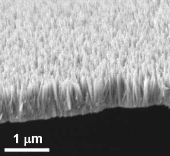
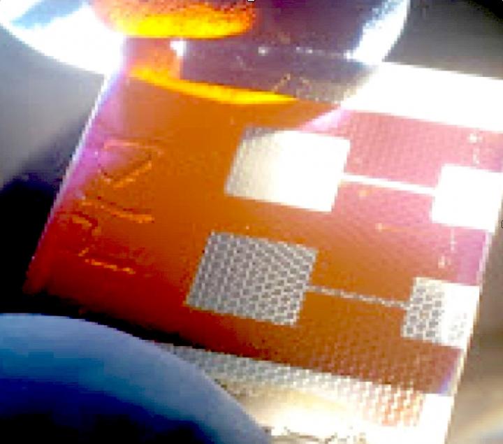
This paper is open access and accompanied by this image illustrating the work,

Courtesy: American Chemical Society and the researchers [Note: The bottom two images of cellulose nanofibres, which are constittuents of an artificial cellulose fibre, appear to be from a scanning tunneling microsscope. Credit: Nitesh Mittal, KTH Stockholm

This news has excited interest at General Electric (GE) (its Wikipedia entry), which has highlighted the work in a May 25, 2018 posting (The 5 Coolest Things On Earth This Week) by Tomas Kellner on the GE Reports blog.

Terminology and spelling

I’ll start with spelling since that’s the easier of the two. In some parts of the world it’s spelled ‘fibres’ and in other parts of the world it’s spelled ‘fibers’. When I write the text in my post, it tends to reflect the spelling used in the news/press releases. In other words, I swing in whichever direction the wind is blowing.

For diehards only

As i understand the terminology situation, nanocellulose and cellulose nanomaterials are interchangeable generic terms. Further, cellulose nanofibres (CNF) seems to be another generic term and it encompasses both cellulose nanocrystals (CNC) and cellulose nanofibrils (CNF). Yes, there appear to be two CNFs. Making matters more interesting is the fact that cellulose nanocrystals were originally christened nanocrystalline cellulose (NCC). For anyone who follows the science and technology scene, it becomes obvious that competing terminologies are the order of the day. Eventually the dust settles and naming conventions are resolved. More or less.

Ordinarily I would reference the Nanocellulose Wikipedia entry in my attempts to clarify the issues but it seems that the writers for the entry have not caught up to the current naming convention for cellulose nanocrystals, still referring to the material as nanocrystalline cellulose. This means, I can’t trust the rest of the entry, which has only one CNF (cellulose nanofibres).

I have paid more attention to the NCC/CNC situation and am not as familiar with the CNF situation. Using, NCC/CNC as an example of a terminology issue, I believe it was first developed in Canada and it was Canadian researchers who were pushing their NCC terminology while the international community pushed back with CNC.

In the end, NCC became a brand name, which was trademarked by CelluForce, a Canadian company in the CNC market. From the CelluForce Products page on Cellulose Nanocrystals,

CNC are not all made equal. The CNC produced by CelluForce is called CelluForce NCCTM and has specific properties and are especially easy to disperse. CelluForce NCCTM is the base material that CelluForce uses in all its products. This base material can be modified and tailored to suit the specific needs in various applications.

These, days CNC is almost universally used but NCC (not as a trademark) is a term still employed on occasion (and, oddly, the researchers are not necessarily Canadian).

Should anyone have better information about terminology issues, please feel free to comment.

Eye implants inspired by glasswing butterflies

Glasswinged butterfly. Greta oto. Credit: David Tiller/CC BY-SA 3.0

My jaw dropped on seeing this image and I still have trouble believing it’s real. (You can find more image of glasswinged butterflies here in an Cot. 25, 2014 posting on thearkinspace. com and there’s a video further down in the post.)

As for the research, an April 30, 2018 news item on phys.org announces work that could improve eye implants,

Inspired by tiny nanostructures on transparent butterfly wings, engineers at Caltech have developed a synthetic analogue for eye implants that makes them more effective and longer-lasting. A paper about the research was published in Nature Nanotechnology.

An April 30, 2018 California Institute of Technology (CalTech) news release (also on EurekAlert) by Robert Perkins, which originated the news item, goes into more detail,

Sections of the wings of a longtail glasswing butterfly are almost perfectly transparent. Three years ago, Caltech postdoctoral researcher Radwanul Hasan Siddique–at the time working on a dissertation involving a glasswing species at Karlsruhe Institute of Technology in Germany–discovered the reason why: the see-through sections of the wings are coated in tiny pillars, each about 100 nanometers in diameter and spaced about 150 nanometers apart. The size of these pillars–50 to 100 times smaller than the width of a human hair–gives them unusual optical properties. The pillars redirect the light that strikes the wings so that the rays pass through regardless of the original angle at which they hit the wings. As a result, there is almost no reflection of the light from the wing’s surface.

In effect, the pillars make the wings clearer than if they were made of just plain glass.

That redirection property, known as angle-independent antireflection, attracted the attention of Caltech’s Hyuck Choo. For the last few years Choo has been developing an eye implant that would improve the monitoring of intra-eye pressure in glaucoma patients. Glaucoma is the second leading cause of blindness worldwide. Though the exact mechanism by which the disease damages eyesight is still under study, the leading theory suggests that sudden spikes in the pressure inside the eye damages the optic nerve. Medication can reduce the increased eye pressure and prevent damage, but ideally it must be taken at the first signs of a spike in eye pressure.

“Right now, eye pressure is typically measured just a couple times a year in a doctor’s office. Glaucoma patients need a way to measure their eye pressure easily and regularly,” says Choo, assistant professor of electrical engineering in the Division of Engineering and Applied Science and a Heritage Medical Research Institute Investigator.

Choo has developed an eye implant shaped like a tiny drum, the width of a few strands of hair. When inserted into an eye, its surface flexes with increasing eye pressure, narrowing the depth of the cavity inside the drum. That depth can be measured by a handheld reader, giving a direct measurement of how much pressure the implant is under.

One weakness of the implant, however, has been that in order to get an accurate measurement, the optical reader has to be held almost perfectly perpendicular–at an angle of 90 degrees (plus or minus 5 degrees)–with respect to the surface of the implant. At other angles, the reader gives an incorrect measurement.

And that’s where glasswing butterflies come into the picture. Choo reasoned that the angle-independent optical property of the butterflies’ nanopillars could be used to ensure that light would always pass perpendicularly through the implant, making the implant angle-insensitive and providing an accurate reading regardless of how the reader is held.

He enlisted Siddique to work in his lab, and the two, working along with Caltech graduate student Vinayak Narasimhan, figured out a way to stud the eye implant with pillars approximately the same size and shape of those on the butterfly’s wings but made from silicon nitride, an inert compound often used in medical implants. Experimenting with various configurations of the size and placement of the pillars, the researchers were ultimately able to reduce the error in the eye implants’ readings threefold.

“The nanostructures unlock the potential of this implant, making it practical for glaucoma patients to test their own eye pressure every day,” Choo says.

The new surface also lends the implants a long-lasting, nontoxic anti-biofouling property.

In the body, cells tend to latch on to the surface of medical implants and, over time, gum them up. One way to avoid this phenomenon, called biofouling, is to coat medical implants with a chemical that discourages the cells from attaching. The problem is that such coatings eventually wear off.

The nanopillars created by Choo’s team, however, work in a different way. Unlike the butterfly’s nanopillars, the lab-made nanopillars are extremely hydrophilic, meaning that they attract water. Because of this, the implant, once in the eye, is soon encased in a coating of water. Cells slide off instead of gaining a foothold.

“Cells attach to an implant by binding with proteins that are adhered to the implant’s surface. The water, however, prevents those proteins from establishing a strong connection on this surface,” says Narasimhan. Early testing suggests that the nanopillar-equipped implant reduces biofouling tenfold compared to previous designs, thanks to this anti-biofouling property.

Being able to avoid biofouling is useful for any implant regardless of its location in the body. The team plans to explore what other medical implants could benefit from their new nanostructures, which can be inexpensively mass produced.

As if the still image wasn’t enough,

Here’s a link to and a citation for the paper,

Multifunctional biophotonic nanostructures inspired by the longtail glasswing butterfly for medical devices by Vinayak Narasimhan, Radwanul Hasan Siddique, Jeong Oen Lee, Shailabh Kumar, Blaise Ndjamen, Juan Du, Natalie Hong, David Sretavan, & Hyuck Choo. Nature Nanotechnology (2018) doi:10.1038/s41565-018-0111-5 Published: 30 April 2018

This paper is behind a paywall.

ETA May 25, 2018:  I’m obsessed. Here’s one more glasswing image,

Caption: The clear wings make this South-American butterfly hard to see in flight, a succesfull defense mechanism. Credit: Eddy Van 3000 from in Flanders fields – Belgiquistan – United Tribes ov Europe Date: 7 October 2007, 14:35 his file is licensed under the Creative Commons Attribution-Share Alike 2.0 Generic license. [downloaded from https://commons.wikimedia.org/wiki/File:E3000_-_the_wings-become-windows_butterfly._(by-sa).jpg]

Beautiful solar cells based on insect eyes

What a gorgeous image!

The compound eye of a fly inspired Stanford researchers to create a compound solar cell consisting of perovskite microcells encapsulated in a hexagon-shaped scaffold. (Image credit: Thomas Shahan/Creative Commons)

An August 31, 2017 news item on Nanowerk describes research into solar cells being performed at Stanford University (Note: A link has been removed),

Packing tiny solar cells together, like micro-lenses in the compound eye of an insect, could pave the way to a new generation of advanced photovoltaics, say Stanford University scientists.

In a new study, the Stanford team used the insect-inspired design to protect a fragile photovoltaic material called perovskite from deteriorating when exposed to heat, moisture or mechanical stress. The results are published in the journal Energy & Environmental Science (“Scaffold-reinforced perovskite compound solar cells”).

An August 31, 2017 Stanford University news release (also on EurekAlert) by Mark Schwartz, which originated the news item,

“Perovskites are promising, low-cost materials that convert sunlight to electricity as efficiently as conventional solar cells made of silicon,” said Reinhold Dauskardt, a professor of materials science and engineering and senior author of the study. “The problem is that perovskites are extremely unstable and mechanically fragile. They would barely survive the manufacturing process, let alone be durable long term in the environment.”

Most solar devices, like rooftop panels, use a flat, or planar, design. But that approach doesn’t work well with perovskite solar cells.

“Perovskites are the most fragile materials ever tested in the history of our lab,” said graduate student Nicholas Rolston, a co-lead author of the E&ES study. “This fragility is related to the brittle, salt-like crystal structure of perovskite, which has mechanical properties similar to table salt.”

Eye of the fly

To address the durability challenge, the Stanford team turned to nature.

“We were inspired by the compound eye of the fly, which consists of hundreds of tiny segmented eyes,” Dauskardt explained. “It has a beautiful honeycomb shape with built-in redundancy: If you lose one segment, hundreds of others will operate. Each segment is very fragile, but it’s shielded by a scaffold wall around it.”

Scaffolds in a compound solar cell filled with perovskite after fracture testing.

Scaffolds in a compound solar cell filled with perovskite after fracture testing. (Image credit: Dauskardt Lab/Stanford University)

Using the compound eye as a model, the researchers created a compound solar cell consisting of a vast honeycomb of perovskite microcells, each encapsulated in a hexagon-shaped scaffold just 0.02 inches (500 microns) wide.

“The scaffold is made of an inexpensive epoxy resin widely used in the microelectronics industry,” Rolston said. “It’s resilient to mechanical stresses and thus far more resistant to fracture.”

Tests conducted during the study revealed that the scaffolding had little effect on how efficiently perovskite converted light into electricity.

“We got nearly the same power-conversion efficiencies out of each little perovskite cell that we would get from a planar solar cell,” Dauskardt said. “So we achieved a huge increase in fracture resistance with no penalty for efficiency.”

Durability

But could the new device withstand the kind of heat and humidity that conventional rooftop solar panels endure?

To find out, the researchers exposed encapsulated perovskite cells to temperatures of 185 F (85 C) and 85 percent relative humidity for six weeks. Despite these extreme conditions, the cells continued to generate electricity at relatively high rates of efficiency.

Dauskardt and his colleagues have filed a provisional patent for the new technology. To improve efficiency, they are studying new ways to scatter light from the scaffold into the perovskite core of each cell.

“We are very excited about these results,” he said. “It’s a new way of thinking about designing solar cells. These scaffold cells also look really cool, so there are some interesting aesthetic possibilities for real-world applications.”

Researchers have also made this image available,

Caption: A compound solar cell illuminated from a light source below. Hexagonal scaffolds are visible in the regions coated by a silver electrode. The new solar cell design could help scientists overcome a major roadblock to the development of perovskite photovoltaics. Credit: Dauskardt Lab/Stanford University

Not quite as weirdly beautiful as the insect eyes.

Here’s a link to and a citation for the paper,

Scaffold-reinforced perovskite compound solar cells by Brian L. Watson, Nicholas Rolston, Adam D. Printz, and Reinhold H. Dauskardt. Energy & Environmental Science 2017 DOI: 10.1039/C7EE02185B first published on 23 Aug 2017

This paper is behind a paywall.

A nano fabrication technique used to create next generation heart valve

I am going to have take the researchers’ word that these somehow lead to healthy heart valve tissue,

In rotary jet spinning technology, a rotating nozzle extrudes a solution of extracellular matrix (ECM) into nanofibers that wrap themselves around heart valve-shaped mandrels. By using a series of mandrels with different sizes, the manufacturing process becomes fully scalable and is able to provide JetValves for all age groups and heart sizes. Credit: Wyss Institute at Harvard University

From a May 18, 2017 news item on ScienceDaily,

The human heart beats approximately 35 million times every year, effectively pumping blood into the circulation via four different heart valves. Unfortunately, in over four million people each year, these delicate tissues malfunction due to birth defects, age-related deteriorations, and infections, causing cardiac valve disease.

Today, clinicians use either artificial prostheses or fixed animal and cadaver-sourced tissues to replace defective valves. While these prostheses can restore the function of the heart for a while, they are associated with adverse comorbidity and wear down and need to be replaced during invasive and expensive surgeries. Moreover, in children, implanted heart valve prostheses need to be replaced even more often as they cannot grow with the child.

A team lead by Kevin Kit Parker, Ph.D. at Harvard University’s Wyss Institute for Biologically Inspired Engineering recently developed a nanofiber fabrication technique to rapidly manufacture heart valves with regenerative and growth potential. In a paper published in Biomaterials, Andrew Capulli, Ph.D. and colleagues fabricated a valve-shaped nanofiber network that mimics the mechanical and chemical properties of the native valve extracellular matrix (ECM). To achieve this, the team used the Parker lab’s proprietary rotary jet spinning technology — in which a rotating nozzle extrudes an ECM solution into nanofibers that wrap themselves around heart valve-shaped mandrels. “Our setup is like a very fast cotton candy machine that can spin a range of synthetic and natural occurring materials. In this study, we used a combination of synthetic polymers and ECM proteins to fabricate biocompatible JetValves that are hemodynamically competent upon implantation and support cell migration and re-population in vitro. Importantly, we can make human-sized JetValves in minutes — much faster than possible for other regenerative prostheses,” said Parker.

A May 18,2017 Wyss Institute for Biologically Inspired Engineering news release (also on EurekAlert), which originated the news item, expands on the theme of Jetvalves,

To further develop and test the clinical potential of JetValves, Parker’s team collaborated with the translational team of Simon P. Hoerstrup, M.D., Ph.D., at the University of Zurich in Switzerland, which is a partner institution with the Wyss Institute. As a leader in regenerative heart prostheses, Hoerstrup and his team in Zurich have previously developed regenerative, tissue-engineered heart valves to replace mechanical and fixed-tissue heart valves. In Hoerstrup’s approach, human cells directly deposit a regenerative layer of complex ECM on biodegradable scaffolds shaped as heart valves and vessels. The living cells are then eliminated from the scaffolds resulting in an “off-the-shelf” human matrix-based prostheses ready for implantation.

In the paper, the cross-disciplinary team successfully implanted JetValves in sheep using a minimally invasive technique and demonstrated that the valves functioned properly in the circulation and regenerated new tissue. “In our previous studies, the cell-derived ECM-coated scaffolds could recruit cells from the receiving animal’s heart and support cell proliferation, matrix remodeling, tissue regeneration, and even animal growth. While these valves are safe and effective, their manufacturing remains complex and expensive as human cells must be cultured for a long time under heavily regulated conditions. The JetValve’s much faster manufacturing process can be a game-changer in this respect. If we can replicate these results in humans, this technology could have invaluable benefits in minimizing the number of pediatric re-operations,” said Hoerstrup.

In support of these translational efforts, the Wyss Institute for Biologically Inspired Engineering and the University of Zurich announced today a cross-institutional team effort to generate a functional heart valve replacement with the capacity for repair, regeneration, and growth. The team is also working towards a GMP-grade version of their customizable, scalable, and cost-effective manufacturing process that would enable deployment to a large patient population. In addition, the new heart valve would be compatible with minimally invasive procedures to serve both pediatric and adult patients.

The project will be led jointly by Parker and Hoerstrup. Parker is a Core Faculty member of the Wyss Institute and the Tarr Family Professor of Bioengineering and Applied Physics at the Harvard John A. Paulson School of Engineering and Applied Sciences (SEAS). Hoerstrup is Chair and Director of the University of Zurich’s Institute for Regenerative Medicine (IREM), Co-Director of the recently founded Wyss Translational Center Zurich and a Wyss Institute Associate Faculty member.

Since JetValves can be manufactured in all desired shapes and sizes, and take seconds to minutes to produce, the team’s goal is to provide customized, ready-to-use, regenerative heart valves much faster and at much lower cost than currently possible.

“Achieving the goal of minimally invasive, low-cost regenerating heart valves could have tremendous impact on patients’ lives across age-, social- and geographical boundaries. Once again, our collaborative team structure that combines unique and leading expertise in bioengineering, regenerative medicine, surgical innovation and business development across the Wyss Institute and our partner institutions, makes it possible for us to advance technology development in ways not possible in a conventional academic laboratory,” said Wyss Institute Founding Director Donald Ingber, M.D., Ph.D., who is also the Judah Folkman Professor of Vascular Biology at HMS and the Vascular Biology Program at Boston Children’s Hospital, as well as Professor of Bioengineering at SEAS.

This scanning electron microscopy image shows how extracellular matrix (ECM) nanofibers generated with JetValve technology are arranged in parallel networks with physical properties comparable to those found in native heart tissue. Credit: Wyss Institute at Harvard University

Here’s a link to and a citation for the paper,

JetValve: Rapid manufacturing of biohybrid scaffolds for biomimetic heart valve replacement by Andrew K. Capulli, Maximillian Y. Emmert, Francesco S. Pasqualini, b, Debora Kehl, Etem Caliskan, Johan U. Lind, Sean P. Sheehy, Sung Jin Park, Seungkuk Ahn, Benedikt Webe, Josue A. Goss. Biomaterials Volume 133, July 2017, Pages 229–241  https://doi.org/10.1016/j.biomaterials.2017.04.033

This paper is behind a paywall.

Controlling the nanostructure of inorganic materials with tumor suppressor proteins

A May 3, 2017 news item on Nanowerk announces research from Japan on using tumor suppressor proteins to control nanostructures,

A new method combining tumor suppressor protein p53 and biomineralization peptide BMPep successfully created hexagonal silver nanoplates, suggesting an efficient strategy for controlling the nanostructure of inorganic materials.

Precise control of nanostructures is a key factor to form functional nanomaterials. Biomimetic approaches are considered effective for fabricating nanomaterials because biomolecules are able to bind with specific targets, self-assemble, and build complex structures. Oligomerization, or the assembly of biomolecules, is a crucial aspect of natural materials that form higher-ordered structures.

A May 3,2017 Hokkaido University research press release, which originated the news item, delves into the details,

Some peptides are known to bind with a specific inorganic substance, such as silver, and enhance its crystal formation. This phenomenon, called peptide-mediated biomineralization, could be used as a biomimetic approach to create functional inorganic structures. Controlling the spatial orientation of the peptides could yield complex inorganic structures, but this has long been a great challenge.

A team of researchers led by Hokkaido University Professor Kazuyasu Sakaguchi has succeeded in controlling the oligomerization of the silver biomineralization peptide (BMPep) which led to the creation of hexagonal silver nanoplates.

The team utilized the well-known tumor suppressor protein p53 which has been known to form tetramers through its tetramerization domain (p53Tet). “The unique symmetry of the p53 tetramer is an attractive scaffold to be used in controlling the overall oligomerization state of the silver BMPep such as its spatial orientation, geometry, and valency,” says Sakaguchi.

In the experiments, the team successfully created silver BMPep fused with p53Tet. This resulted in the formation of BMPep tetramers which yielded hexagonal silver nanoplates. They also found that the BMPep tetramers have enhanced specificity to the structured silver surface, apparently regulating the direction of crystal growth to form hexagonal nanoplates. Furthermore, the tetrameric peptide acted as a catalyst, controlling the silver’s crystal growth without consuming the peptide.

“Our novel method can be applied to other biomineralization peptides and oligomerization proteins, thus providing an efficient and versatile strategy for controlling nanostructures of various inorganic materials. The production of tailor-made nanomaterials is now more feasible,” Sakaguchi commented.

monomeric and tetrameric biomineralization peptides

(Left panels) Schematic illustrations of monomeric and tetrameric biomineralization peptides fused with p53Tet and electron microscopy images of silver nanostructures formed by the biomineralization peptides. Scale bar = 100 nm. (Right) The proposed model in which tetrameric biomineralization peptides regulate the direction of crystal growth and therefore its nanostructure.

Here’s a link to and a citation for the paper,

Oligomerization enhances the binding affinity of a silver biomineralization peptide and catalyzes nanostructure formation by Tatsuya Sakaguchi, Jose Isagani B. Janairo, Mathieu Lussier-Price, Junya Wada, James G. Omichinski, & Kazuyasu Sakaguchi. Scientific Reports 7, Article number: 1400 (2017)  doi:10.1038/s41598-017-01442-8 Published online: 03 May 2017

This paper is open access.