Tag Archives: blood clotting

Injectable bandages for internal bleeding and hydrogel for the brain

This injectable bandage could be a gamechanger (as they say) if it can be taken beyond the ‘in vitro’ (i.e., petri dish) testing stage. A May 22, 2018 news item on Nanowerk makes the announcement (Note: A link has been removed),

While several products are available to quickly seal surface wounds, rapidly stopping fatal internal bleeding has proven more difficult. Now researchers from the Department of Biomedical Engineering at Texas A&M University are developing an injectable hydrogel bandage that could save lives in emergencies such as penetrating shrapnel wounds on the battlefield (Acta Biomaterialia, “Nanoengineered injectable hydrogels for wound healing application”).

A May 22, 2018 US National Institute of Biomedical Engineering and Bioengiineering news release, which originated the news item, provides more detail (Note: Links have been removed),

The researchers combined a hydrogel base (a water-swollen polymer) and nanoparticles that interact with the body’s natural blood-clotting mechanism. “The hydrogel expands to rapidly fill puncture wounds and stop blood loss,” explained Akhilesh Gaharwar, Ph.D., assistant professor and senior investigator on the work. “The surface of the nanoparticles attracts blood platelets that become activated and start the natural clotting cascade of the body.”

Enhanced clotting when the nanoparticles were added to the hydrogel was confirmed by standard laboratory blood clotting tests. Clotting time was reduced from eight minutes to six minutes when the hydrogel was introduced into the mixture. When nanoparticles were added, clotting time was significantly reduced, to less than three minutes.

In addition to the rapid clotting mechanism of the hydrogel composite, the engineers took advantage of special properties of the nanoparticle component. They found they could use the electric charge of the nanoparticles to add growth factors that efficiently adhered to the particles. “Stopping fatal bleeding rapidly was the goal of our work,” said Gaharwar. “However, we found that we could attach growth factors to the nanoparticles. This was an added bonus because the growth factors act to begin the body’s natural wound healing process—the next step needed after bleeding has stopped.”

The researchers were able to attach vascular endothelial growth factor (VEGF) to the nanoparticles. They tested the hydrogel/nanoparticle/VEGF combination in a cell culture test that mimics the wound healing process. The test uses a petri dish with a layer of endothelial cells on the surface that create a solid skin-like sheet. The sheet is then scratched down the center creating a rip or hole in the sheet that resembles a wound.

When the hydrogel containing VEGF bound to the nanoparticles was added to the damaged endothelial cell wound, the cells were induced to grow back and fill-in the scratched region—essentially mimicking the healing of a wound.

“Our laboratory experiments have verified the effectiveness of the hydrogel for initiating both blood clotting and wound healing,” said Gaharwar. “We are anxious to begin tests in animals with the hope of testing and eventual use in humans where we believe our formulation has great potential to have a significant impact on saving lives in critical situations.”

The work was funded by grant EB023454 from the National Institute of Biomedical Imaging and Bioengineering (NIBIB), and the National Science Foundation. The results were reported in the February issue of the journal Acta Biomaterialia.

The paper was published back in April 2018 and there was an April 2, 2018 Texas A&M University news release on EurekAlert making the announcement (and providing a few unique details),

A penetrating injury from shrapnel is a serious obstacle in overcoming battlefield wounds that can ultimately lead to death.Given the high mortality rates due to hemorrhaging, there is an unmet need to quickly self-administer materials that prevent fatality due to excessive blood loss.

With a gelling agent commonly used in preparing pastries, researchers from the Inspired Nanomaterials and Tissue Engineering Laboratory have successfully fabricated an injectable bandage to stop bleeding and promote wound healing.

In a recent article “Nanoengineered Injectable Hydrogels for Wound Healing Application” published in Acta Biomaterialia, Dr. Akhilesh K. Gaharwar, assistant professor in the Department of Biomedical Engineering at Texas A&M University, uses kappa-carrageenan and nanosilicates to form injectable hydrogels to promote hemostasis (the process to stop bleeding) and facilitate wound healing via a controlled release of therapeutics.

“Injectable hydrogels are promising materials for achieving hemostasis in case of internal injuries and bleeding, as these biomaterials can be introduced into a wound site using minimally invasive approaches,” said Gaharwar. “An ideal injectable bandage should solidify after injection in the wound area and promote a natural clotting cascade. In addition, the injectable bandage should initiate wound healing response after achieving hemostasis.”

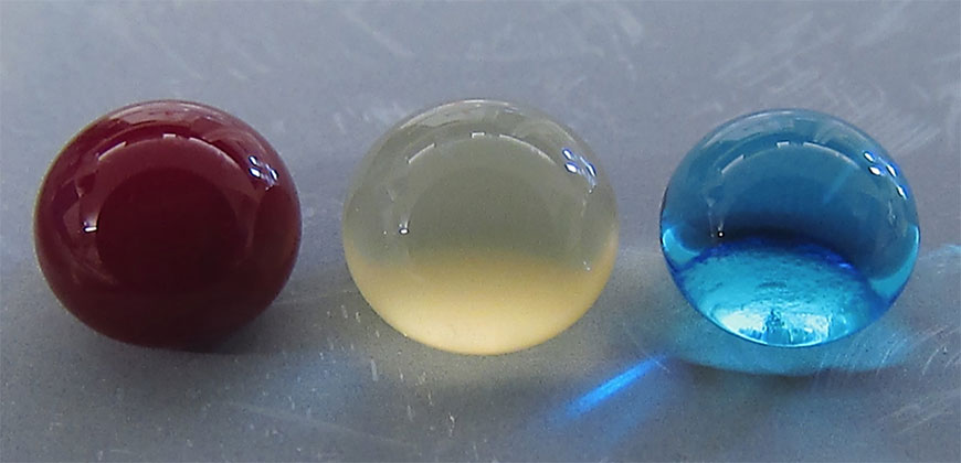
The study uses a commonly used thickening agent known as kappa-carrageenan, obtained from seaweed, to design injectable hydrogels. Hydrogels are a 3-D water swollen polymer network, similar to Jell-O, simulating the structure of human tissues.

When kappa-carrageenan is mixed with clay-based nanoparticles, injectable gelatin is obtained. The charged characteristics of clay-based nanoparticles provide hemostatic ability to the hydrogels. Specifically, plasma protein and platelets form blood adsorption on the gel surface and trigger a blood clotting cascade.

“Interestingly, we also found that these injectable bandages can show a prolonged release of therapeutics that can be used to heal the wound” said Giriraj Lokhande, a graduate student in Gaharwar’s lab and first author of the paper. “The negative surface charge of nanoparticles enabled electrostatic interactions with therapeutics thus resulting in the slow release of therapeutics.”

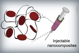
Nanoparticles that promote blood clotting and wound healing (red discs), attached to the wound-filling hydrogel component (black) form a nanocomposite hydrogel. The gel is designed to be self-administered to stop bleeding and begin wound-healing in emergency situations. Credit: Lokhande, et al. 1

Here’s a link to and a citation for the paper,

Nanoengineered injectable hydrogels for wound healing application by Giriraj Lokhande, James K. Carrow, Teena Thakur, Janet R. Xavier, Madasamy Parani, Kayla J. Bayless, Akhilesh K. Gaharwar. Acta Biomaterialia Volume 70, 1 April 2018, Pages 35-47
https://doi.org/10.1016/j.actbio.2018.01.045

This paper is behind a paywall.

Hydrogel and the brain

It’s been an interesting week for hydrogels. On May 21, 2018 there was a news item on ScienceDaily about a bioengineered hydrogel which stimulated brain tissue growth after a stroke (mouse model),

In a first-of-its-kind finding, a new stroke-healing gel helped regrow neurons and blood vessels in mice with stroke-damaged brains, UCLA researchers report in the May 21 issue of Nature Materials.

“We tested this in laboratory mice to determine if it would repair the brain in a model of stroke, and lead to recovery,” said Dr. S. Thomas Carmichael, Professor and Chair of neurology at UCLA. “This study indicated that new brain tissue can be regenerated in what was previously just an inactive brain scar after stroke.”

The brain has a limited capacity for recovery after stroke and other diseases. Unlike some other organs in the body, such as the liver or skin, the brain does not regenerate new connections, blood vessels or new tissue structures. Tissue that dies in the brain from stroke is absorbed, leaving a cavity, devoid of blood vessels, neurons or axons, the thin nerve fibers that project from neurons.

After 16 weeks, stroke cavities in mice contained regenerated brain tissue, including new neural networks — a result that had not been seen before. The mice with new neurons showed improved motor behavior, though the exact mechanism wasn’t clear.

Remarkable stuff.

‘Superhemophobic’ medical implants

Counterintuitively, repelling blood is the concept behind a new type of medical implant according to a Jan. 18, 2017 news item on ScienceDaily,

Medical implants like stents, catheters and tubing introduce risk for blood clotting and infection — a perpetual problem for many patients.

Colorado State University engineers offer a potential solution: A specially grown, “superhemophobic” titanium surface that’s extremely repellent to blood. The material could form the basis for surgical implants with lower risk of rejection by the body.

Blood, plasma and water droplets beading on a superomniphobic surface. CSU researchers have created a superhemophobic titanium surface, repellent to blood, that has potential applications for biocompatible medical devices. Courtesy: Colorado State University

A Jan. 18, 2017 Colorado State University news release by Anne Ju Manning, which originated the news item, explains more,

t’s an outside-the-box innovation achieved at the intersection of two disciplines: biomedical engineering and materials science. The work, recently published in Advanced Healthcare Materials, is a collaboration between the labs of Arun Kota, assistant professor of mechanical engineering and biomedical engineering; and Ketul Popat, associate professor in the same departments.

Kota, an expert in novel, “superomniphobic” materials that repel virtually any liquid, joined forces with Popat, an innovator in tissue engineering and bio-compatible materials. Starting with sheets of titanium, commonly used for medical devices, their labs grew chemically altered surfaces that act as perfect barriers between the titanium and blood. Their teams conducted experiments showing very low levels of platelet adhesion, a biological process that leads to blood clotting and eventual rejection of a foreign material.

Chemical compatibility

A material “phobic” (repellent) to blood might seem counterintuitive, the researchers say, as often biomedical scientists use materials “philic” (with affinity) to blood to make them biologically compatible. “What we are doing is the exact opposite,” Kota said. “We are taking a material that blood hates to come in contact with, in order to make it compatible with blood.” The key innovation is that the surface is so repellent, that blood is tricked into believing there’s virtually no foreign material there at all.

The undesirable interaction of blood with foreign materials is an ongoing problem in medical research, Popat said. Over time, stents can form clots, obstructions, and lead to heart attacks or embolisms. Often patients need blood-thinning medications for the rest of their lives – and the drugs aren’t foolproof.

“The reason blood clots is because it finds cells in the blood to go to and attach,” Popat said. “Normally, blood flows in vessels. If we can design materials where blood barely contacts the surface, there is virtually no chance of clotting, which is a coordinated set of events. Here, we’re targeting the prevention of the first set of events.”

nanotubes

Fluorinated nanotubes provided the best superhemophobic surface in the researchers’ experiments.

The researchers analyzed variations of titanium surfaces, including different textures and chemistries, and they compared the extent of platelet adhesion and activation. Fluorinated nanotubes offered the best protection against clotting, and they plan to conduct follow-up experiments.

Growing a surface and testing it in the lab is only the beginning, the researchers say. They want to continue examining other clotting factors, and eventually, to test real medical devices.

Here’s a link to and a citation for the paper,

Hemocompatibility of Superhemophobic Titania Surfaces by Sanli Movafaghi, Victoria Leszczak, Wei Wang, Jonathan A. Sorkin, Lakshmi P. Dasi, Ketul C. Popat, and Arun K. Kota. Advanced Healthcare Materials DOI: 10.1002/adhm.201600717 Version of Record online: 21 DEC 2016

© 2016 WILEY-VCH Verlag GmbH & Co. KGaA, Weinheim

Nanoparticles could make blood clot faster

It was the 252nd meeting for the American Chemical Society from Aug. 21 – 25, 2016 and that meant a flurry of news about the latest research. From an Aug. 23, 2016 news item on Nanowerk,

Whether severe trauma occurs on the battlefield or the highway, saving lives often comes down to stopping the bleeding as quickly as possible. Many methods for controlling external bleeding exist, but at this point, only surgery can halt blood loss inside the body from injury to internal organs. Now, researchers have developed nanoparticles that congregate wherever injury occurs in the body to help it form blood clots, and they’ve validated these particles in test tubes and in vivo [animal testing].

The researchers will present their work today [Aug. 22, 2016] at the 252nd National Meeting & Exposition of the American Chemical Society (ACS). ACS, the world’s largest scientific society, is holding the meeting here through Thursday. It features more than 9,000 presentations on a wide range of science topics.

An Aug. 22, 2016 American Chemical Society (ACS) news release (also on EurekAlert), which originated the news item, provided more detail,

“When you have uncontrolled internal bleeding, that’s when these particles could really make a difference,” says Erin B. Lavik, Sc.D. “Compared to injuries that aren’t treated with the nanoparticles, we can cut bleeding time in half and reduce total blood loss.”

Trauma remains a top killer of children and younger adults, and doctors have few options for treating internal bleeding. To address this great need, Lavik’s team developed a nanoparticle that acts as a bridge, binding to activated platelets and helping them join together to form clots. To do this, the nanoparticle is decorated with a molecule that sticks to a glycoprotein found only on the activated platelets.

Initial studies suggested that the nanoparticles, delivered intravenously, helped keep rodents from bleeding out due to brain and spinal injury, Lavik says. But, she acknowledges, there was still one key question: “If you are a rodent, we can save your life, but will it be safe for humans?”

As a step toward assessing whether their approach would be safe in humans, they tested the immune response toward the particles in pig’s blood. If a treatment triggers an immune response, it would indicate that the body is mounting a defense against the nanoparticle and that side effects are likely. The team added their nanoparticles to pig’s blood and watched for an uptick in complement, a key indicator of immune activation. The particles triggered complement in this experiment, so the researchers set out to engineer around the problem.

“We made a battery of particles with different charges and tested to see which ones didn’t have this immune-response effect,” Lavik explains. “The best ones had a neutral charge.” But neutral nanoparticles had their own problems. Without repulsive charge-charge interactions, the nanoparticles have a propensity to aggregate even before being injected. To fix this issue, the researchers tweaked their nanoparticle storage solution, adding a slippery polymer to keep the nanoparticles from sticking to each other.

Lavik also developed nanoparticles that are stable at higher temperatures, up to 50 degrees Celsius (122 degrees Fahrenheit). This would allow the particles to be stored in a hot ambulance or on a sweltering battlefield.

In future studies, the researchers will test whether the new particles activate complement in human blood. Lavik also plans to identify additional critical safety studies they can perform to move the research forward. For example, the team needs to be sure that the nanoparticles do not cause non-specific clotting, which could lead to a stroke. Lavik is hopeful though that they could develop a useful clinical product in the next five to 10 years.

It’s not unusual for scientists to give an estimate of 5 – 10 years before their science reaches the market.  Another popular range is 3 – 5 years.

Multi-walled carbon nanotubes and blood clotting

There’s been a lot of interest in using carbon nanotubes (CNTs) for biomedical applications such as drug delivery. New research from Trinity College Dublin (TCD) suggests that multi-walled carbon nanotubes (MWCNTs) may have some limitations when applied to biomedical uses. From a Jan. 20, 2014 news item on Nanowerk (Note: A link has been removed),

Scientists in the School of Pharmacy and Pharmaceutical Sciences in Trinity College Dublin, have made an important discovery about the safety issues of using carbon nanotubes as biomaterials which come into contact with blood. The significance of their findings is reflected in their paper being published as the feature story and front page cover of the international, peer-reviewed journal Nanomedicine (“Blood biocompatibility of surface-bound multi-walled carbon nanotubes”).

A Jan. 19, 2015 TCD press release, which originated the news item, offers a good description of the issues around blood clotting and the research problem (nonfunctionalized CNTs and blood compartibility) the scientists were addressing (Note: Links have been removed),

When blood comes into contact with foreign surfaces the blood’s platelets are activated which in turn leads to blood clots being formed. This can be catastrophic in clinical settings where extracorporeal circulation technologies are used such as during heart-lung bypass, in which the blood is circulated in PVC tubing outside the body. More than one million cardiothoracic surgeries are performed each year and while new circulation surfaces that prevent platelet activation are urgently needed, effective technologies have remained elusive.

One hope has been that carbon nanotubes, which are enormously important as potentially useful biomedical materials, might provide a solution to this challenge and this led the scientists from the School of Pharmacy and Pharmaceutical Sciences in collaboration with Trinity’s School of Chemistry and with colleagues from UCD and the University of Michigan in Ann Arbour to test the blood biocompatibility of carbon nanotubes. They found that the carbon nanotubes did actually stimulate blood platelet activation, subsequently leading to serious and devastating blood clotting. The findings have implications for the design of medical devices which contain nanoparticles and which are used in conjunction with flowing blood.

Speaking about their findings, Professor Marek Radomski, Chair of Pharmacology, Trinity and the paper’s senior author said: “Our results bear significance for the design of blood-facing medical devices, surface-functionalised with nanoparticles or containing surface-shedding nanoparticles. We feel that the risk/benefit ratio with particular attention to blood compatibility should be carefully evaluated during the development of such devices. Furthermore, it is clear that non-functionalised carbon nanotubes both soluble and surface-bound are not blood-compatible”.

The press release also quotes a TCD graduate,

Speaking about the significance of these findings for Nanomedicine research, the paper’s first author Dr Alan Gaffney, a Trinity PhD graduate who is now Assistant Professor of Anaesthesiology in Columbia University Medical Centre, New York said: “When new and exciting technologies with enormous potential benefits for medicine are being studied, there is often a bias towards the publication of positive findings. [emphasis mine] The ultimate successful and safe application of nanotechnology in medicine requires a complete understanding of the negative as well as positive effects so that un-intended side effects can be prevented. Our study is an important contribution to the field of nanomedicine and nanotoxicology research and will help to ensure that nanomaterials that come in contact with blood are thoroughly tested for their interaction with blood platelets before they are used in patients.”

Point well taken Dr. Gaffney. Too often there’s an almost euphoric quality to the nanomedicine discussion where nanoscale treatments are described as if they are perfectly benign in advance of any real testing. For example, I wrote about surgical nanobots being used in a human clinical trial in a Jan. 7, 2015 post which features a video of the researcher ‘selling’ his idea. The enthusiasm is laudable and necessary (researchers work for years trying to develop new treatments) but as Gaffney notes there needs to be some counter-ballast and recognition of the ‘positive bias’ issue.

Getting back to the TCD research, here’s a link to and a citation for the paper (or counter-ballast),

Blood biocompatibility of surface-bound multi-walled carbon nanotubes by Alan M. Gaffney, MD, PhD, Maria J. Santos-Martinez, MD, Amro Satti, Terry C. Major, Kieran J. Wynne, Yurii K. Gun’ko, PhD, Gail M. Annich, Giuliano Elia, Marek W. Radomski, MD. January 2015 Volume 11, Issue 1, Pages 39–46 DOI: http://dx.doi.org/10.1016/j.nano.2014.07.005 Published Online: July 26, 2014

This paper is open access.