Tag Archives: brain cells

Cortical spheroids (like mini-brains) could unlock (larger) brain’s mysteries

A March 19, 2021 Northwestern University news release on EurekAlert announces the creation of a device designed to monitor brain organoids (for anyone unfamiliar with brain organoids there’s more information after the news),

A team of scientists, led by researchers at Northwestern University, Shirley Ryan AbilityLab and the University of Illinois at Chicago (UIC), has developed novel technology promising to increase understanding of how brains develop, and offer answers on repairing brains in the wake of neurotrauma and neurodegenerative diseases.

Their research is the first to combine the most sophisticated 3-D bioelectronic systems with highly advanced 3-D human neural cultures. The goal is to enable precise studies of how human brain circuits develop and repair themselves in vitro. The study is the cover story for the March 19 [March 17, 2021 according to the citation] issue of Science Advances.

The cortical spheroids used in the study, akin to “mini-brains,” were derived from human-induced pluripotent stem cells. Leveraging a 3-D neural interface system that the team developed, scientists were able to create a “mini laboratory in a dish” specifically tailored to study the mini-brains and collect different types of data simultaneously. Scientists incorporated electrodes to record electrical activity. They added tiny heating elements to either keep the brain cultures warm or, in some cases, intentionally overheated the cultures to stress them. They also incorporated tiny probes — such as oxygen sensors and small LED lights — to perform optogenetic experiments. For instance, they introduced genes into the cells that allowed them to control the neural activity using different-colored light pulses.

This platform then enabled scientists to perform complex studies of human tissue without directly involving humans or performing invasive testing. In theory, any person could donate a limited number of their cells (e.g., blood sample, skin biopsy). Scientists can then reprogram these cells to produce a tiny brain spheroid that shares the person’s genetic identity. The authors believe that, by combining this technology with a personalized medicine approach using human stem cell-derived brain cultures, they will be able to glean insights faster and generate better, novel interventions.

“The advances spurred by this research will offer a new frontier in the way we study and understand the brain,” said Shirley Ryan AbilityLab’s Dr. Colin Franz, co-lead author on the paper who led the testing of the cortical spheroids. “Now that the 3-D platform has been developed and validated, we will be able to perform more targeted studies on our patients recovering from neurological injury or battling a neurodegenerative disease.”

Yoonseok Park, postdoctoral fellow at Northwestern University and co-lead author, added, “This is just the beginning of an entirely new class of miniaturized, 3-D bioelectronic systems that we can construct to expand the capacity of the regenerative medicine field. For example, our next generation of device will support the formation of even more complex neural circuits from brain to muscle, and increasingly dynamic tissues like a beating heart.”

Current electrode arrays for tissue cultures are 2-D, flat and unable to match the complex structural designs found throughout nature, such as those found in the human brain. Moreover, even when a system is 3-D, it is extremely challenging to incorporate more than one type of material into a small 3-D structure. With this advance, however, an entire class of 3-D bioelectronics devices has been tailored for the field of regenerative medicine.

“Now, with our small, soft 3-D electronics, the capacity to build devices that mimic the complex biological shapes found in the human body is finally possible, providing a much more holistic understanding of a culture,” said Northwestern’s John Rogers, who led the technology development using technology similar to that found in phones and computers. “We no longer have to compromise function to achieve the optimal form for interfacing with our biology.”

As a next step, scientists will use the devices to better understand neurological disease, test drugs and therapies that have clinical potential, and compare different patient-derived cell models. This understanding will then enable a better grasp of individual differences that may account for the wide variation of outcomes seen in neurological rehabilitation.

“As scientists, our goal is to make laboratory research as clinically relevant as possible,” said Kristen Cotton, research assistant in Dr. Franz’s lab. “This 3-D platform opens the door to new experiments, discovery and scientific advances in regenerative neurorehabilitation medicine that have never been possible.”
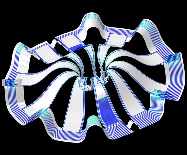
Caption: Three dimensional multifunctional neural interfaces for cortical spheroids and engineered assembloids Credit: Northwestern University

As for what brain ogranoids might be, Carl Zimmer in an Aug. 29, 2019 article for the New York Times provides an explanation,

Organoids Are Not Brains. How Are They Making Brain Waves?

Two hundred and fifty miles over Alysson Muotri’s head, a thousand tiny spheres of brain cells were sailing through space.

The clusters, called brain organoids, had been grown a few weeks earlier in the biologist’s lab here at the University of California, San Diego. He and his colleagues altered human skin cells into stem cells, then coaxed them to develop as brain cells do in an embryo.

The organoids grew into balls about the size of a pinhead, each containing hundreds of thousands of cells in a variety of types, each type producing the same chemicals and electrical signals as those cells do in our own brains.

In July, NASA packed the organoids aboard a rocket and sent them to the International Space Station to see how they develop in zero gravity.

Now the organoids were stowed inside a metal box, fed by bags of nutritious broth. “I think they are replicating like crazy at this stage, and so we’re going to have bigger organoids,” Dr. Muotri said in a recent interview in his office overlooking the Pacific.

What, exactly, are they growing into? That’s a question that has scientists and philosophers alike scratching their heads.

On Thursday, Dr. Muotri and his colleagues reported that they  have recorded simple brain waves in these organoids. In mature human brains, such waves are produced by widespread networks of neurons firing in synchrony. Particular wave patterns are linked to particular forms of brain activity, like retrieving memories and dreaming.

As the organoids mature, the researchers also found, the waves change in ways that resemble the changes in the developing brains of premature babies.

“It’s pretty amazing,” said Giorgia Quadrato, a neurobiologist at the University of Southern California who was not involved in the new study. “No one really knew if that was possible.”

But Dr. Quadrato stressed it was important not to read too much into the parallels. What she, Dr. Muotri and other brain organoid experts build are clusters of replicating brain cells, not actual brains.

If you have the time, I recommend reading Zimmer’s article in its entirety. Perhaps not coincidentally, Zimmer has an excerpt titled “Lab-Grown Brain Organoids Aren’t Alive. But They’re Not Not Alive, Either.” published in Slate.com,

From Life’s Edge: The Search For What It Means To Be Alive by Carl Zimmer, published by Dutton, an imprint of Penguin Publishing Group, a division of Penguin Random House, LLC. Copyright © 2021 by Carl Zimmer.

Cleber Trujillo led me to a windowless room banked with refrigerators, incubators, and microscopes. He extended his blue-gloved hands to either side and nearly touched the walls. “This is where we spend half our day,” he said.

In that room Trujillo and a team of graduate students raised a special kind of life. He opened an incubator and picked out a clear plastic box. Raising it above his head, he had me look up at it through its base. Inside the box were six circular wells, each the width of a cookie and filled with what looked like watered-down grape juice. In each well 100 pale globes floated, each the size of a housefly head.

Getting back to the research about monitoring brain organoids, here’s a link to and a citation for the paper about cortical spheroids,

Three-dimensional, multifunctional neural interfaces for cortical spheroids and engineered assembloids by Yoonseok Park, Colin K. Franz, Hanjun Ryu, Haiwen Luan, Kristen Y. Cotton, Jong Uk Kim, Ted S. Chung, Shiwei Zhao, Abraham Vazquez-Guardado, Da Som Yang, Kan Li, Raudel Avila, Jack K. Phillips, Maria J. Quezada, Hokyung Jang, Sung Soo Kwak, Sang Min Won, Kyeongha Kwon, Hyoyoung Jeong, Amay J. Bandodkar, Mengdi Han, Hangbo Zhao, Gabrielle R. Osher, Heling Wang, KunHyuck Lee, Yihui Zhang, Yonggang Huang, John D. Finan and John A. Rogers. Science Advances 17 Mar 2021: Vol. 7, no. 12, eabf9153 DOI: 10.1126/sciadv.abf9153

This paper appears to be open access.

According to a March 22, 2021 posting on the Shirley Riley AbilityLab website, the paper is featured on the front cover of Science Advances (vol. 7 no. 12).
Could synergistic action of engineered nanoparticles have a health impact?

Synergistic action can be difficult to study especially when you’re looking at nanoparticles which could be naturally occurring and/or engineered. I believe this study is focused on engineered nanoparticles (ENPs) and I think it’s the first one I’ve seen that examines synergistic action of any kind. So, bravo to the scientists for tackling a very ambitious project.

An October 1, 2020 news item on phys.org describes this work from Denmark,

Nanoparticles are used in a wide range of products and manufacturing processes because the properties of a material can change dramatically when the material comes in nano-form.

They can be used, for example, to purify wastewater and to transport medicine around the body. They are also added to, for example, socks, pillows, mattresses, phone covers and refrigerators to supply the items with an antibacterial surface.

Much research has been done on how nanoparticles affect humans and the environment and a number of studies have shown that nanoparticles can disrupt or damage our cells.

This is confirmed by a new study that has also looked at how cells react when exposed to more than one kind of nano particle at the same time.

An October 1, 2020 University of Southern Denmark press release (also on EurekAlert) by Birgitte Svennevig, which originated the news item, provides more insight into the research,

The lead author of the study is Barbara Korzeniowska from the Department of Biochemistry and Molecular Biology at SDU. The head of research is Professor Frank Kjeldsen from the same department.

His research into metal nanoparticles is supported by a European Research Grant of DKK 14 million.

“Throughout a lifetime, we are exposed to many different kinds of nano-particles, and we should investigate how the combination of different nano-particles affects us and also whether an accumulation through life can harm us,” says Barbara Korzeniowska.

She herself became interested in the subject when her little daughter one day was going in the bathtub and got a rubber duck as a toy.

– It turned out that it had been treated with nano-silver, probably to keep it free of bacteria, but small children put their toys in their mouths, and she could thus ingest nano-silver. That is highly worrying when research shows that nano-silver can damage human cells, she says.

In her new study, she looked at nano-silver and nano-platinum. She has investigated their individual effect and whether exposure of both types of nanoparticles results in a synergy effect in two types of brain cells.

– There are almost no studies of the synergy effect of nano particles, so it is important to get started with these studies, she says.

She chose nano-silver because it is already known to be able to damage cells and nano-platinum, because nano-platinum is considered to be so-called bio-inert; i.e. has a minimal interaction with human tissue.

The nanoparticles were tested on two types of brain cells: astrocytes and endothelial cells. Astrocytes are supporter cells in the central nervous system, which i.a. helps to supply the nervous system with nutrients and repair damage to the brain. Endothelial cells sit on the inside of the blood vessels and transport substances from the bloodstream to the brain.

When the endothelial cells were exposed to nano-platinum, nothing happened. When exposed to nano-silver, their ability to divide deteriorated. When exposed to both nano-silver and nano-platinum, the effect was amplified, and they died in large numbers. Furthermore, their defense mechanisms decreased, and they had difficulty communicating with each other.

– So even though nano-platinum alone does not do harm, something drastic happens when they are combined with a different kind of nano-particle, says Frank Kjeldsen.

The astrocytes were more hardy and reacted “only” with impaired ability to divide when exposed to both types of nano-particles.

An earlier study, conducted by Frank Kjeldsen, has shown a dramatic synergy effect of silver nanoparticles and cadmium ions, which are found naturally all around us on Earth.

In that study, 72 % of the cells died (in this study it was intestinal cells) as they were exposed to both nano-silver and cadmium ions. When they were only exposed to nano-silver, 25% died. When exposed to cadmium ions only, 12% died.

We are involuntarily exposed

– Little is known about how large concentrations of nano-particles are used in industrial products. We also do not know what size particles they use – size also has an effect on whether they can enter a cell, says Barbara Korzeniowska and continues:

– But we know that a lot of people are involuntarily exposed to nano-particles, and that there can be lifelong exposure.

There are virtually no restrictions on adding nanoparticles to products. In the EU, however, manufacturers must have an approval if they want to use nanoparticles in products with antibacterial properties. In Denmark, they must also declare nano-content in such products on the label.

Here’s a link to and a citation for the paper,

The Cytotoxicity of Metal Nanoparticles Depends on Their Synergistic Interactions by Barbara Korzeniowska, Micaella P. Fonseca, Vladimir Gorshkov, Lilian Skytte, Kaare L. Rasmussen, Henrik D. Schrøder, Frank Kjeldsen. Particle Volume 37, Issue 8, August 2020,. 2000135 DOI: https://doi.org/10.1002/ppsc.202000135 First published: 06 July 2020

This paper is behind a paywall.

Revival of dead pig brains raises moral questions about life and death

The line between life and death may not be what we thought it was according to some research that was reported in April 2019. Ed Wong’s April 17, 2019 article (behind a paywall) for The Atlantic was my first inkling about the life-death questions raised by some research performed at Yale University, (Note: Links have been removed)

The brain, supposedly, cannot long survive without blood. Within seconds, oxygen supplies deplete, electrical activity fades, and unconsciousness sets in. If blood flow is not restored, within minutes, neurons start to die in a rapid, irreversible, and ultimately fatal wave.

But maybe not? According to a team of scientists led by Nenad Sestan at Yale School of Medicine, this process might play out over a much longer time frame, and perhaps isn’t as inevitable or irreparable as commonly believed. Sestan and his colleagues showed this in dramatic fashion—by preserving and restoring signs of activity in the isolated brains of pigs that had been decapitated four hours earlier.

The team sourced 32 pig brains from a slaughterhouse, placed them in spherical chambers, and infused them with nutrients and protective chemicals, using pumps that mimicked the beats of a heart. This system, dubbed BrainEx, preserved the overall architecture of the brains, preventing them from degrading. It restored flow in their blood vessels, which once again became sensitive to dilating drugs. It stopped many neurons and other cells from dying, and reinstated their ability to consume sugar and oxygen. Some of these rescued neurons even started to fire. “Everything was surprising,” says Zvonimir Vrselja, who performed most of the experiments along with Stefano Daniele.

… “I don’t see anything in this report that should undermine confidence in brain death as a criterion of death,” says Winston Chiong, a neurologist at the University of California at San Francisco. The matter of when to declare someone dead has become more controversial since doctors began relying more heavily on neurological signs, starting around 1968, when the criteria for “brain death” were defined. But that diagnosis typically hinges on the loss of brainwide activity—a line that, at least for now, is still final and irreversible. After MIT Technology Review broke the news of Sestan’s work a year ago, he started receiving emails from people asking whether he could restore brain function to their loved ones. He very much cannot. BrainEx isn’t a resurrection chamber.

“It’s not going to result in human brain transplants,” adds Karen Rommelfanger, who directs Emory University’s neuroethics program. “And I don’t think this means that the singularity is coming, or that radical life extension is more possible than before.”

So why do the study? “There’s potential for using this method to develop innovative treatments for patients with strokes or other types of brain injuries, and there’s a real need for those kinds of treatments,” says L. Syd M Johnson, a neuroethicist at Michigan Technological University. The BrainEx method might not be able to fully revive hours-dead brains, but Yama Akbari, a critical-care neurologist at the University of California at Irvine, wonders whether it would be more successful if applied minutes after death. Alternatively, it could help to keep oxygen-starved brains alive and intact while patients wait to be treated. “It’s an important landmark study,” Akbari says.

Yong notes that the study still needs to be replicated in his article which also probes some of the ethical issues associated with the latest neuroscience research.

Nature published the Yale study,

Restoration of brain circulation and cellular functions hours post-mortem by Zvonimir Vrselja, Stefano G. Daniele, John Silbereis, Francesca Talpo, Yury M. Morozov, André M. M. Sousa, Brian S. Tanaka, Mario Skarica, Mihovil Pletikos, Navjot Kaur, Zhen W. Zhuang, Zhao Liu, Rafeed Alkawadri, Albert J. Sinusas, Stephen R. Latham, Stephen G. Waxman & Nenad Sestan. Nature 568, 336–343 (2019) DOI: https://doi.org/10.1038/s41586-019-1099-1 Published 17 April 2019 Issue Date 18 April 2019

This paper is behind a paywall.

Two neuroethicists had this to say (link to their commentary in Nature follows) as per an April 71, 2019 news release from Case Western Reserve University (also on EurekAlert), Note: Links have been removed,

The brain is more resilient than previously thought. In a groundbreaking experiment published in this week’s issue of Nature, neuroscientists created an artificial circulation system that successfully restored some functions and structures in donated pig brains–up to four hours after the pigs were butchered at a USDA food processing facility. Though there was no evidence of restored consciousness, brains from the pigs were without oxygen for hours, yet could still support key functions provided by the artificial system. The result challenges the notion that mammalian brains are fully and irreversibly damaged by a lack of oxygen.

“The assumptions have always been that after a couple minutes of anoxia, or no oxygen, the brain is ‘dead,'” says Stuart Youngner, MD, who co-authored a commentary accompanying the study with Insoo Hyun, PhD, both professors in the Department of Bioethics at Case Western Reserve University School of Medicine. “The system used by the researchers begs the question: How long should we try to save people?”

In the pig experiment, researchers used an artificial perfusate (a type of cell-free “artificial blood”), which helped brain cells maintain their structure and some functions. Resuscitative efforts in humans, like CPR, are also designed to get oxygen to the brain and stave off brain damage. After a period of time, if a person doesn’t respond to resuscitative efforts, emergency medical teams declare them dead.

The acceptable duration of resuscitative efforts is somewhat uncertain. “It varies by country, emergency medical team, and hospital,” Youngner said. Promising results from the pig experiment further muddy the waters about the when to stop life-saving efforts.

At some point, emergency teams must make a critical switch from trying to save a patient, to trying to save organs, said Youngner. “In Europe, when emergency teams stop resuscitation efforts, they declare a patient dead, and then restart the resuscitation effort to circulate blood to the organs so they can preserve them for transplantation.”

The switch can involve extreme means. In the commentary, Youngner and Hyun describe how some organ recovery teams use a balloon to physically cut off blood circulation to the brain after declaring a person dead, to prepare the organs for transplantation.

The pig experiment implies that sophisticated efforts to perfuse the brain might maintain brain cells. If technologies like those used in the pig experiment could be adapted for humans (a long way off, caution Youngner and Hyun), some people who, today, are typically declared legally dead after a catastrophic loss of oxygen could, tomorrow, become candidates for brain resuscitation, instead of organ donation.

Said Youngner, “As we get better at resuscitating the brain, we need to decide when are we going to save a patient, and when are we going to declare them dead–and save five or more who might benefit from an organ.”

Because brain resuscitation strategies are in their infancy and will surely trigger additional efforts, the scientific and ethics community needs to begin discussions now, says Hyun. “This study is likely to raise a lot of public concerns. We hoped to get ahead of the hype and offer an early, reasoned response to this scientific advance.”

Both Youngner and Hyun praise the experiment as a “major scientific advancement” that is overwhelmingly positive. It raises the tantalizing possibility that the grave risks of brain damage caused by a lack of oxygen could, in some cases, be reversible.
“Pig brains are similar in many ways to human brains, which makes this study so compelling,” Hyun said. “We urge policymakers to think proactively about what this line of research might mean for ongoing debates around organ donation and end of life care.”

Here’s a link to and a citation to the Nature commentary,

Pig experiment challenges assumptions around brain damage in people by Stuart Youngner and Insoo Hyun. Nature 568, 302-304 (2019) DOI: 10.1038/d41586-019-01169-8 April 17, 2019

This paper is open access.

I was hoping to find out more about BrainEx, but this April 17, 2019 US National Institute of Mental Health news release is all I’ve been able to find in my admittedly brief online search. The news release offers more celebration than technical detail.

Quick comment

Interestingly, there hasn’t been much of a furor over this work. Not yet.

The roles mathematics and light play in cellular communication

These are two entirely different types of research but taken together they help build a picture about how the cells in our bodies function.

Cells and light

An April 30, 2018 news item on phys.org describes work on controlling biology with light,

Over the past five years, University of Chicago chemist Bozhi Tian has been figuring out how to control biology with light.

A longterm science goal is devices to serve as the interface between researcher and body—both as a way to understand how cells talk among each other and within themselves, and eventually, as a treatment for brain or nervous system disorders [emphasis mine] by stimulating nerves to fire or limbs to move. Silicon—a versatile, biocompatible material used in both solar panels and surgical implants—is a natural choice.

In a paper published April 30 in Nature Biomedical Engineering, Tian’s team laid out a system of design principles for working with silicon to control biology at three levels—from individual organelles inside cells to tissues to entire limbs. The group has demonstrated each in cells or mice models, including the first time anyone has used light to control behavior without genetic modification.

“We want this to serve as a map, where you can decide which problem you would like to study and immediately find the right material and method to address it,” said Tian, an assistant professor in the Department of Chemistry.

Researchers built this thin layer of silicon lace to modulate neural signals when activated by light. Courtesy of Yuanwen Jiang and Bozhi Tian

An April 30, 2018 University of Chicago news release by Louise Lerner, which originated the news item, describes the work in greater detail,

The scientists’ map lays out best methods to craft silicon devices depending on both the intended task and the scale—ranging from inside a cell to a whole animal.

For example, to affect individual brain cells, silicon can be crafted to respond to light by emitting a tiny ionic current, which encourages neurons to fire. But in order to stimulate limbs, scientists need a system whose signals can travel farther and are stronger—such as a gold-coated silicon material in which light triggers a chemical reaction.

The mechanical properties of the implant are important, too. Say researchers would like to work with a larger piece of the brain, like the cortex, to control motor movement. The brain is a soft, squishy substance, so they’ll need a material that’s similarly soft and flexible, but can bind tightly against the surface. They’d want thin and lacy silicon, say the design principles.

The team favors this method because it doesn’t require genetic modification or a power supply wired in, since the silicon can be fashioned into what are essentially tiny solar panels. (Many other forms of monitoring or interacting with the brain need to have a power supply, and keeping a wire running into a patient is an infection risk.)

They tested the concept in mice and found they could stimulate limb movements by shining light on brain implants. Previous research tested the concept in neurons.

“We don’t have answers to a number of intrinsic questions about biology, such as whether individual mitochondria communicate remotely through bioelectric signals,” said Yuanwen Jiang, the first author on the paper, then a graduate student at UChicago and now a postdoctoral researcher at Stanford. “This set of tools could address such questions as well as pointing the way to potential solutions for nervous system disorders.”

Other UChicago authors were Assoc. Profs. Chin-Tu Chen and Chien-Min Kao, Asst. Prof Xiaoyang, postdoctoral researchers Jaeseok Yi, Yin Fang, Xiang Gao, Jiping Yue, Hsiu-Ming Tsai, Bing Liu and Yin Fang, graduate students Kelliann Koehler, Vishnu Nair, and Edward Sudzilovsky, and undergraduate student George Freyermuth.

Other researchers on the paper hailed from Northwestern University, the University of Illinois at Chicago and Hong Kong Polytechnic University.

The researchers have also made this video illustrating their work,

via Gfycat Tiny silicon nanowires (in blue), activated by light, trigger activity in neurons. (Courtesy Yuanwen Jiang and Bozhi Tian)

Here’s a link to and a citation for the paper,

Rational design of silicon structures for optically controlled multiscale biointerfaces by Yuanwen Jiang, Xiaojian Li, Bing Liu, Jaeseok Yi, Yin Fang, Fengyuan Shi, Xiang Gao, Edward Sudzilovsky, Ramya Parameswaran, Kelliann Koehler, Vishnu Nair, Jiping Yue, KuangHua Guo, Yin Fang, Hsiu-Ming Tsai, George Freyermuth, Raymond C. S. Wong, Chien-Min Kao, Chin-Tu Chen, Alan W. Nicholls, Xiaoyang Wu, Gordon M. G. Shepherd, & Bozhi Tian. Nature Biomedical Engineering (2018) doi:10.1038/s41551-018-0230-1 Published: 30 April 2018

This paper is behind a paywall.

Mathematics and how living cells ‘think’

This May 2, 2018 Queensland University of Technology (QUT; Australia) press release is also on EurekAlert,

How does the ‘brain’ of a living cell work, allowing an organism to function and thrive in changing and unfavourable environments?

Queensland University of Technology (QUT) researcher Dr Robyn Araujo has developed new mathematics to solve a longstanding mystery of how the incredibly complex biological networks within cells can adapt and reset themselves after exposure to a new stimulus.

Her findings, published in Nature Communications, provide a new level of understanding of cellular communication and cellular ‘cognition’, and have potential application in a variety of areas, including new targeted cancer therapies and drug resistance.

Dr Araujo, a lecturer in applied and computational mathematics in QUT’s Science and Engineering Faculty, said that while we know a great deal about gene sequences, we have had extremely limited insight into how the proteins encoded by these genes work together as an integrated network – until now.

“Proteins form unfathomably complex networks of chemical reactions that allow cells to communicate and to ‘think’ – essentially giving the cell a ‘cognitive’ ability, or a ‘brain’,” she said. “It has been a longstanding mystery in science how this cellular ‘brain’ works.

“We could never hope to measure the full complexity of cellular networks – the networks are simply too large and interconnected and their component proteins are too variable.

“But mathematics provides a tool that allows us to explore how these networks might be constructed in order to perform as they do.

“My research is giving us a new way to look at unravelling network complexity in nature.”

Dr Araujo’s work has focused on the widely observed function called perfect adaptation – the ability of a network to reset itself after it has been exposed to a new stimulus.

“An example of perfect adaptation is our sense of smell,” she said. “When exposed to an odour we will smell it initially but after a while it seems to us that the odour has disappeared, even though the chemical, the stimulus, is still present.

“Our sense of smell has exhibited perfect adaptation. This process allows it to remain sensitive to further changes in our environment so that we can detect both very feint and very strong odours.

“This kind of adaptation is essentially what takes place inside living cells all the time. Cells are exposed to signals – hormones, growth factors, and other chemicals – and their proteins will tend to react and respond initially, but then settle down to pre-stimulus levels of activity even though the stimulus is still there.

“I studied all the possible ways a network can be constructed and found that to be capable of this perfect adaptation in a robust way, a network has to satisfy an extremely rigid set of mathematical principles. There are a surprisingly limited number of ways a network could be constructed to perform perfect adaptation.

“Essentially we are now discovering the needles in the haystack in terms of the network constructions that can actually exist in nature.

“It is early days, but this opens the door to being able to modify cell networks with drugs and do it in a more robust and rigorous way. Cancer therapy is a potential area of application, and insights into how proteins work at a cellular level is key.”

Dr Araujo said the published study was the result of more than “five years of relentless effort to solve this incredibly deep mathematical problem”. She began research in this field while at George Mason University in Virginia in the US.

Her mentor at the university’s College of Science and co-author of the Nature Communications paper, Professor Lance Liotta, said the “amazing and surprising” outcome of Dr Araujo’s study is applicable to any living organism or biochemical network of any size.

“The study is a wonderful example of how mathematics can have a profound impact on society and Dr Araujo’s results will provide a set of completely fresh approaches for scientists in a variety of fields,” he said.

“For example, in strategies to overcome cancer drug resistance – why do tumours frequently adapt and grow back after treatment?

“It could also help understanding of how our hormone system, our immune defences, perfectly adapt to frequent challenges and keep us well, and it has future implications for creating new hypotheses about drug addiction and brain neuron signalling adaptation.”

Hre’s a link to and a citation for the paper,

The topological requirements for robust perfect adaptation in networks of any size by Robyn P. Araujo & Lance A. Liotta. Nature Communicationsvolume 9, Article number: 1757 (2018) doi:10.1038/s41467-018-04151-6 Published: 01 May 2018

This paper is open access.

Getting your brain cells to glow in the dark

The extraordinary effort to colonize our brains continues apace with a new sensor from Vanderbilt University. From an Oct. 27, 2016 news item on ScienceDaily,

A new kind of bioluminescent sensor causes individual brain cells to imitate fireflies and glow in the dark.

The probe, which was developed by a team of Vanderbilt scientists, is a genetically modified form of luciferase, the enzyme that a number of other species including fireflies use to produce light. …

The scientists created the technique as a new and improved method for tracking the interactions within large neural networks in the brain.

“For a long time neuroscientists relied on electrical techniques for recording the activity of neurons. These are very good at monitoring individual neurons but are limited to small numbers of neurons. The new wave is to use optical techniques to record the activity of hundreds of neurons at the same time,” said Carl Johnson, Stevenson Professor of Biological Sciences, who headed the effort.

Individual neuron glowing with bioluminescent light produced by a new genetically engineered sensor. (Johnson Lab / Vanderbilt University)

Individual neuron glowing with bioluminescent light produced by a new genetically engineered sensor. (Johnson Lab / Vanderbilt University)

An Oct. 27, 2016 Vanderbilt University news release (also on EurekAlert) by David Salisbury, which originated the news item, explains the work in more detail,

“Most of the efforts in optical recording use fluorescence, but this requires a strong external light source which can cause the tissue to heat up and can interfere with some biological processes, particularly those that are light sensitive,” he [Carl Johnson] said.

Based on their research on bioluminescence in “a scummy little organism, the green alga Chlamydomonas, that nobody cares much about” Johnson and his colleagues realized that if they could combine luminescence with optogenetics – a new biological technique that uses light to control cells, particularly neurons, in living tissue – they could create a powerful new tool for studying brain activity.

“There is an inherent conflict between fluorescent techniques and optogenetics. The light required to produce the fluorescence interferes with the light required to control the cells,” said Johnson. “Luminescence, on the other hand, works in the dark!”

Johnson and his collaborators – Associate Professor Donna Webb, Research Assistant Professor Shuqun Shi, post-doctoral student Jie Yang and doctoral student Derrick Cumberbatch in biological sciences and Professor Danny Winder and postdoctoral student Samuel Centanni in molecular physiology and biophysics – genetically modified a type of luciferase obtained from a luminescent species of shrimp so that it would light up when exposed to calcium ions. Then they hijacked a virus that infects neurons and attached it to their sensor molecule so that the sensors are inserted into the cell interior.

The researchers picked calcium ions because they are involved in neuron activation. Although calcium levels are high in the surrounding area, normally they are very low inside the neurons. However, the internal calcium level spikes briefly when a neuron receives an impulse from one of its neighbors.

They tested their new calcium sensor with one of the optogenetic probes (channelrhodopsin) that causes the calcium ion channels in the neuron’s outer membrane to open, flooding the cell with calcium. Using neurons grown in culture they found that the luminescent enzyme reacted visibly to the influx of calcium produced when the probe was stimulated by brief light flashes of visible light.

To determine how well their sensor works with larger numbers of neurons, they inserted it into brain slices from the mouse hippocampus that contain thousands of neurons. In this case they flooded the slices with an increased concentration of potassium ions, which causes the cell’s ion channels to open. Again, they found that the sensor responded to the variations in calcium concentrations by brightening and dimming.

“We’ve shown that the approach works,” Johnson said. “Now we have to determine how sensitive it is. We have some indications that it is sensitive enough to detect the firing of individual neurons, but we have to run more tests to determine if it actually has this capability.”

Here’s a link to and a citation for the paper,

Coupling optogenetic stimulation with NanoLuc-based luminescence (BRET) Ca++ sensing by Jie Yang, Derrick Cumberbatch, Samuel Centanni, Shu-qun Shi, Danny Winder, Donna Webb, & Carl Hirschie Johnson. Nature Communications 7, Article number: 13268 (2016)  doi:10.1038/ncomms13268 Published online: 27 October 2016

This paper is open access.

A bionic hybrid neurochip from the University of Calgary (Canada)

The University of Calgary is publishing some very exciting work these days as can be seen in my Sept. 21, 2016 posting about quantum teleportation. Today, the university announced this via an Oct. 26, 2016 news item on Nanowerk (Note: A link has been removed),

Brain functions are controlled by millions of brain cells. However, in order to understand how the brain controls functions, such as simple reflexes or learning and memory, we must be able to record the activity of large networks and groups of neurons. Conventional methods have allowed scientists to record the activity of neurons for minutes, but a new technology, developed by University of Calgary researchers, known as a bionic hybrid neuro chip, is able to record activity in animal brain cells for weeks at a much higher resolution. The technological advancement was published in the journal Scientific Reports(“A novel bio-mimicking, planar nano-edge microelectrode enables enhanced long-term neural recording”).

There’s more from an Oct. 26, 2016 University of Calgary news release on EurekAlert, which originated the news item,

“These chips are 15 times more sensitive than conventional neuro chips,” says Naweed Syed, PhD, scientific director of the University of Calgary, Cumming School of Medicine’s Alberta Children’s Hospital Research Institute, member of the Hotchkiss Brain Institute and senior author on the study. “This allows brain cell signals to be amplified more easily and to see real time recordings of brain cell activity at a resolution that has never been achieved before.”

The development of this technology will allow researchers to investigate and understand in greater depth, in animal models, the origins of neurological diseases and conditions such as epilepsy, as well as other cognitive functions such as learning and memory.

“Recording this activity over a long period of time allows you to see changes that occur over time, in the activity itself,” says Pierre Wijdenes, a PhD student in the Biomedical Engineering Graduate Program and the study’s first author. “This helps to understand why certain neurons form connections with each other and why others won’t.”

The cross-faculty team created the chip to mimic the natural biological contact between brain cells, essentially tricking the brain cells into believing that they are connecting with other brain cells. As a result, the cells immediately connect with the chip, thereby allowing researchers to view and record the two-way communication that would go on between two normal functioning brain cells.

“We simulated what mother-nature does in nature and provided brain cells with an environment where they feel as if they are at home,” says Syed. “This has allowed us to increase the sensitivity of our readings and help neurons build a long-term relationship with our electronic chip.”

While the chip is currently used to analyze animal brain cells, this increased resolution and the ability to make long-term recordings is bringing the technology one step closer to being effective in the recording of human brain cell activity.

“Human brain cell signals are smaller and therefore require more sensitive electronic tools to be designed to pick up the signals,” says Colin Dalton, Adjunct Professor in the Department of Electrical and Computer Engineering at the Schulich School of Engineering and a co-author on this study. Dalton is also the Facility Manager of the University of Calgary’s Advanced Micro/nanosystems Integration Facility (AMIF), where the chips were designed and fabricated.

Researchers hope the technology will one day be used as a tool to bring personalized therapeutic options to patients facing neurological disease.

Here’s a link to and a citation for the paper,

A novel bio-mimicking, planar nano-edge microelectrode enables enhanced long-term neural recording by Pierre Wijdenes, Hasan Ali, Ryden Armstrong, Wali Zaidi, Colin Dalton & Naweed I. Syed. Scientific Reports 6, Article number: 34553 (2016) doi:10.1038/srep34553
Published online: 12 October 2016

This paper is  open access.

Listening to an individual brain cell using a carbon nanotube ‘harpoon’

Apparently, the prime motivation for listening to individual neurons or brain cells is to “better understand the computational complexity of the brain,” according to a June 20,  2013 news item on Azonano,

The new brain cell spear is a millimeter long, only a few nanometers wide and harnesses the superior electromechanical properties of carbon nanotubes to capture electrical signals from individual neurons.

“To our knowledge, this is the first time scientists have used carbon nanotubes to record signals from individual neurons, what we call intracellular recordings, in brain slices or intact brains of vertebrates,” said Bruce Donald, a professor of computer science and biochemistry at Duke University who helped developed the probe.

The June 19, 2013 Duke University news release by Ashley Yeager, which originated the news item, provides some insight into the current state of the art and how this new technique is an improvement,

Currently, they use two main types of electrodes, metal and glass, to record signals from brain cells. Metal electrodes record spikes from a population of brain cells and work well in live animals. Glass electrodes also measure spikes, as well as the computations individual cells perform, but are delicate and break easily.”The new carbon nanotubes combine the best features of both metal and glass electrodes. They record well both inside and outside brain cells, and they are quite flexible. Because they won’t shatter, scientists could use them to record signals from individual brain cells of live animals,” said Duke neurobiologist Michael Platt, who was not involved in the study.

This is not the first time researchers have tried to use carbon nanotubes for this purpose, from the news release,

In the past, other scientists have experimented with carbon nanotube probes. But the electrodes were thick, causing tissue damage, or they were short, limiting how far they could penetrate into brain tissue. They could not probe inside individual neurons.

To change this, Donald began working on a harpoon-like carbon-nanotube probe with Duke neurobiologist Richard Mooney five years ago. The two met during their first year at Yale in the 1976, kept in touch throughout graduate school and began meeting to talk about their research after they both came to Duke.

Mooney told Donald about his work recording brain signals from live zebra finches and mice. The work was challenging, he said, because the probes and machinery to do the studies were large and bulky on the small head of a mouse or bird.

With Donald’s expertise in nanotechnology and robotics and Mooney’s in neurobiology, the two thought they could work together to shrink the machinery and improve the probes with nano-materials.

To make the probe, graduate student Inho Yoon and Duke physicist Gleb Finkelstein used the tip of an electrochemically sharpened tungsten wire as the base and extended it with self-entangled multi-wall carbon nanotubes to create a millimeter-long rod. The scientists then sharpened the nanotubes into a tiny harpoon using a focused ion beam at North Carolina State University.

Yoon then took the nano-harpoon to Mooney’s lab and jabbed it into slices of mouse brain tissue and then into the brains of anesthetized mice. The results show that the probe transmits brain signals as well as, and sometimes better than, conventional glass electrodes and is less likely to break off in the tissue. The new probe also penetrates individual neurons, recording the signals of a single cell rather than the nearest population of them.

Based on the results, the team has applied for a patent on the nano-harpoon.  Platt said scientists might use the probes in a range of applications, from basic science to human brain-computer interfaces and brain prostheses.

Donald said the new probe makes advances in those directions, but the insulation layers, electrical recording abilities and geometry of the device still need improvement.

The research paper is available in the open access journal PLoS ONE,

Intracellular Neural Recording with Pure Carbon Nanotube Probes by Inho Yoon, Kosuke Hamaguchi, Ivan V. Borzenets, Gleb Finkelstein, Richard Mooney, and Bruce R. Donald. 2013. PLOS ONE. DOI: 10.1371/journal.pone.0065715

As for calling this a ‘harpoon’, these carbon nanotube probes really do resemble harpoons,

This image, taken with a scanning electron microscope, shows a new brain electrode that tapers to a point as thick as a single carbon nanotube. Credit: Inho Yoon and Bruce Donald, Duke.  [downloaded from http://today.duke.edu/2013/06/brainharpoon]

This image, taken with a scanning electron microscope, shows a new brain electrode that tapers to a point as thick as a single carbon nanotube. Credit: Inho Yoon and Bruce Donald, Duke. [downloaded from http://today.duke.edu/2013/06/brainharpoon]

You can compare it to this harpoon from The Specialists Prop House, Traditional harpoon page,

[downloaded from The Specialists Prop House, Traditional harpoon page, http://thespecialistsltd.com/traditional-harpoon]

[downloaded from The Specialists Prop House, Traditional harpoon page, http://thespecialistsltd.com/traditional-harpoon]

I have written about some of the neuroscience work coming out of Duke University in the past, e.g., my March 4, 2013 posting about Miguel Nicolelis’ work on brain-to-brain communication.