Tag Archives: Brigham and Women’s Hospital

Colo(u)r-changing bandage for better compression

This is a structural colo(u)r story, from a May 29, 2018 news item on Nanowerk,

Compression therapy is a standard form of treatment for patients who suffer from venous ulcers and other conditions in which veins struggle to return blood from the lower extremities. Compression stockings and bandages, wrapped tightly around the affected limb, can help to stimulate blood flow. But there is currently no clear way to gauge whether a bandage is applying an optimal pressure for a given condition.

Now engineers at MIT {Massachusetts Institute of Technology] have developed pressure-sensing photonic fibers that they have woven into a typical compression bandage. As the bandage is stretched, the fibers change color. Using a color chart, a caregiver can stretch a bandage until it matches the color for a desired pressure, before, say, wrapping it around a patient’s leg.

The photonic fibers can then serve as a continuous pressure sensor — if their color changes, caregivers or patients can use the color chart to determine whether and to what degree the bandage needs loosening or tightening.

A May 29, 2018 MIT news release (also on EurekAlert), which originated the news item, provides more detail,

“Getting the pressure right is critical in treating many medical conditions including venous ulcers, which affect several hundred thousand patients in the U.S. each year,” says Mathias Kolle, assistant professor of mechanical engineering at MIT. “These fibers can provide information about the pressure that the bandage exerts. We can design them so that for a specific desired pressure, the fibers reflect an easily distinguished color.”

Kolle and his colleagues have published their results in the journal Advanced Healthcare Materials. Co-authors from MIT include first author Joseph Sandt, Marie Moudio, and Christian Argenti, along with J. Kenji Clark of the Univeristy of Tokyo, James Hardin of the United States Air Force Research Laboratory, Matthew Carty of Brigham and Women’s Hospital-Harvard Medical School, and Jennifer Lewis of Harvard University.

Natural inspiration

The color of the photonic fibers arises not from any intrinsic pigmentation, but from their carefully designed structural configuration. Each fiber is about 10 times the diameter of a human hair. The researchers fabricated the fiber from ultrathin layers of transparent rubber materials, which they rolled up to create a jelly-roll-type structure. Each layer within the roll is only a few hundred nanometers thick.

In this rolled-up configuration, light reflects off each interface between individual layers. With enough layers of consistent thickness, these reflections interact to strengthen some colors in the visible spectrum, for instance red, while diminishing the brightness of other colors. This makes the fiber appear a certain color, depending on the thickness of the layers within the fiber.

“Structural color is really neat, because you can get brighter, stronger colors than with inks or dyes just by using particular arrangements of transparent materials,” Sandt says. “These colors persist as long as the structure is maintained.”

The fibers’ design relies upon an optical phenomenon known as “interference,” in which light, reflected from a periodic stack of thin, transparent layers, can produce vibrant colors that depend on the stack’s geometric parameters and material composition. Optical interference is what produces colorful swirls in oily puddles and soap bubbles. It’s also what gives peacocks and butterflies their dazzling, shifting shades, as their feathers and wings are made from similarly periodic structures.

“My interest has always been in taking interesting structural elements that lie at the origin of nature’s most dazzling light manipulation strategies, to try recreating and employing them in useful applications,” Kolle says.

A multilayered approach

The team’s approach combines known optical design concepts with soft materials, to create dynamic photonic materials.

While a postdoc at Harvard in the group of Professor Joanna Aizenberg, Kolle was inspired by the work of Pete Vukusic, professor of biophotonics at the University of Exeter in the U.K., on Margaritaria nobilis, a tropical plant that produces extremely shiny blue berries. The fruits’ skin is made up of cells with a periodic cellulose structure, through which light can reflect to give the fruit its signature metallic blue color.

Together, Kolle and Vukusic sought ways to translate the fruit’s photonic architecture into a useful synthetic material. Ultimately, they fashioned multilayered fibers from stretchable materials, and assumed that stretching the fibers would change the individual layers’ thicknesses, enabling them to tune the fibers’ color. The results of these first efforts were published in Advanced Materials in 2013.

When Kolle joined the MIT faculty in the same year, he and his group, including Sandt, improved on the photonic fiber’s design and fabrication. In their current form, the fibers are made from layers of commonly used and widely available transparent rubbers, wrapped around highly stretchable fiber cores. Sandt fabricated each layer using spin-coating, a technique in which a rubber, dissolved into solution, is poured onto a spinning wheel. Excess material is flung off the wheel, leaving a thin, uniform coating, the thickness of which can be determined by the wheel’s speed.

For fiber fabrication, Sandt formed these two layers on top of a water-soluble film on a silicon wafer. He then submerged the wafer, with all three layers, in water to dissolve the water-soluble layer, leaving the two rubbery layers floating on the water’s surface. Finally, he carefully rolled the two transparent layers around a black rubber fiber, to produce the final colorful photonic fiber.

Reflecting pressure

The team can tune the thickness of the fibers’ layers to produce any desired color tuning, using standard optical modeling approaches customized for their fiber design.

“If you want a fiber to go from yellow to green, or blue, we can say, ‘This is how we have to lay out the fiber to give us this kind of [color] trajectory,'” Kolle says. “This is powerful because you might want to have something that reflects red to show a dangerously high strain, or green for ‘ok.’ We have that capacity.”

The team fabricated color-changing fibers with a tailored, strain-dependent color variation using the theoretical model, and then stitched them along the length of a conventional compression bandage, which they previously characterized to determine the pressure that the bandage generates when it’s stretched by a certain amount.

The team used the relationship between bandage stretch and pressure, and the correlation between fiber color and strain, to draw up a color chart, matching a fiber’s color (produced by a certain amount of stretching) to the pressure that is generated by the bandage.

To test the bandage’s effectiveness, Sandt and Moudio enlisted over a dozen student volunteers, who worked in pairs to apply three different compression bandages to each other’s legs: a plain bandage, a bandage threaded with photonic fibers, and a commercially-available bandage printed with rectangular patterns. This bandage is designed so that when it is applying an optimal pressure, users should see that the rectangles become squares.

Overall, the bandage woven with photonic fibers gave the clearest pressure feedback. Students were able to interpret the color of the fibers, and based on the color chart, apply a corresponding optimal pressure more accurately than either of the other bandages.

The researchers are now looking for ways to scale up the fiber fabrication process. Currently, they are able to make fibers that are several inches long. Ideally, they would like to produce meters or even kilometers of such fibers at a time.

“Currently, the fibers are costly, mostly because of the labor that goes into making them,” Kolle says. “The materials themselves are not worth much. If we could reel out kilometers of these fibers with relatively little work, then they would be dirt cheap.”

Then, such fibers could be threaded into bandages, along with textiles such as athletic apparel and shoes as color indicators for, say, muscle strain during workouts. Kolle envisions that they may also be used as remotely readable strain gauges for infrastructure and machinery.

“Of course, they could also be a scientific tool that could be used in a broader context, which we want to explore,” Kolle says.

Here’s what the bandage looks like,

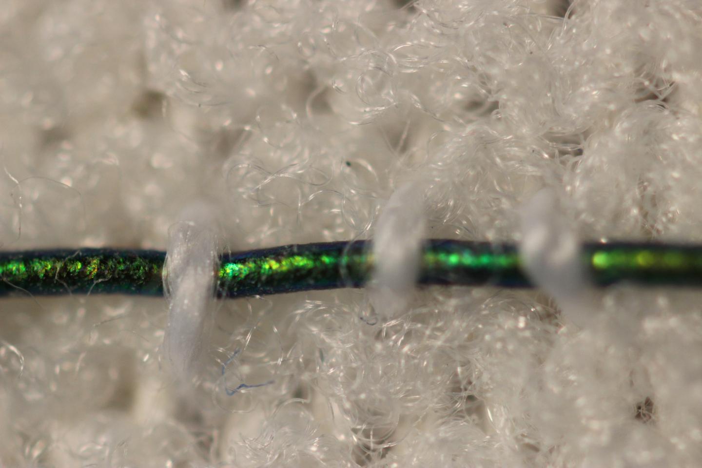
Caption: Engineers at MIT have developed pressure-sensing photonic fibers that they have woven into a typical compression bandage. Credit Courtesy of the researchers

Here’s a link to and a citation for the paper,

Stretchable Optomechanical Fiber Sensors for Pressure Determination in Compressive Medical Textiles by Joseph D. Sandt, Marie Moudio, J. Kenji Clark, James Hardin, Christian Argenti, Matthew Carty, Jennifer A. Lewis, Mathias Kolle. Advanced Healthcare Materials https://doi.org/10.1002/adhm.201800293 First published: 29 May 2018

This paper is behind a paywall.

Sutures that can gather data wirelessly

Are sutures which gather data hackable? It’s a little early to start thinking about that issue as this seems to be brand new research. A July 18, 2016 news item on ScienceDaily tells more,

For the first time, researchers led by Tufts University engineers have integrated nano-scale sensors, electronics and microfluidics into threads — ranging from simple cotton to sophisticated synthetics — that can be sutured through multiple layers of tissue to gather diagnostic data wirelessly in real time, according to a paper published online July 18 [2016] in Microsystems & Nanoengineering. The research suggests that the thread-based diagnostic platform could be an effective substrate for a new generation of implantable diagnostic devices and smart wearable systems.

A July 18, 2016 Tufts University news release (also on EurekAlert), which originated the news item, provides more detail,

The researchers used a variety of conductive threads that were dipped in physical and chemical sensing compounds and connected to wireless electronic circuitry to create a flexible platform that they sutured into tissue in rats as well as in vitro. The threads collected data on tissue health (e.g. pressure, stress, strain and temperature), pH and glucose levels that can be used to determine such things as how a wound is healing, whether infection is emerging, or whether the body’s chemistry is out of balance. The results were transmitted wirelessly to a cell phone and computer.

The three-dimensional platform is able to conform to complex structures such as organs, wounds or orthopedic implants.

While more study is needed in a number of areas, including investigation of long-term biocompatibility, researchers said initial results raise the possibility of optimizing patient-specific treatments.

“The ability to suture a thread-based diagnostic device intimately in a tissue or organ environment in three dimensions adds a unique feature that is not available with other flexible diagnostic platforms,” said Sameer Sonkusale, Ph.D., corresponding author on the paper and director of the interdisciplinary Nano Lab in the Department of Electrical and Computer Engineering at Tufts School of Engineering. “We think thread-based devices could potentially be used as smart sutures for surgical implants, smart bandages to monitor wound healing, or integrated with textile or fabric as personalized health monitors and point-of-care diagnostics.”

Until now, the structure of substrates for implantable devices has essentially been two-dimensional, limiting their usefulness to flat tissue such as skin, according to the paper. Additionally, the materials in those substrates are expensive and require specialized processing.

Here’s a link to and a citation for the paper,

A toolkit of thread-based microfluidics, sensors, and electronics for 3D tissue embedding for medical diagnostics by Pooria Mostafalu, Mohsen Akbari, Kyle A. Alberti, Qiaobing Xu, Ali Khademhosseini, & Sameer R. Sonkusale. Microsystems & Nanoengineering 2, Article number: 16039 (2016) doi:10.1038/micronano.2016.39 Published online 18 July 2016

This paper is open access.

Nanotechnology, math, cancer, and a boxing metaphor

Violent metaphors in medicine are not unusual although the reference is often to war rather than boxing as it is in this news from the University of Waterloo (Canada). Still, it seems counter-intuitive to closely link violence with healing but the practice is well entrenched and it seems attempts to counteract it are a ‘losing battle’ (pun intended).

Credit: Gabriel Picolo "2-in-1 punch." Courtesy: University of Waterloo

Credit: Gabriel Picolo “2-in-1 punch.” Courtesy: University of Waterloo

A June 23, 2016 news item on ScienceDaily describes a new approach to cancer therapy,

Math, biology and nanotechnology are becoming strange, yet effective bed-fellows in the fight against cancer treatment resistance. Researchers at the University of Waterloo and Harvard Medical School have engineered a revolutionary new approach to cancer treatment that pits a lethal combination of drugs together into a single nanoparticle.

Their work, published online on June 3, 2016 in the leading nanotechnology journal ACS Nano, finds a new method of shrinking tumors and prevents resistance in aggressive cancers by activating two drugs within the same cell at the same time.

A June 23, 2016 University of Waterloo news release (also on EurekAlert), which originated the news item, provides more information,

Every year thousands of patients die from recurrent cancers that have become resistant to therapy, resulting in one of the greatest unsolved challenges in cancer treatment. By tracking the fate of individual cancer cells under pressure of chemotherapy, biologists and bioengineers at Harvard Medical School studied a network of signals and molecular pathways that allow the cells to generate resistance over the course of treatment.

Using this information, a team of applied mathematicians led by Professor Mohammad Kohandel at the University of Waterloo, developed a mathematical model that incorporated algorithms that define the phenotypic cell state transitions of cancer cells in real-time while under attack by an anticancer agent. The mathematical simulations enabled them to define the exact molecular behavior and pathway of signals, which allow cancer cells to survive treatment over time.

They discovered that the PI3K/AKT kinase, which is often over-activated in cancers, enables cells to undergo a resistance program when pressured with the cytotoxic chemotherapy known as Taxanes, which are conventionally used to treat aggressive breast cancers. This revolutionary window into the life of a cell reveals that vulnerabilities to small molecule PI3K/AKT kinase inhibitors exist, and can be targeted if they are applied in the right sequence with combinations of other drugs.

Previously theories of drug resistance have relied on the hypothesis that only certain, “privileged” cells can overcome therapy. The mathematical simulations demonstrate that, under the right conditions and signaling events, any cell can develop a resistance program.

“Only recently have we begun to appreciate how important mathematics and physics are to understanding the biology and evolution of cancer,” said Professor Kohandel. “In fact, there is now increasing synergy between these disciplines, and we are beginning to appreciate how critical this information can be to create the right recipes to treat cancer.”

Although previous studies explored the use of drug combinations to treat cancer, the one-two punch approach is not always successful. In the new study, led by Professor Aaron Goldman, a faculty member in the division of Engineering in Medicine at Brigham and Women’s Hospital, the scientists realized a major shortcoming of the combination therapy approach is that both drugs need to be active in the same cell, something that current delivery methods can’t guarantee.

“We were inspired by the mathematical understanding that a cancer cell rewires the mechanisms of resistance in a very specific order and time-sensitive manner,” said Professor Goldman. “By developing a 2-in-1 nanomedicine, we could ensure the cell that was acquiring this new resistance saw the lethal drug combination, shutting down the survival program and eliminating the evidence of resistance. This approach could redefine how clinicians deliver combinations of drugs in the clinic.”

The approach the bioengineers took was to build a single nanoparticle, inspired by computer models, that exploit a technique known as supramolecular chemistry. This nanotechnology enables scientists to build cholesterol-tethered drugs together from “tetris-like” building blocks that self-assemble, incorporating multiple drugs into stable, individual nano-vehicles that target tumors through the leaky vasculature. This 2-in-1 strategy ensures that resistance to therapy never has a chance to develop, bringing together the right recipe to destroy surviving cancer cells.

Using mouse models of aggressive breast cancer, the scientists confirmed the predictions from the mathematical model that both drugs must be deterministically delivered to the same cell.

Here’s a link to and a citation for the paper,

Rationally Designed 2-in-1 Nanoparticles Can Overcome Adaptive Resistance in Cancer by Aaron Goldman, Ashish Kulkarni, Mohammad Kohandel, Prithvi Pandey, Poornima Rao, Siva Kumar Natarajan, Venkata Sabbisetti, and Shiladitya Sengupta. ACS Nano, Article ASAP DOI: 10.1021/acsnano.6b00320 Publication Date (Web): June 03, 2016

Copyright © 2016 American Chemical Society

This paper is behind a paywall.

The researchers have made this illustration of their work available,

Courtesy: American Chemical Society

Courtesy: American Chemical Society

Evolution is the best problem-solver

Dr. Jeffrey Karp of Brigham and Women’s Hospital says, “I truly believe evolution is the best problem-solver,” when discussing his medical biomimcry work in this video,

You can find the video and more in a Mar. 20, 2013 news item on Nanowerk which was originated by a Mar. 6, 2013 article by Alisa Zapp Machalek for the US National Institutes of Health (NIH) National Institute of General Medical Sciences (NGMS) Inside Life Science webpage,

Velcro® was inspired by the grappling hooks of burrs. Supersonic jets have structures that work like the nostrils of peregrine falcons in a speed dive. Full-body swimsuits, now banned from the Olympics, lend athletes a smooth, streamlined shape like fish.

Nature’s designs are also giving researchers funded by the National Institutes of Health ideas for new technologies that could help wounds heal, make injections less painful and provide new materials for a variety of purposes.

… scientists [Jeffrey Karp and Robert Langer] discovered, to their surprise, that a [porcupine] quill’s puncture power comes from its barbed tip. Barbs seem to work like the points on a serrated knife, concentrating pressure onto small areas to aid penetration. Because they require significantly less force to puncture skin, barbed shafts don’t hurt as much when they enter flesh as their smooth-tipped counterparts do.

Zapp Machalek goes on to detail work inspired by gecko feet and spider webs, as well as, porcupine quills.

Inflammation isn’t all bad but sometimes you need to reduce it with nanomedicines

Researchers from Brigham and Women’s Hospital (BWH), Columbia University Medical Center, Icahn School of Medicine at Mount Sinai, and Massachusetts Institute of Technology (MIT) have published about a study about their use of nano-sized particles to release therapeutic drugs that are designed to relieve chronic inflammation. From the Mar. 18, 2013 news release on EurekAlert,

Inflammation is the body’s natural defense mechanism against invading organisms and tissue injury. In acute inflammation, the pathogen or inflammatory mediators are cleared away and homeostasis is reached, however in chronic inflammatory states, this resolving response is impaired, leading to chronic inflammation and tissue damage. It is now widely believed that an impaired resolution of inflammation is a major contributing factor to the progression of a number of devastating diseases such as atherosclerosis, arthritis, and neurodegenerative diseases, in addition to cancer. Since the level of inflammation in these diseases is very high—targeted therapeutic solutions are required to help keep inflammation contained.

A new study from researchers at Brigham and Women’s Hospital (BWH), Columbia University Medical Center, Icahn School of Medicine at Mount Sinai, and Massachusetts Institute of Technology presents the development of tiny nanomedicines in the sub 100 nm range (100,000 times smaller than the diameter of a human hair strand) that are capable of encapsulating and releasing an inflammation-resolving peptide drug. The authors showed that these nanoparticles are potent pro-resolving nanomedicines, capable of selectively homing to sites of tissue injury in mice, and releasing their therapeutic payload in a controlled manner over time. Uniquely, these nanoparticles are designed to target the extracellular microenvironment of inflamed tissues. The particles then slowly release their potent inflammation-resolving payload such that it can diffuse through the inflamed tissue. There the drug binds to receptors on the plasma membrane of activated white blood cells and causes them to become more quiescent.

The research will be published some time this week (week of Mar. 18, 2013) by the Proceedings of the National Academy of Science. The news release offers more detail about the work,

“The beauty of this approach is that it takes advantage of nature’s own design for preventing inflammation-induced damage, which, unlike many other anti-inflammatory strategies, does not compromise host defense and promotes tissue repair,” said Ira Tabas, MD, PhD, physician-scientist at Columbia University Medical Center and co-senior author of this study.

“The development of self-assembled targeted nanoparticles which are capable of resolving inflammation has broad application in medicine including the treatment of atherosclerosis,” said Omid Farokhzad, MD, physician-scientist at BWH, and a co-senior author of this study.

Polymers consisting of three chains attached end-to-end were developed as building blocks for the engineering of self-assembled targeted nanoparticles; one chain enabled the entrapment and controlled release of the therapeutic payload, in this case a peptide which mimics the pro-resolving properties of the Annexin A1 protein. Another chain conferred stealth properties to the nanoparticles, enabling their long-circulation after systemic administration. Yet a third chain gave homing capability to the nanoparticles to target the collagen IV protein to the vascular wall. As such these nanoparticles are capable of selectively sticking to injured vasculature allowing their therapeutic anti-inflammatory cargo to be released where it is needed to effectively promote inflammation resolution in a deliberate and targeted manner.

“These targeted polymeric nanoparticles are capable of stopping neutrophils, which are the most abundant form of white blood cells, from infiltrating sites of disease or injury at very small doses. This action stops the neutrophils from secreting further signaling molecules which can lead to a constant hyper-inflammatory state and further disease complications,” said Nazila Kamaly, PhD, a postdoctoral fellow at BWH and co-lead author of this study.

“Nanoparticles that selectively bind to injured vasculature could have a profound impact in prevalent diseases, such as atherosclerosis, where damaged or comprised vasculature underlie the pathology. This work offers a novel targeted nanomedicine to the burgeoning field of inflammation-resolution, a field previously pioneered by BWH’s Dr. Charles Serhan,” said Gabrielle Fredman, PhD, a post-doctoral fellow at Columbia University Medical Center and co-lead author of this study.

These new developments have led the researchers to start investigating the potential of these pro-resolving nanomedicines for their effects on shrinking atherosclerotic plaques, and these studies are currently underway.

This news release does not offer any information as to what type of studies might be underway. My guess is that we are still years away from human clinical trials. Azonano also features this work in a Mar. 19, 2013 news item.

Organ chips for DARPA (Defense Advanced Research Projects Agency)

The Wyss Institute will receive up to  $37M US for a project that integrates ten different organ-on-a-chip projects into one system. From the July 24, 2012 news release on EurekAlert,

With this new DARPA funding, Institute researchers and a multidisciplinary team of collaborators seek to build 10 different human organs-on-chips, to link them together to more closely mimic whole body physiology, and to engineer an automated instrument that will control fluid flow and cell viability while permitting real-time analysis of complex biochemical functions. As an accurate alternative to traditional animal testing models that often fail to predict human responses, this instrumented “human-on-a-chip” will be used to rapidly assess responses to new drug candidates, providing critical information on their safety and efficacy.

This unique platform could help ensure that safe and effective therapeutics are identified sooner, and ineffective or toxic ones are rejected early in the development process. As a result, the quality and quantity of new drugs moving successfully through the pipeline and into the clinic may be increased, regulatory decision-making could be better informed, and patient outcomes could be improved.

Jesse Goodman, FDA Chief Scientist and Deputy Commissioner for Science and Public Health, commented that the automated human-on-chip instrument being developed “has the potential to be a better model for determining human adverse responses. FDA looks forward to working with the Wyss Institute in its development of this model that may ultimately be used in therapeutic development.”

Wyss Founding Director, Donald Ingber, M.D., Ph.D., and Wyss Core Faculty member, Kevin Kit Parker, Ph.D., will co-lead this five-year project.

I note that Kevin Kit Parker was mentioned in an earlier posting today (July 26, 2012) titled, Medusa, jellyfish, and tissue engineering, and Donald Ingber in my Dec.1e, 2011 posting about Shrilk and insect skeletons.

As for the Wyss Institute, here’s a description from the news release,

The Wyss Institute for Biologically Inspired Engineering at Harvard University (http://wyss.harvard.edu) uses Nature’s design principles to develop bioinspired materials and devices that will transform medicine and create a more sustainable world. Working as an alliance among Harvard’s Schools of Medicine, Engineering, and Arts & Sciences, and in partnership with Beth Israel Deaconess Medical Center, Boston Children’s Hospital, Brigham and Women’s Hospital, , Dana Farber Cancer Institute, Massachusetts General Hospital, the University of Massachusetts Medical School, Spaulding Rehabilitation Hospital, Tufts University, and Boston University, the Institute crosses disciplinary and institutional barriers to engage in high-risk research that leads to transformative technological breakthroughs. By emulating Nature’s principles for self-organizing and self-regulating, Wyss researchers are developing innovative new engineering solutions for healthcare, energy, architecture, robotics, and manufacturing. These technologies are translated into commercial products and therapies through collaborations with clinical investigators, corporate alliances, and new start-ups.

I hadn’t thought of an organ-on-a-chip as particularly bioinspired so I’ll have to think about that one for a while.