Tag Archives: cancer

Wearable technology: two types of sensors one from the University of Glasgow (Scotland) and the other from the University of British Columbia (Canada)

Sometimes it’s good to try and pull things together.

University of Glasgow and monitoring chronic conditions

A February 23, 2018 news item on phys.org describes the latest wearable tech from the University of Glasgow,

A new type of flexible, wearable sensor could help people with chronic conditions like diabetes avoid the discomfort of regular pin-prick blood tests by monitoring the chemical composition of their sweat instead.

In a new paper published in the journal Biosensors and Bioelectronics, a team of scientists from the University of Glasgow’s School of Engineering outline how they have built a stretchable, wireless system which is capable of measuring the pH level of users’ sweat.

A February 22, 2018 University of Glasgow press release, which originated the news item, expands on the theme,

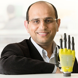
Ravinder Dahiya

 Courtesy: University of Glasgow


Sweat, like blood, contains chemicals generated in the human body, including glucose and urea. Monitoring the levels of those chemicals in sweat could help clinicians diagnose and monitor chronic conditions such as diabetes, kidney disease and some types of cancers without invasive tests which require blood to be drawn from patients.

However, non-invasive, wearable systems require consistent contact with skin to offer the highest-quality monitoring. Current systems are made from rigid materials, making it more difficult to ensure consistent contact, and other potential solutions such as adhesives can irritate skin. Wireless systems which use Bluetooth to transmit their information are also often bulky and power-hungry, requiring frequent recharging.

The University of Glasgow team’s new system is built around an inexpensively-produced sensor capable of measuring pH levels which can stretch and flex to better fit the contours of users’ bodies. Made from a graphite-polyurethane composite and measuring around a single square centimetre, it can stretch up to 53% in length without compromising performance. It will also continue to work after being subjected to flexes of 30% up to 500 times, which the researchers say will allow it to be used comfortably on human skin with minimal impact on the performance of the sensor.

The sensor can transmit its data wirelessly, and without external power, to an accompanying smartphone app called ‘SenseAble’, also developed by the team. The transmissions use near-field communication, a data transmission system found in many current smartphones which is used most often for smartphone payments like ApplePay, via a stretchable RFID antenna integrated into the system – another breakthrough innovation from the research team.

The smartphone app allows users to track pH levels in real time and was demonstrated in the lab using a chemical solution created by the researchers which mimics the composition of human sweat.

The research was led by Professor Ravinder Dahiya, head of the University of Glasgow’s School of Engineering’s Bendable Electronics and Sensing Technologies (BEST) group.

Professor Dahiya said: “Human sweat contains much of the same physiological information that blood does, and its use in diagnostic systems has the significant advantage of not needing to break the skin in order to administer tests.

“Now that we’ve demonstrated that our stretchable system can be used to monitor pH levels, we’ve already begun additional research to expand the capabilities of the sensor and make it a more complete diagnostic system. We’re planning to add sensors capable of measuring glucose, ammonia and urea, for example, and ultimately we’d like to see a system ready for market in the next few years.”

The team’s paper, titled ‘Stretchable Wireless System for Sweat pH Monitoring’, is published in Biosensors and Bioelectronics. The research was supported by funding from the European Commission and the Engineering and Physical Sciences Research Council (EPSRC).

Here’s a link to and a citation for the paper,

Stretchable wireless system for sweat pH monitoring by Wenting Dang, Libu Manjakkal, William Taube Navaraj, Leandro Lorenzelli, Vincenzo Vinciguerra. Biosensors and Bioelectronics Volume 107, 1 June 2018, Pages 192–202 [Available online February 2018] https://doi.org/10.1016/j.bios.2018.02.025

This paper is behind a paywall.

University of British Columbia (UBC; Okanagan) and monitor bio-signals

This is a completely other type of wearable tech monitor, from a February 22, 2018 UBC news release (also on EurekAlert) by Patty Wellborn (A link has been removed),

Creating the perfect wearable device to monitor muscle movement, heart rate and other tiny bio-signals without breaking the bank has inspired scientists to look for a simpler and more affordable tool.

Now, a team of researchers at UBC’s Okanagan campus have developed a practical way to monitor and interpret human motion, in what may be the missing piece of the puzzle when it comes to wearable technology.

What started as research to create an ultra-stretchable sensor transformed into a sophisticated inter-disciplinary project resulting in a smart wearable device that is capable of sensing and understanding complex human motion, explains School of Engineering Professor Homayoun Najjaran.

The sensor is made by infusing graphene nano-flakes (GNF) into a rubber-like adhesive pad. Najjaran says they then tested the durability of the tiny sensor by stretching it to see if it can maintain accuracy under strains of up to 350 per cent of its original state. The device went through more than 10,000 cycles of stretching and relaxing while maintaining its electrical stability.

“We tested this sensor vigorously,” says Najjaran. “Not only did it maintain its form but more importantly it retained its sensory functionality. We have further demonstrated the efficacy of GNF-Pad as a haptic technology in real-time applications by precisely replicating the human finger gestures using a three-joint robotic finger.”

The goal was to make something that could stretch, be flexible and a reasonable size, and have the required sensitivity, performance, production cost, and robustness. Unlike an inertial measurement unit—an electronic unit that measures force and movement and is used in most step-based wearable technologies—Najjaran says the sensors need to be sensitive enough to respond to different and complex body motions. That includes infinitesimal movements like a heartbeat or a twitch of a finger, to large muscle movements from walking and running.

School of Engineering Professor and study co-author Mina Hoorfar says their results may help manufacturers create the next level of health monitoring and biomedical devices.

“We have introduced an easy and highly repeatable fabrication method to create a highly sensitive sensor with outstanding mechanical and electrical properties at a very low cost,” says Hoorfar.

To demonstrate its practicality, researchers built three wearable devices including a knee band, a wristband and a glove. The wristband monitored heartbeats by sensing the pulse of the artery. In an entirely different range of motion, the finger and knee bands monitored finger gestures and larger scale muscle movements during walking, running, sitting down and standing up. The results, says Hoorfar, indicate an inexpensive device that has a high-level of sensitivity, selectivity and durability.

Hoorfar and Najjaran are both members of the Okanagan node of UBC’s STITCH (SmarT Innovations for Technology Connected Health) Institute that creates and investigates advanced wearable devices.

The research, partially funded by the Natural Sciences and Engineering Research Council, was recently published in the Journal of Sensors and Actuators A: Physical.

Here’s a link to and a citation for the paper,

Low-cost ultra-stretchable strain sensors for monitoring human motion and bio-signals by Seyed Reza Larimi, Hojatollah Rezaei Nejad, Michael Oyatsi, Allen O’Brien, Mina Hoorfar, Homayoun Najjaran. Sensors and Actuators A: Physical Volume 271, 1 March 2018, Pages 182-191 [Published online February 2018] https://doi.org/10.1016/j.sna.2018.01.028

This paper is behind a paywall.

Final comments

The term ‘wearable tech’ covers a lot of ground. In addition to sensors, there are materials that harvest energy, detect poisons, etc.  making for a diverse field.
CRISPR-Cas12a as a new diagnostic tool

Similar to Cas9, Cas12a is has an added feature as noted in this February 15, 2018 news item on ScienceDaily,

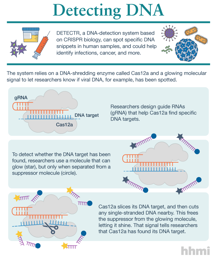
Utilizing an unsuspected activity of the CRISPR-Cas12a protein, researchers created a simple diagnostic system called DETECTR to analyze cells, blood, saliva, urine and stool to detect genetic mutations, cancer and antibiotic resistance and also diagnose bacterial and viral infections. The scientists discovered that when Cas12a binds its double-stranded DNA target, it indiscriminately chews up all single-stranded DNA. They then created reporter molecules attached to single-stranded DNA to signal when Cas12a finds its target.

A February 15, 2018 University of California at Berkeley (UC Berkeley) news release by Robert Sanders and which originated the news item, provides more detail and history,

CRISPR-Cas12a, one of the DNA-cutting proteins revolutionizing biology today, has an unexpected side effect that makes it an ideal enzyme for simple, rapid and accurate disease diagnostics.

blood in test tube

(iStock)

Cas12a, discovered in 2015 and originally called Cpf1, is like the well-known Cas9 protein that UC Berkeley’s Jennifer Doudna and colleague Emmanuelle Charpentier turned into a powerful gene-editing tool in 2012.

CRISPR-Cas9 has supercharged biological research in a mere six years, speeding up exploration of the causes of disease and sparking many potential new therapies. Cas12a was a major addition to the gene-cutting toolbox, able to cut double-stranded DNA at places that Cas9 can’t, and, because it leaves ragged edges, perhaps easier to use when inserting a new gene at the DNA cut.

But co-first authors Janice Chen, Enbo Ma and Lucas Harrington in Doudna’s lab discovered that when Cas12a binds and cuts a targeted double-stranded DNA sequence, it unexpectedly unleashes indiscriminate cutting of all single-stranded DNA in a test tube.

Most of the DNA in a cell is in the form of a double-stranded helix, so this is not necessarily a problem for gene-editing applications. But it does allow researchers to use a single-stranded “reporter” molecule with the CRISPR-Cas12a protein, which produces an unambiguous fluorescent signal when Cas12a has found its target.

“We continue to be fascinated by the functions of bacterial CRISPR systems and how mechanistic understanding leads to opportunities for new technologies,” said Doudna, a professor of molecular and cell biology and of chemistry and a Howard Hughes Medical Institute investigator.

DETECTR diagnostics

The new DETECTR system based on CRISPR-Cas12a can analyze cells, blood, saliva, urine and stool to detect genetic mutations, cancer and antibiotic resistance as well as diagnose bacterial and viral infections. Target DNA is amplified by RPA to make it easier for Cas12a to find it and bind, unleashing indiscriminate cutting of single-stranded DNA, including DNA attached to a fluorescent marker (gold star) that tells researchers that Cas12a has found its target.

The UC Berkeley researchers, along with their colleagues at UC San Francisco, will publish their findings Feb. 15 [2018] via the journal Science’s fast-track service, First Release.

The researchers developed a diagnostic system they dubbed the DNA Endonuclease Targeted CRISPR Trans Reporter, or DETECTR, for quick and easy point-of-care detection of even small amounts of DNA in clinical samples. It involves adding all reagents in a single reaction: CRISPR-Cas12a and its RNA targeting sequence (guide RNA), fluorescent reporter molecule and an isothermal amplification system called recombinase polymerase amplification (RPA), which is similar to polymerase chain reaction (PCR). When warmed to body temperature, RPA rapidly multiplies the number of copies of the target DNA, boosting the chances Cas12a will find one of them, bind and unleash single-strand DNA cutting, resulting in a fluorescent readout.

The UC Berkeley researchers tested this strategy using patient samples containing human papilloma virus (HPV), in collaboration with Joel Palefsky’s lab at UC San Francisco. Using DETECTR, they were able to demonstrate accurate detection of the “high-risk” HPV types 16 and 18 in samples infected with many different HPV types.

“This protein works as a robust tool to detect DNA from a variety of sources,” Chen said. “We want to push the limits of the technology, which is potentially applicable in any point-of-care diagnostic situation where there is a DNA component, including cancer and infectious disease.”

The indiscriminate cutting of all single-stranded DNA, which the researchers discovered holds true for all related Cas12 molecules, but not Cas9, may have unwanted effects in genome editing applications, but more research is needed on this topic, Chen said. During the transcription of genes, for example, the cell briefly creates single strands of DNA that could accidentally be cut by Cas12a.

The activity of the Cas12 proteins is similar to that of another family of CRISPR enzymes, Cas13a, which chew up RNA after binding to a target RNA sequence. Various teams, including Doudna’s, are developing diagnostic tests using Cas13a that could, for example, detect the RNA genome of HIV.

infographic about DETECTR system

(Infographic by the Howard Hughes Medical Institute)

These new tools have been repurposed from their original role in microbes where they serve as adaptive immune systems to fend off viral infections. In these bacteria, Cas proteins store records of past infections and use these “memories” to identify harmful DNA during infections. Cas12a, the protein used in this study, then cuts the invading DNA, saving the bacteria from being taken over by the virus.

The chance discovery of Cas12a’s unusual behavior highlights the importance of basic research, Chen said, since it came from a basic curiosity about the mechanism Cas12a uses to cleave double-stranded DNA.

“It’s cool that, by going after the question of the cleavage mechanism of this protein, we uncovered what we think is a very powerful technology useful in an array of applications,” Chen said.

Here’s a link to and a citation for the paper,

CRISPR-Cas12a target binding unleashes indiscriminate single-stranded DNase activity by Janice S. Chen, Enbo Ma, Lucas B. Harrington, Maria Da Costa, Xinran Tian, Joel M. Palefsky, Jennifer A. Doudna. Science 15 Feb 2018: eaar6245 DOI: 10.1126/science.aar6245

This paper is behind a paywall.

Colliding organic nanoparticles caught on camera for the first time

There is high excitement about this development in a November 17, 2017 news item on Nanowerk,

A Northwestern University research team is the first to capture on video organic nanoparticles colliding and fusing together. This unprecedented view of “chemistry in motion” will aid Northwestern nanoscientists developing new drug delivery methods as well as demonstrate to researchers around the globe how an emerging imaging technique opens a new window on a very tiny world.

A November 17, 2017 Northwestern University news release (also on EurekAlert) by Megan Fellman, which originated the news item, further illuminates the matter,

This is a rare example of particles in motion. The dynamics are reminiscent of two bubbles coming together and merging into one: first they join and have a membrane between them, but then they fuse and become one larger bubble.

“I had an image in my mind, but the first time I saw these fusing nanoparticles in black and white was amazing,” said professor Nathan C. Gianneschi, who led the interdisciplinary study and works at the intersection of nanotechnology and biomedicine.

“To me, it’s literally a window opening up to this world you have always known was there, but now you’ve finally got an image of it. I liken it to the first time I saw Jupiter’s moons through a telescope. Nothing compares to actually seeing,” he said.

Gianneschi is the Jacob and Rosaline Cohn Professor in the department of chemistry in the Weinberg College of Arts and Sciences and in the departments of materials science and engineering and of biomedical engineering in the McCormick School of Engineering.

The study, which includes videos of different nanoparticle fusion events, was published today (Nov. 1 [2017]7) by the Journal of the American Chemical Society.

The research team used liquid-cell transmission electron microscopy to directly image how polymer-based nanoparticles, or micelles, that Gianneschi’s lab is developing for treating cancer and heart attacks change over time. The powerful new technique enabled the scientists to directly observe the particles’ transformation and characterize their dynamics.

“We can see on the molecular level how the polymeric matter rearranges when the particles fuse into one object,” said Lucas R. Parent, first author of the paper and a National Institutes of Health Postdoctoral Fellow in Gianneschi’s research group. “This is the first study of many to come in which researchers will use this method to look at all kinds of dynamic phenomena in organic materials systems on the nanoscale.”

In the Northwestern study, organic particles in water bounce off each other, and some collide and merge, undergoing a physical transformation. The researchers capture the action by shining an electron beam through the sample. The tiny particles — the largest are only approximately 200 nanometers in diameter — cast shadows that are captured directly by a camera below.

“We’ve observed classical fusion behavior on the nanoscale,” said Gianneschi, a member of Northwestern’s International Institute for Nanotechnology. “Capturing the fundamental growth and evolution processes of these particles in motion will help us immensely in our work with synthetic materials and their interactions with biological systems.”

The National Institutes of Health, the National Science Foundation, the Air Force Office of Scientific Research and the Army Research Office supported the research.

Here’s a link to and a citation for the paper,

Directly Observing Micelle Fusion and Growth in Solution by Liquid-Cell Transmission Electron Microscopy by Lucas R. Parent, Evangelos Bakalis, Abelardo Ramírez-Hernández, Jacquelin K. Kammeyer, Chiwoo Park, Juan de Pablo, Francesco Zerbetto, Joseph P. Patterson, and Nathan C. Gianneschi. J. Am. Chem. Soc., Article ASAP DOI: 10.1021/jacs.7b09060 Publication Date (Web): November 17, 2017

Copyright © 2017 American Chemical Society

This paper is behind a paywall.

Hit and run gene therapy?

The approach looks promising but there’s a still long way to go before this ‘simpler, gentler’ approach to gene therapy will make its way into any treatments. From an August 30, 2017 news item on Nanowerk,

A new biomedical tool using nanoparticles that deliver transient gene changes to targeted cells could make therapies for a variety of diseases — including cancer, diabetes and HIV — faster and cheaper to develop, and more customizable.

The tool, developed by researchers at Fred Hutchinson Cancer Research Center and tested in preclinical models, is described in a paper published August 30 [2017] in Nature Communications.

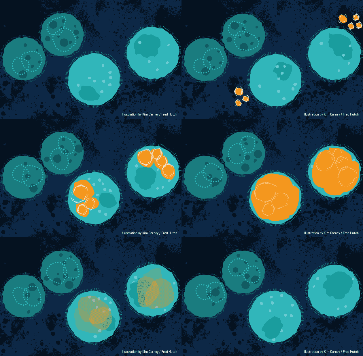
This animation demonstrates the approach,

Biodegradable nanoparticles (orange) carry short-lived gene therapy to specific cells (light teal). Animation by Kimberly Carney / Fred Hutch News Service

An August 30, 2017 Fred Hutchinson Cancer Research Center (Fred Hutch) news release (from news release received via email; also on EurekAlert) by Sabrina Richards, which originated the news item, elucidates further (Note: Some links and notes have been removed),

“Our goal is to streamline the manufacture of cell-based therapies,” said lead author DR. MATTHIAS STEPHAN [6], a faculty member in the Fred Hutch Clinical Research Division and an expert in developing biomaterials. “In this study, we created a product where you just add it to cultured cells and that’s it — no additional manufacturing steps.”

Stephan and his colleagues developed a nanoparticle delivery system to extend the therapeutic potential of messenger RNA, which delivers molecular instructions from DNA to cells in the body, directing them to make proteins to prevent or fight disease.

The researchers’ approach was designed to zero in on specific cell types — T cells of the immune system and blood stem cells — and deliver mRNA directly to the cells, triggering short-term gene expression. It’s called “hit-and-run” genetic programming because the transient effect of mRNA does not change the DNA, but it is enough to make a permanent impact on the cells’ therapeutic potential.

Stephan and colleagues used three examples in the Nature Communications paper to demonstrate their technology:

* Nanoparticles carried a gene-editing tool to T cells of the immune system that snipped out their natural T-cell receptors, and then was paired with genes encoding a “chimeric antigen receptor” or CAR, a synthetic molecule designed to attack cancer.
* Targeted to blood stem cells, nanoparticles were equipped with mRNA that enabled the stem cells to multiply and replace blood cancer cells with healthy cells when used in bone marrow transplants.
* Nanoparticles targeted to CAR-T cells and containing foxo1 mRNA, which signals the anti-cancer T cells to develop into a type of “memory” cell that is more aggressive and destroys tumor cells more effectively and maintains anti-tumor activity longer.

Other attempts to engineer mRNA into disease-fighting cells have been tricky. The large messenger molecule degrades quickly before it can have an effect, and the body’s immune system recognizes it as foreign — not coming from DNA in the nucleus of the cell — and destroys it.

Stephan and his Fred Hutch collaborators devised a workaround to those hurdles.

“We developed a nanocarrier that binds and condenses synthetic mRNA and protects it from degradation,” Stephan said. The researchers surrounded the nanoparticle with a negatively charged envelope with a targeting ligand attached to the surface so that the particle selectively homes in and binds to a particular cell type.

The cells swallow up the tiny carrier, which can be loaded with different types of manmade mRNA. “If you know from the scientific literature that a signaling pathway works in synergy, you could co-deliver mRNA in a single nanoparticle,” Stephan said. “Every cell that takes up the nanoparticle can express both.”

The approach involves mixing the freeze-dried nanoparticles with water and a sample of cells. Within four hours, cells start showing signs that the editing has taken effect. Boosters can be given if needed. Made from a dissolving biomaterial, the nanoparticles are removed from the body like other cell waste.

“Just add water to our freeze-dried product,” Stephan said. Since it’s built on existing technologies and doesn’t require knowledge of nanotechnology, he intends for it to be an off-the-shelf way for cell-therapy engineers to develop new approaches to treating a variety of diseases.

The approach could replace labor-intensive electroporation, a multistep cell-manufacturing technique that requires specialized equipment and clean rooms. All the handling ends up destroying many of the cells, which limits the amount that can be used in treatments for patients.

Gentler to cells, the nanoparticle system developed by the Fred Hutch team showed that up to 60 times more cells survive the process compared with electroporation. This is a critical feature for ensuring enough cells are viable when transferred to patients.

“You can imagine taking the nanoparticles, injecting them into a patient and then you don’t have to culture cells at all anymore,” he said.

Stephan has tested the technology is cultured cells in the lab, and it’s not yet available as a treatment. Stephan is looking for commercial partners to move the technology toward additional applications and into clinical trials where it could be developed into a therapy.

Here’s a link to and a citation for the paper,

Hit-and-run programming of therapeutic cytoreagents using mRNA nanocarriers by H. F. Moffett, M. E. Coon, S. Radtke, S. B. Stephan, L. McKnight, A. Lambert, B. L. Stoddard, H. P. Kiem, & M. T. Stephan. Nature Communications 8, Article number: 389 (2017) doi:10.1038/s41467-017-00505-8 Published online: 30 August 2017

This paper is open access.

Training drugs

This summarizes some of what’s happening in nanomedicine and provides a plug (boost) for the  University of Cambridge’s nanotechnology programmes (from a June 26, 2017 news item on Nanowerk),

Nanotechnology is creating new opportunities for fighting disease – from delivering drugs in smart packaging to nanobots powered by the world’s tiniest engines.

Chemotherapy benefits a great many patients but the side effects can be brutal.
When a patient is injected with an anti-cancer drug, the idea is that the molecules will seek out and destroy rogue tumour cells. However, relatively large amounts need to be administered to reach the target in high enough concentrations to be effective. As a result of this high drug concentration, healthy cells may be killed as well as cancer cells, leaving many patients weak, nauseated and vulnerable to infection.

One way that researchers are attempting to improve the safety and efficacy of drugs is to use a relatively new area of research known as nanothrapeutics to target drug delivery just to the cells that need it.

Professor Sir Mark Welland is Head of the Electrical Engineering Division at Cambridge. In recent years, his research has focused on nanotherapeutics, working in collaboration with clinicians and industry to develop better, safer drugs. He and his colleagues don’t design new drugs; instead, they design and build smart packaging for existing drugs.

The University of Cambridge has produced a video interview (referencing a 1966 movie ‘Fantastic Voyage‘ in its title)  with Sir Mark Welland,

A June 23, 2017 University of Cambridge press release, which originated the news item, delves further into the topic of nanotherapeutics (nanomedicine) and nanomachines,

Nanotherapeutics come in many different configurations, but the easiest way to think about them is as small, benign particles filled with a drug. They can be injected in the same way as a normal drug, and are carried through the bloodstream to the target organ, tissue or cell. At this point, a change in the local environment, such as pH, or the use of light or ultrasound, causes the nanoparticles to release their cargo.

Nano-sized tools are increasingly being looked at for diagnosis, drug delivery and therapy. “There are a huge number of possibilities right now, and probably more to come, which is why there’s been so much interest,” says Welland. Using clever chemistry and engineering at the nanoscale, drugs can be ‘taught’ to behave like a Trojan horse, or to hold their fire until just the right moment, or to recognise the target they’re looking for.

“We always try to use techniques that can be scaled up – we avoid using expensive chemistries or expensive equipment, and we’ve been reasonably successful in that,” he adds. “By keeping costs down and using scalable techniques, we’ve got a far better chance of making a successful treatment for patients.”

In 2014, he and collaborators demonstrated that gold nanoparticles could be used to ‘smuggle’ chemotherapy drugs into cancer cells in glioblastoma multiforme, the most common and aggressive type of brain cancer in adults, which is notoriously difficult to treat. The team engineered nanostructures containing gold and cisplatin, a conventional chemotherapy drug. A coating on the particles made them attracted to tumour cells from glioblastoma patients, so that the nanostructures bound and were absorbed into the cancer cells.

Once inside, these nanostructures were exposed to radiotherapy. This caused the gold to release electrons that damaged the cancer cell’s DNA and its overall structure, enhancing the impact of the chemotherapy drug. The process was so effective that 20 days later, the cell culture showed no evidence of any revival, suggesting that the tumour cells had been destroyed.

While the technique is still several years away from use in humans, tests have begun in mice. Welland’s group is working with MedImmune, the biologics R&D arm of pharmaceutical company AstraZeneca, to study the stability of drugs and to design ways to deliver them more effectively using nanotechnology.

“One of the great advantages of working with MedImmune is they understand precisely what the requirements are for a drug to be approved. We would shut down lines of research where we thought it was never going to get to the point of approval by the regulators,” says Welland. “It’s important to be pragmatic about it so that only the approaches with the best chance of working in patients are taken forward.”

The researchers are also targeting diseases like tuberculosis (TB). With funding from the Rosetrees Trust, Welland and postdoctoral researcher Dr Íris da luz Batalha are working with Professor Andres Floto in the Department of Medicine to improve the efficacy of TB drugs.

Their solution has been to design and develop nontoxic, biodegradable polymers that can be ‘fused’ with TB drug molecules. As polymer molecules have a long, chain-like shape, drugs can be attached along the length of the polymer backbone, meaning that very large amounts of the drug can be loaded onto each polymer molecule. The polymers are stable in the bloodstream and release the drugs they carry when they reach the target cell. Inside the cell, the pH drops, which causes the polymer to release the drug.

In fact, the polymers worked so well for TB drugs that another of Welland’s postdoctoral researchers, Dr Myriam Ouberaï, has formed a start-up company, Spirea, which is raising funding to develop the polymers for use with oncology drugs. Ouberaï is hoping to establish a collaboration with a pharma company in the next two years.

“Designing these particles, loading them with drugs and making them clever so that they release their cargo in a controlled and precise way: it’s quite a technical challenge,” adds Welland. “The main reason I’m interested in the challenge is I want to see something working in the clinic – I want to see something working in patients.”

Could nanotechnology move beyond therapeutics to a time when nanomachines keep us healthy by patrolling, monitoring and repairing the body?

Nanomachines have long been a dream of scientists and public alike. But working out how to make them move has meant they’ve remained in the realm of science fiction.

But last year, Professor Jeremy Baumberg and colleagues in Cambridge and the University of Bath developed the world’s tiniest engine – just a few billionths of a metre [nanometre] in size. It’s biocompatible, cost-effective to manufacture, fast to respond and energy efficient.

The forces exerted by these ‘ANTs’ (for ‘actuating nano-transducers’) are nearly a hundred times larger than those for any known device, motor or muscle. To make them, tiny charged particles of gold, bound together with a temperature-responsive polymer gel, are heated with a laser. As the polymer coatings expel water from the gel and collapse, a large amount of elastic energy is stored in a fraction of a second. On cooling, the particles spring apart and release energy.

The researchers hope to use this ability of ANTs to produce very large forces relative to their weight to develop three-dimensional machines that swim, have pumps that take on fluid to sense the environment and are small enough to move around our bloodstream.

Working with Cambridge Enterprise, the University’s commercialisation arm, the team in Cambridge’s Nanophotonics Centre hopes to commercialise the technology for microfluidics bio-applications. The work is funded by the Engineering and Physical Sciences Research Council and the European Research Council.

“There’s a revolution happening in personalised healthcare, and for that we need sensors not just on the outside but on the inside,” explains Baumberg, who leads an interdisciplinary Strategic Research Network and Doctoral Training Centre focused on nanoscience and nanotechnology.

“Nanoscience is driving this. We are now building technology that allows us to even imagine these futures.”

I have featured Welland and his work here before and noted his penchant for wanting to insert nanodevices into humans as per this excerpt from an April 30, 2010 posting,
Getting back to the Cambridge University video, do go and watch it on the Nanowerk site. It is fun and very informative and approximately 17 mins. I noticed that they reused part of their Nokia morph animation (last mentioned on this blog here) and offered some thoughts from Professor Mark Welland, the team leader on that project. Interestingly, Welland was talking about yet another possibility. (Sometimes I think nano goes too far!) He was suggesting that we could have chips/devices in our brains that would allow us to think about phoning someone and an immediate connection would be made to that person. Bluntly—no. Just think what would happen if the marketers got access and I don’t even want to think what a person who suffers psychotic breaks (i.e., hearing voices) would do with even more input. Welland starts to talk at the 11 minute mark (I think). For an alternative take on the video and more details, visit Dexter Johnson’s blog, Nanoclast, for this posting. Hint, he likes the idea of a phone in the brain much better than I do.

I’m not sure what could have occasioned this latest press release and related video featuring Welland and nanotherapeutics other than guessing that it was a slow news period.

Curcumin: a scientific literature review concludes health benefits may be overstated

Given the number of times I’ve featured ‘curcumin research’, it seems only right to include this latest work. A Jan. 11, 2017 American Chemical Society (ACS) news release (also on EurekAlert) describes the results of a review of the scientific literature on curcumin’s (a constituent of turmeric) medicinal effectiveness,

Curcumin, a compound in turmeric, continues to be hailed as a natural treatment for a wide range of health conditions, including cancer and Alzheimer’s disease. But a new review of the scientific literature on curcumin has found it’s probably not all it’s ground up to be. The report in ACS’ Journal of Medicinal Chemistry instead cites evidence that, contrary to numerous reports, the compound has limited — if any — therapeutic benefit.

Turmeric, a spice often added to curries and mustards because of its distinct flavor and color, has been used for centuries in traditional medicine. Since the early 1990’s, scientists have zeroed in on curcumin, which makes up about 3 to 5 percent of turmeric, as the potential constituent that might give turmeric its health-boosting properties. More than 120 clinical trials to test these claims have been or are in the process of being run by clinical investigators. To get to the root of curcumin’s essential medicinal chemistry, the research groups of Michael A. Walters and Guido F. Pauli teamed up to extract key findings from thousands of scientific articles on the topic.

The researchers’ review of the vast curcumin literature provides evidence that curcumin is unstable under physiological conditions and not readily absorbed by the body, properties that make it a poor therapeutic candidate. Additionally, they could find no evidence of a double-blind, placebo-controlled clinical trial on curcumin to support its status as a potential cure-all. But, the authors say, this doesn’t necessarily mean research on turmeric should halt [emphasis mine]. Turmeric extracts and preparations could have health benefits, although probably not for the number of conditions currently touted. The researchers suggest that future studies should take a more holistic approach to account for the spice’s chemically diverse constituents that may synergistically contribute to its potential benefits.

Here’s a link to and citation for the paper,

The Essential Medicinal Chemistry of Curcumin by Kathryn M. Nelson, Jayme L. Dahlin, Jonathan Bisson, James Graham, Guido F. Pauli, and Michael A. Walters. J. Med. Chem., Article ASAP DOI: 10.1021/acs.jmedchem.6b00975 Publication Date (Web): January 11, 2017

Copyright © 2017 American Chemical Society

This paper is open access.

Sniffing out disease (Na-Nose)

The ‘artificial nose’ is not a newcomer to this blog. The most recent post prior to this is a March 15, 2016 piece about Disney using an artificial nose for art conservation. Today’s (Jan. 9, 2016) piece concerns itself with work from Israel and ‘sniffing out’ disease, according to a Dec. 30, 2016 news item in Sputnik News,

A team from the Israel Institute of Technology has developed a device that from a single breath can identify diseases such as multiple forms of cancer, Parkinson’s disease, and multiple sclerosis. While the machine is still in the experimental stages, it has a high degree of promise for use in non-invasive diagnoses of serious illnesses.

The international team demonstrated that a medical theory first proposed by the Greek physician Hippocrates some 2400 years ago is true, certain diseases leave a “breathprint” on the exhalations of those afflicted. The researchers created a prototype for a machine that can pick up on those diseases using the outgoing breath of a patient. The machine, called the Na-Nose, tests breath samples for the presence of trace amounts of chemicals that are indicative of 17 different illnesses.

A Dec. 22, 2016 Technion Israel Institute of Technology press release offers more detail about the work,

An international team of 56 researchers in five countries has confirmed a hypothesis first proposed by the ancient Greeks – that different diseases are characterized by different “chemical signatures” identifiable in breath samples. …

Diagnostic techniques based on breath samples have been demonstrated in the past, but until now, there has not been scientific proof of the hypothesis that different and unrelated diseases are characterized by distinct chemical breath signatures. And technologies developed to date for this type of diagnosis have been limited to detecting a small number of clinical disorders, without differentiation between unrelated diseases.

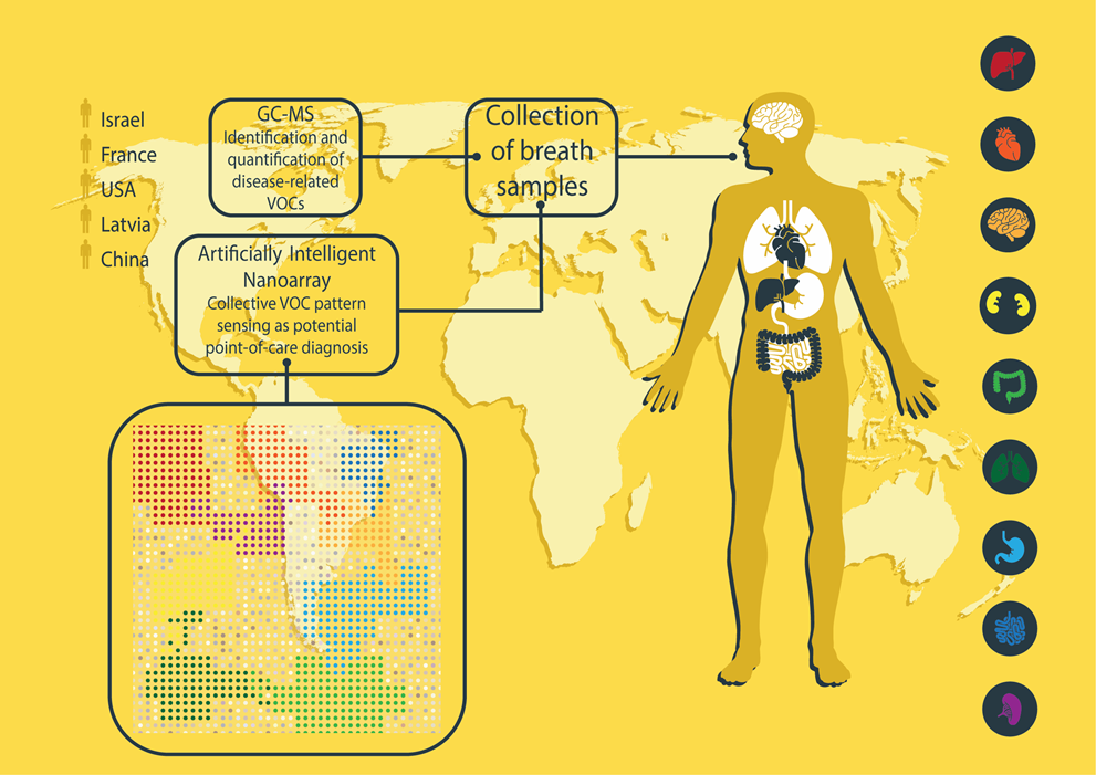
The study of more than 1,400 patients included 17 different and unrelated diseases: lung cancer, colorectal cancer, head and neck cancer, ovarian cancer, bladder cancer, prostate cancer, kidney cancer, stomach cancer, Crohn’s disease, ulcerative colitis, irritable bowel syndrome, Parkinson’s disease (two types), multiple sclerosis, pulmonary hypertension, preeclampsia and chronic kidney disease. Samples were collected between January 2011 and June 2014 from in 14 departments at 9 medical centers in 5 countries: Israel, France, the USA, Latvia and China.

The researchers tested the chemical composition of the breath samples using an accepted analytical method (mass spectrometry), which enabled accurate quantitative detection of the chemical compounds they contained. 13 chemical components were identified, in different compositions, in all 17 of the diseases.

According to Prof. Haick, “each of these diseases is characterized by a unique fingerprint, meaning a different composition of these 13 chemical components.  Just as each of us has a unique fingerprint that distinguishes us from others, each disease has a chemical signature that distinguishes it from other diseases and from a normal state of health. These odor signatures are what enables us to identify the diseases using the technology that we developed.”

With a new technology called “artificially intelligent nanoarray,” developed by Prof. Haick, the researchers were able to corroborate the clinical efficacy of the diagnostic technology. The array enables fast and inexpensive diagnosis and classification of diseases, based on “smelling” the patient’s breath, and using artificial intelligence to analyze the data obtained from the sensors. Some of the sensors are based on layers of gold nanoscale particles and others contain a random network of carbon nanotubes coated with an organic layer for sensing and identification purposes.

The study also assessed the efficiency of the artificially intelligent nanoarray in detecting and classifying various diseases using breath signatures. To verify the reliability of the system, the team also examined the effect of various factors (such as gender, age, smoking habits and geographic location) on the sample composition, and found their effect to be negligible, and without impairment on the array’s sensitivity.

“Each of the sensors responds to a wide range of exhalation components,” explain Prof. Haick and his previous Ph.D student, Dr. Morad Nakhleh, “and integration of the information provides detailed data about the unique breath signatures characteristic of the various diseases. Our system has detected and classified various diseases with an average accuracy of 86%.

This is a new and promising direction for diagnosis and classification of diseases, which is characterized not only by considerable accuracy but also by low cost, low electricity consumption, miniaturization, comfort and the possibility of repeating the test easily.”

“Breath is an excellent raw material for diagnosis,” said Prof. Haick. “It is available without the need for invasive and unpleasant procedures, it’s not dangerous, and you can sample it again and again if necessary.”

Here’s a schematic of the study, which the researchers have made available,

Diagram: A schematic view of the study. Two breath samples were taken from each subject, one was sent for chemical mapping using mass spectrometry, and the other was analyzed in the new system, which produced a clinical diagnosis based on the chemical fingerprint of the breath sample. Courtesy: Tech;nion

There is also a video, which covers much of the same ground as the press release but also includes information about the possible use of the Na-Nose technology in the European Union’s SniffPhone project,

Here’s a link to and a citation for the paper,

Diagnosis and Classification of 17 Diseases from 1404 Subjects via Pattern Analysis of Exhaled Molecules by Morad K. Nakhleh, Haitham Amal, Raneen Jeries, Yoav Y. Broza, Manal Aboud, Alaa Gharra, Hodaya Ivgi, Salam Khatib, Shifaa Badarneh, Lior Har-Shai, Lea Glass-Marmor, Izabella Lejbkowicz, Ariel Miller, Samih Badarny, Raz Winer, John Finberg, Sylvia Cohen-Kaminsky, Frédéric Perros, David Montani, Barbara Girerd, Gilles Garcia, Gérald Simonneau, Farid Nakhoul, Shira Baram, Raed Salim, Marwan Hakim, Maayan Gruber, Ohad Ronen, Tal Marshak, Ilana Doweck, Ofer Nativ, Zaher Bahouth, Da-you Shi, Wei Zhang, Qing-ling Hua, Yue-yin Pan, Li Tao, Hu Liu, Amir Karban, Eduard Koifman, Tova Rainis, Roberts Skapars, Armands Sivins, Guntis Ancans, Inta Liepniece-Karele, Ilze Kikuste, Ieva Lasina, Ivars Tolmanis, Douglas Johnson, Stuart Z. Millstone, Jennifer Fulton, John W. Wells, Larry H. Wilf, Marc Humbert, Marcis Leja, Nir Peled, and Hossam Haick. ACS Nano, Article ASAP DOI: 10.1021/acsnano.6b04930 Publication Date (Web): December 21, 2016

Copyright © 2017 American Chemical Society

This paper appears to be open access.

As for SniffPhone, they’re hoping that Na-Nose or something like it will allow them to modify smartphones in a way that will allow diseases to be detected.

I can’t help wondering who will own the data if your smartphone detects a disease. If you think that’s an idle question, here’s an excerpt from Sue Halpern’s Dec. 22, 2016 review of two books (“Weapons of Math Destruction: How Big Data Increases Inequality and Threatens Democracy” by Cathy O’Neil and “Virtual Competition: The Promise and Perils of the Algorithm-Driven Economy” by Ariel Ezrachi and Maurice E. Stucke) for the New York Times Review of Books,

We give our data away. We give it away in drips and drops, not thinking that data brokers will collect it and sell it, let alone that it will be used against us. There are now private, unregulated DNA databases culled, in part, from DNA samples people supply to genealogical websites in pursuit of their ancestry. These samples are available online to be compared with crime scene DNA without a warrant or court order. (Police are also amassing their own DNA databases by swabbing cheeks during routine stops.) In the estimation of the Electronic Frontier Foundation, this will make it more likely that people will be implicated in crimes they did not commit.

Or consider the data from fitness trackers, like Fitbit. As reported in The Intercept:

During a 2013 FTC panel on “Connected Health and Fitness,” University of Colorado law professor Scott Peppet said, “I can paint an incredibly detailed and rich picture of who you are based on your Fitbit data,” adding, “That data is so high quality that I can do things like price insurance premiums or I could probably evaluate your credit score incredibly accurately.”

Halpern’s piece is well worth reading in its entirety.

Nanoparticle ‘caterpillars’ and immune system ‘crows’

This University of Colorado work fits in nicely with other efforts to ensure that nanoparticle medical delivery systems get to their destinations. From a Dec. 19, 2016 news item on phys.org,

In the lab, doctors can attach chemotherapy to nanoparticles that target tumors, and can use nanoparticles to enhance imaging with MRI, PET and CT scans. Unfortunately, nanoparticles look a lot like pathogens – introducing nanoparticles to the human body can lead to immune system activation in which, at best, nanoparticles are cleared before accomplishing their purpose, and at worst, the onset of dangerous allergic reaction. A University of Colorado Cancer Center paper published today [Dec. 19, 2016] in the journal Nature Nanotechnology details how the immune system recognizes nanoparticles, potentially paving the way to counteract or avoid this detection.

Specifically, the study worked with dextran-coated iron oxide nanoparticles, a promising and versatile class of particles used as drug-delivery vehicles and MRI contrast enhancers in many studies. As their name implies, the particles are tiny flecks of iron oxide encrusted with sugar chains.

“We used several sophisticated microscopy approaches to understand that the particles basically look like caterpillars,” says Dmitri Simberg, PhD, investigator at the CU Cancer Center and assistant professor in the Skaggs School of Pharmacy and Pharmaceutical Sciences, the paper’s senior author.

The comparison is striking: the iron oxide particle is the caterpillar’s body, which is surrounded by fine hairs of dextran.

Caption: University of Colorado Cancer Study shows how nanoparticles activate the complement system, potentially paving the way for expanded use of these technologies.
Credit: University of Colorado Cancer Center

A Dec. 19, 2016 University of Colorado news release on EurekAlert, which originated the news item, describes the work in more detail,

If Simberg’s dextran-coated iron oxide nanoparticles are caterpillars, then the immune system is a fat crow that would eat them – that is, if it can find them. In fact, the immune system has evolved for exactly this purpose – to find and “eat” foreign particles – and rather than one homogenous entity is actually composed of a handful of interrelated systems, each specialized to counteract a specific form of invading particle.

Simberg’s previous work shows that it is the immune subcomponent called the complement system that most challenges nanoparticles. Basically, the complement system is a group of just over 30 proteins that circulate through the blood and attach to invading particles and pathogens. In humans, complement system activation requires that three proteins come together on a particle -C3b, Bb and properdin – which form a stable complex called C3-convertase.

“The whole complement system activation starts with the assembly of C3-convertase,” Simberg says. “In this paper, we ask the question of how the complement proteins actually recognize the nanoparticle surface. How is this whole reaction triggered?”

First, it was clear that the dextran coating that was supposed to protect the nanoparticles from human complement attack was not doing its job. Simberg and colleagues could see complement proteins literally invade the barrier of dextran hairs.

“Electron microscopy images show protein getting inside the particle to touch the iron oxide core,” Simberg says.

In fact, as long as the nanoparticle coating allowed the nanoparticle to absorb proteins from blood, the C3 convertase was assembled and activated on these proteins. The composition of the coating was irrelevant – if any blood protein was able to bind to nanoparticles, it always led to complement activation. Moreover, Simberg and colleagues also showed that complement system activation is a dynamic and ongoing process – blood proteins and C3 convertase constantly dissociate from nanoparticles, and new proteins and C3 convertases bind to the particles, continuing the cascade of immune system activation. The group also demonstrated that this dynamic assembly of complement proteins occurs not only in the test tubes but also in living organisms as particles circulate in blood.

Simberg suggests that the work points to challenges and three possible strategies to avoid complement system activation by nanoparticles: “First, we could try to change the nanoparticle coating so that it can’t absorb proteins, which is a difficult task; second, we could better understand the composition of proteins absorbed from blood on the particle surface that allow it to bind complement proteins; and third, there are natural inhibitors of complement activation – for example blood Factor H – but in the context of nanoparticles, it’s not strong enough to stop complement activation. Perhaps we could get nanoparticles to attract more Factor H to decrease this activation.”

At one point, the concept of nanomedicine seemed as if it would be simple – engineers and chemists would make a nanoparticle with affinity for tumor tissue and then attach a drug molecule to it. Or they would inject nanoparticles into patients that would improve the resolution of diagnostic imaging. When the realities associated with the use of nanoparticles in the landscape of the human immune system proved more challenging, many researchers realized the need to step back from possible clinical use to better understand the mechanisms that challenge nanoparticle use.

“This basic groundwork is absolutely necessary,” says Seyed Moein Moghimi, PhD, nanotechnologist at Durham University, UK, and the coauthor of the Simberg paper. “It’s essential that we learn to control the process of immune recognition so that we can bridge between the promise that nanoparticles demonstrate in the lab and their use with real patients in the real world.”

Here’s a link to and a citation for the paper,

Complement proteins bind to nanoparticle protein corona and undergo dynamic exchange in vivo by Fangfang Chen, Guankui Wang, James I. Griffin, Barbara Brenneman, Nirmal K. Banda, V. Michael Holers, Donald S. Backos, LinPing Wu, Seyed Moein Moghimi, & Dmitri Simberg. Nature Nanotechnology  (2016) doi:10.1038/nnano.2016.269 19 December 2016

This paper is behind a paywall.

I have a few previous postings about nanoparticles as drug delivery systems which have yet to fulfill their promise. There’s the April 27, 2016 posting (How many nanoparticle-based drugs does it take to kill a cancer tumour? More than 1%) and the Sept. 9, 2016 posting (Discovering how the liver prevents nanoparticles from reaching cancer cells).

Liquid biopsy chip that uses carbon nanotubes in place of microfluidics

They’re calling this a breakthrough technology in a Dec. 15, 2016 news item on ScienceDaily,

A chip developed by mechanical engineers at Worcester Polytechnic Institute (WPI) [UK] can trap and identify metastatic cancer cells in a small amount of blood drawn from a cancer patient. The breakthrough technology uses a simple mechanical method that has been shown to be more effective in trapping cancer cells than the microfluidic approach employed in many existing devices.

The WPI device uses antibodies attached to an array of carbon nanotubes at the bottom of a tiny well. Cancer cells settle to the bottom of the well, where they selectively bind to the antibodies based on their surface markers (unlike other devices, the chip can also trap tiny structures called exosomes produced by cancers cells). This “liquid biopsy,” described in a recent issue of the journal Nanotechnology, could become the basis of a simple lab test that could quickly detect early signs of metastasis and help physicians select treatments targeted at the specific cancer cells identified.

A Dec. 15, 2016 WPI press release (also on EurekAlert), which originated the news item, explains the breakthrough in more detail (Note: Links have been removed),

Metastasis is the process by which a cancer can spread from one organ to other parts of the body, typically by entering the bloodstream. Different types of tumors show a preference for specific organs and tissues; circulating breast cancer cells, for example, are likely to take root in bones, lungs, and the brain. The prognosis for metastatic cancer (also called stage IV cancer) is generally poor, so a technique that could detect these circulating tumor cells before they have a chance to form new colonies of tumors at distant sites could greatly increase a patient’s survival odds.

“The focus on capturing circulating tumor cells is quite new,” said Balaji Panchapakesan, associate professor of mechanical engineering at WPI and director of the Small Systems Laboratory. “It is a very difficult challenge, not unlike looking for a needle in a haystack. There are billions of red blood cells, tens of thousands of white blood cells, and, perhaps, only a small number of tumor cells floating among them. We’ve shown how those cells can be captured with high precision.”

The device developed by Panchapakesan’s team includes an array of tiny elements, each about a tenth of an inch (3 millimeters) across. Each element has a well, at the bottom of which are antibodies attached to carbon nanotubes. Each well holds a specific antibody that will bind selectively to one type of cancer cell type, based on genetic markers on its surface. By seeding elements with an assortment of antibodies, the device could be set up to capture several different cancer cells types using a single blood sample. In the lab, the researchers were able to fill a total of 170 wells using just under 0.3 fluid ounces (0.85 milliliter) of blood. Even with that small sample, they captured between one and a thousand cells per device, with a capture efficiency of between 62 and 100 percent.

In a paper published in the journal Nanotechnology [“Static micro-array isolation, dynamic time series classification, capture and enumeration of spiked breast cancer cells in blood: the nanotube–CTC chip”], Panchapakesan’s team, which includes postdoctoral researcher Farhad Khosravi, the paper’s lead author, and researchers at the University of Louisville and Thomas Jefferson University, describe a study in which antibodies specific for two markers of metastatic breast cancer, EpCam and Her2, were attached to the carbon nanotubes in the chip. When a blood sample that had been “spiked” with cells expressing those markers was placed on the chip, the device was shown to reliably capture only the marked cells.

In addition to capturing tumor cells, Panchapakesan says the chip will also latch on to tiny structures called exosomes, which are produced by cancers [sic] cells and carry the same markers. “These highly elusive 3-nanometer structures are too small to be captured with other types of liquid biopsy devices, such as microfluidics, due to shear forces that can potentially destroy them,” he noted. “Our chip is currently the only device that can potentially capture circulating tumor cells and exosomes directly on the chip, which should increase its ability to detect metastasis. This can be important because emerging evidence suggests that tiny proteins excreted with exosomes can drive reactions that may become major barriers to effective cancer drug delivery and treatment.”

Panchapakesan said the chip developed by his team has additional advantages over other liquid biopsy devices, most of which use microfluidics to capture cancer cells. In addition to being able to capture circulating tumor cells far more efficiently than microfluidic chips (in which cells must latch onto anchored antibodies as they pass by in a stream of moving liquid), the WPI device is also highly effective in separating cancer cells from the other cells and material in the blood through differential settling.

While the initial tests with the chip have focused on breast cancer, Panchapakesan says the device could be set up to detect a wide range of tumor types, and plans are already in the works for development of an advanced device as well as testing for other cancer types, including lung and pancreas cancer. He says he envisions a day when a device like his could be employed not only for regular follow ups for patients who have had cancer, but in routine cancer screening.

“Imagine going to the doctor for your yearly physical,” he said. “You have blood drawn and that one blood sample can be tested for a comprehensive array of cancer cell markers. Cancers would be caught at their earliest stage and other stages of development, and doctors would have the necessary protein or genetic information from these captured cells to customize your treatment based on the specific markers for your cancer. This would really be a way to put your health in your own hands.”

“White blood cells, in particular, are a problem, because they are quite numerous in blood and they can be mistaken for cancer cells,” he said. “Our device uses what is called a passive leukocyte depletion strategy. Because of density differences, the cancer cells tend to settle to the bottom of the wells (and this only happens in a narrow window), where they encounter the antibodies. The remainder of the blood contents stays at the top of the wells and can simply be washed away.”

Here’s a link to and a citation for the paper,

Static micro-array isolation, dynamic time series classification, capture and enumeration of spiked breast cancer cells in blood: the nanotube–CTC chip by Farhad Khosravi, Patrick J Trainor, Christopher Lambert, Goetz Kloecker, Eric Wickstrom, Shesh N Rai, and Balaji Panchapakesan. Nanotechnology, Volume 27, Number 44 DOI http://dx.doi.org/10.1088/0957-4484/27/44/44LT03 Published 29 September 2016

© 2016 IOP Publishing Ltd

This paper is open access.

A ‘vascular running of the bulls’; nanoparticles in your bloodstream

An Oct. 5, 2016 news item on phys.org announces research into how nanoparticles behave in the bloodstream (Note: A link has been removed),

Researchers at the University of Connecticut have uncovered new information about how particles behave in our bloodstream, an important advancement that could help pharmaceutical scientists develop more effective cancer drugs.

Making sure cancer medications reach the leaky blood vessels surrounding most tumor sites is one of the critical aspects of treatment and drug delivery. While surface chemistry, molecular interactions, and other factors come into play once drug-carrying particles arrive at a tumor, therapeutic medication doesn’t do very much good if it never reaches its intended target.

Anson Ma, an assistant professor of chemical and biomolecular engineering at UConn, used a microfluidic channel device to observe, track, and measure how individual particles behaved in a simulated blood vessel.

The research team’s goal: to learn more about the physics influencing a particle’s behavior as it travels in our blood and to determine which particle size might be the most effective for delivering drugs to their targets. The team’s experimental findings mark the first time such quantitative data has been gathered. …

“Even before particles reach a target site, you have to worry about what is going to happen with them after they get injected into the bloodstream,” Ma says. “Are they going to clump together? How are they going to move around? Are they going to get swept away and flushed out of our bodies?”

Using a high-powered fluorescence microscope in UConn’s Complex Fluids Lab, Ma was able to observe particles being carried along in the simulated blood vessel in what could be described as a vascular Running of the Bulls [emphasis mine]. Red blood cells race through the middle of the channel as the particles – highlighted under the fluorescent light – get carried along in the rush, bumping and bouncing off the blood cells until they are pushed to open spaces – called the cell-free layer – along the vessel’s walls.

Nanocarrier particles injected into the bloodstream bounce off red and white blood cells and platelets, and are pushed toward the blood vessel walls. This physical interaction, measured and quantified for the first time by engineering professor Anson Ma’s lab, provides important information for drug developers. (Image courtesy of Anson Ma)

Nanocarrier particles injected into the bloodstream bounce off red and white blood cells and platelets, and are pushed toward the blood vessel walls. This physical interaction, measured and quantified for the first time by engineering professor Anson Ma’s lab, provides important information for drug developers. (Image courtesy of Anson Ma)

An Oct. 4, 2016 University of Connecticut news release, which originated the news item, provides more detail about the research,

What Ma found was that larger particles – the optimum size appeared to be about 2 microns – were most likely to get pushed to the cell-free layer, where their chances of carrying medication into a tumor site are greatest. The research team also determined that 2 microns was the largest size that should be used if particles are going to have any chance of going through the leaky blood vessel walls into the tumor site.

“When it comes to using particles for the delivery of cancer drugs, size matters,” Ma says. “When you have a bigger particle, the chance of it bumping into blood cells is much higher, there are a lot more collisions, and they tend to get pushed to the blood vessel walls.”

The results were somewhat surprising. In preparing their hypothesis, the research team estimated that smaller particles were probably the most effective since they would move the most in collisions with blood cells, much like what happens when a small ball bounces off a larger one. But just the opposite proved true. The smaller particles appeared to skirt through the mass of moving blood cells and were less likely to experience the “trampoline” effect and get bounced to the cell-free layer, says Ma.

Ma proposed the study after talking to a UConn pharmaceutical scientist about drug development at a campus event five years ago.

“We had a great conversation about how drugs are made and then I asked, ‘But how can you be sure where the particles go?’” Ma recalls, laughing. “I’m an engineer. That’s how we think. I was curious. This was an engineering question. So I said, ‘Let’s write a proposal!’”

The proposal was funded by the National Science Foundation’s Early-concept Grants for Exploratory Research or EAGER program, which supports exploratory work in its early stages on untested, but potentially transformative, research ideas or approaches.

Knowing how particles behave in our circulatory system should help improve targeted drug delivery, Ma says, which in turn will further reduce the toxic side effects caused by potent cancer drugs missing their target and impacting the body’s healthy tissue.

The findings were particularly meaningful for Ma, who lost two of his grandparents to cancer and who has long wanted to contribute to cancer research in a meaningful way as an engineer.

Measuring how particles of different sizes move in the bloodstream may also be beneficial in bioimaging, where scientists and doctors want to keep particles circulating in the bloodstream long enough for imaging to occur. In that case, smaller particles would be better, says Ma.

Moving forward, Ma would like to explore other aspects of particle flow in our circulatory system, such as how particles behave when they pass through a constricted area, such as from a blood vessel to a capillary. Capillaries are only about 7 microns in diameter. The average human hair is 100 microns.  Ma says he would like to know how that constricted space might impact particle flow or the ability of particles to accumulate near the vessel walls.

“We have all of this complex geometry in our bodies,” says Ma. “Most people just assume there is no impact when a particle moves from a bigger channel to a smaller channel because they haven’t quantified it. Our plan is to do some experiments to look at this more carefully, building on the work that we just published.”

Here’s a link to and a citation for the paper,

Direct Tracking of Particles and Quantification of Margination in Blood Flow by Erik J. Carbon, Brice H. Bognet, Grant M. Bouchillon, Andrea L. Kadilak, Leslie M. Shor, Michael D. Ward, Anson W.K. Ma. Biophysical Journal Volume 111, Issue 7, p1487–1495, 4 October 2016  DOI: http://dx.doi.org/10.1016/j.bpj.2016.08.026

This paper is behind a paywall.