Tag Archives: cardiac pacemakers

Powered by light: a battery-free pacemaker

I think it looks more like a potato than a heart but it does illustrate how this new battery-free pacemaker would wrap around a heart,
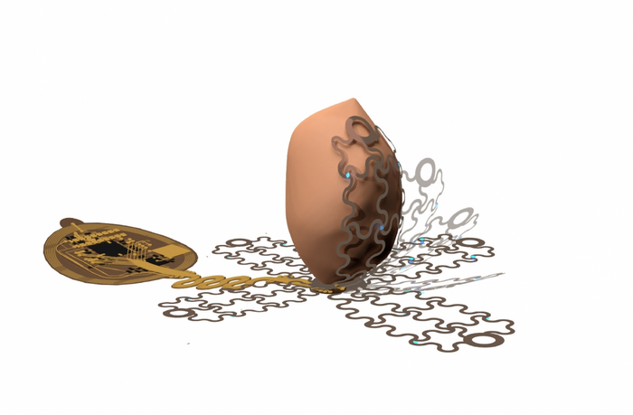
Caption: An artist’s rendering shows how a new pacemaker, designed by a UArizona-led team of researchers, is able to envelop the heart. The wireless, battery-free pacemaker could be implanted with a less invasive procedure than currently possible and would cause patients less pain. Credit: Philipp Gutruff

An October 27, 2022 news item on ScienceDaily announces a technology that could make life much easier for people with pacemakers (Comment: In the image, that looks more like a potato than a heart, to me),

University of Arizona engineers lead a research team that is developing a new kind of pacemaker, which envelops the heart and uses precise targeting capabilities to bypass pain receptors and reduce patient discomfort.

An October 27, 2022 University of Arizona news release (also on EurekAlert) by Emily Dieckman, which originated the news item, explains the reasons for the research and provides some technical details (Note: Links have been removed),

Pacemakers are lifesaving devices that regulate the heartbeats of people with chronic heart diseases like atrial fibrillation and other forms of arrhythmia. However, pacemaker implantation is an invasive procedure, and the lifesaving pacing the devices provide can be extremely painful. Additionally, pacemakers can only be used to treat a few specific types of disease.

In a paper published Wednesday [October 26, 2022] in Science Advances, a University of Arizona-led team of researchers detail the workings of a wireless, battery-free pacemaker they designed that could be implanted with a less invasive procedure than currently possible and would cause patients less pain. The study was helmed by researchers in the Gutruf Lab, led by biomedical engineering assistant professor and Craig M. Berge Faculty Fellow Philipp Gutruf.

Currently available pacemakers work by implanting one or two leads, or points of contact, into the heart with hooks or screws. If the sensors on these leads detect a dangerous irregularity, they send an electrical shock through the heart to reset the beat.

“All of the cells inside the heart get hit at one time, including the pain receptors, and that’s what makes pacing or defibrillation painful,” Gutruf said. “It affects the heart muscle as a whole.”

The device Gutruf’s team has developed, which has not yet been tested in humans, would allow pacemakers to send much more targeted signals using a new digitally manufactured mesh design that encompasses the entire heart. The device uses light and a technique called optogenetics.

Optogenetics modifies cells, usually neurons, sensitive to light, then uses light to affect the behavior of those cells. This technique only targets cardiomyocytes, the cells of the muscle that trigger contraction and make up the beat of the heart. This precision will not only reduce pain for pacemaker patients by bypassing the heart’s pain receptors, it will also allow the pacemaker to respond to different kinds of irregularities in more appropriate ways. For example, during atrial fibrillation, the upper and lower chambers of the heart beat asynchronously, and a pacemaker’s role is to get the two parts back in line.

“Whereas right now, we have to shock the whole heart to do this, these new devices can do much more precise targeting, making defibrillation both more effective and less painful,” said Igor Efimov, professor of biomedical engineering and medicine at Northwestern University, where the devices were lab-tested. “This technology could make life easier for patients all over the world, while also helping scientists and physicians learn more about how to monitor and treat the disease.”

Flexible mesh encompasses the heart

To ensure the light signals can reach many different parts of the heart, the team created a design that involves encompassing the organ, rather than implanting leads that provide limited points of contact.

The new pacemaker model consists of four petallike structures made of thin, flexible film, which contain light sources and a recording electrode. The petals, specially designed to accommodate the way the heart changes shape as it beats, fold up around the sides of the organ to envelop it, like a flower closing up at night.

“Current pacemakers record basically a simple threshold, and they will tell you, ‘This is going into arrhythmia, now shock!'” Gutruf said. “But this device has a computer on board where you can input different algorithms that allow you to pace in a more sophisticated way. It’s made for research.”

Because the system uses light to affect the heart, rather than electrical signals, the device can continue recording information even when the pacemaker needs to defibrillate. In current pacemakers, the electrical signal from the defibrillation can interfere with recording capabilities, leaving physicians with an incomplete picture of cardiac episodes. Additionally, the device does not require a battery, which could save pacemaker patients from needing to replace the battery in their device every five to seven years, as is currently the norm.

Gutruf’s team collaborated with researchers at Northwestern University on the project. While the current version of the device has been successfully demonstrated in animal models, the researchers look forward to furthering their work, which could improve the quality of life for millions of people.

The prototype looks like this,
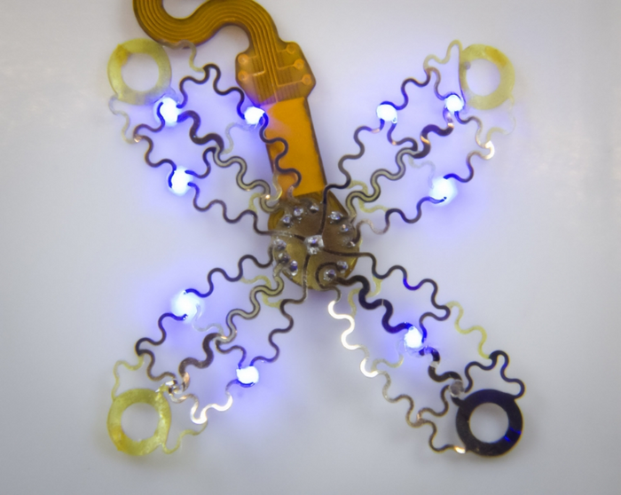
Caption: The device uses light and a technique called optogenetics, which modifies cells that are sensitive to light, then uses light to affect the behavior of those cells.. Credit: Philipp Gutruff

Here’s a link to and a citation for the paper,

Wireless, fully implantable cardiac stimulation and recording with on-device computation for closed-loop pacing and defibrillation by Jokubas Ausra, Micah Madrid, Rose T. Yin, Jessica Hanna, Suzanne Arnott, Jaclyn A. Brennan, Roberto Peralta, David Clausen, Jakob A. Bakall, Igor R. Efimov, and Philipp Gutruf. Science Advances 26 Oct 2022 Vol 8, Issue 43 DOI: 10.1126/sciadv.abq7469

This paper is open access.

A biocompatible (implantable) micromachine (microrobot)

I appreciate the detail and information in this well written Jan. 4, 2017 Columbia University news release (h/t Jan. 4, 2016 Nanowerk; Note: Links have been removed),

A team of researchers led by Biomedical Engineering Professor Sam Sia has developed a way to manufacture microscale-sized machines from biomaterials that can safely be implanted in the body. Working with hydrogels, which are biocompatible materials that engineers have been studying for decades, Sia has invented a new technique that stacks the soft material in layers to make devices that have three-dimensional, freely moving parts. The study, published online January 4, 2017, in Science Robotics, demonstrates a fast manufacturing method Sia calls “implantable microelectromechanical systems” (iMEMS).

By exploiting the unique mechanical properties of hydrogels, the researchers developed a “locking mechanism” for precise actuation and movement of freely moving parts, which can provide functions such as valves, manifolds, rotors, pumps, and drug delivery. They were able to tune the biomaterials within a wide range of mechanical and diffusive properties and to control them after implantation without a sustained power supply such as a toxic battery. They then tested the “payload” delivery in a bone cancer model and found that the triggering of release of doxorubicin from the device over 10 days showed high treatment efficacy and low toxicity, at 1/10 of the standard systemic chemotherapy dose.

“Overall, our iMEMS platform enables development of biocompatible implantable microdevices with a wide range of intricate moving components that can be wirelessly controlled on demand and solves issues of device powering and biocompatibility,” says Sia, also a member of the Data Science Institute. “We’re really excited about this because we’ve been able to connect the world of biomaterials with that of complex, elaborate medical devices. Our platform has a large number of potential applications, including the drug delivery system demonstrated in our paper which is linked to providing tailored drug doses for precision medicine.”

I particularly like this bit about hydrogels being a challenge to work with and the difficulties of integrating both rigid and soft materials,

Most current implantable microdevices have static components rather than moving parts and, because they require batteries or other toxic electronics, have limited biocompatibility. Sia’s team spent more than eight years working on how to solve this problem. “Hydrogels are difficult to work with, as they are soft and not compatible with traditional machining techniques,” says Sau Yin Chin, lead author of the study who worked with Sia. “We have tuned the mechanical properties and carefully matched the stiffness of structures that come in contact with each other within the device. Gears that interlock have to be stiff in order to allow for force transmission and to withstand repeated actuation. Conversely, structures that form locking mechanisms have to be soft and flexible to allow for the gears to slip by them during actuation, while at the same time they have to be stiff enough to hold the gears in place when the device is not actuated. We also studied the diffusive properties of the hydrogels to ensure that the loaded drugs do not easily diffuse through the hydrogel layers.”

The team used light to polymerize sheets of gel and incorporated a stepper mechanization to control the z-axis and pattern the sheets layer by layer, giving them three-dimensionality. Controlling the z-axis enabled the researchers to create composite structures within one layer of the hydrogel while managing the thickness of each layer throughout the fabrication process. They were able to stack multiple layers that are precisely aligned and, because they could polymerize a layer at a time, one right after the other, the complex structure was built in under 30 minutes.

Sia’s iMEMS technique addresses several fundamental considerations in building biocompatible microdevices, micromachines, and microrobots: how to power small robotic devices without using toxic batteries, how to make small biocompatible moveable components that are not silicon which has limited biocompatibility, and how to communicate wirelessly once implanted (radio frequency microelectronics require power, are relatively large, and are not biocompatible). The researchers were able to trigger the iMEMS device to release additional payloads over days to weeks after implantation. They were also able to achieve precise actuation by using magnetic forces to induce gear movements that, in turn, bend structural beams made of hydrogels with highly tunable properties. (Magnetic iron particles are commonly used and FDA-approved for human use as contrast agents.)

In collaboration with Francis Lee, an orthopedic surgeon at Columbia University Medical Center at the time of the study, the team tested the drug delivery system on mice with bone cancer. The iMEMS system delivered chemotherapy adjacent to the cancer, and limited tumor growth while showing less toxicity than chemotherapy administered throughout the body.

“These microscale components can be used for microelectromechanical systems, for larger devices ranging from drug delivery to catheters to cardiac pacemakers, and soft robotics,” notes Sia. “People are already making replacement tissues and now we can make small implantable devices, sensors, or robots that we can talk to wirelessly. Our iMEMS system could bring the field a step closer in developing soft miniaturized robots that can safely interact with humans and other living systems.”

Here’s a link to and a citation for the paper,

Additive manufacturing of hydrogel-based materials for next-generation implantable medical devices by Sau Yin Chin, Yukkee Cheung Poh, Anne-Céline Kohler, Jocelyn T. Compton, Lauren L. Hsu, Kathryn M. Lau, Sohyun Kim, Benjamin W. Lee, Francis Y. Lee, and Samuel K. Sia. Science Robotics  04 Jan 2017: Vol. 2, Issue 2, DOI: 10.1126/scirobotics.aah6451

This paper appears to be open access.

The researchers have provided a video demonstrating their work (you may want to read the caption below before watching),

Magnetic actuation of the Geneva drive device. A magnet is placed about 1cm below and without contact with the device. The rotating magnet results in the rotational movement of the smaller driving gear. With each full rotation of this driving gear, the larger driven gear is engaged and rotates by 60º, exposing the next reservoir to the aperture on the top layer of the device.

—Video courtesy of Sau Yin Chin/Columbia Engineering

You can hear some background conversation but it doesn’t seem to have been included for informational purposes.

Cardiac pacemakers: Korea’s in vivo demonstration of a self-powered one* and UK’s breath-based approach

As i best I can determine ,the last mention of a self-powered pacemaker and the like on this blog was in a Nov. 5, 2012 posting (Developing self-powered batteries for pacemakers). This latest news from The Korea Advanced Institute of Science and Technology (KAIST) is, I believe, the first time that such a device has been successfully tested in vivo. From a June 23, 2014 news item on ScienceDaily,

As the number of pacemakers implanted each year reaches into the millions worldwide, improving the lifespan of pacemaker batteries has been of great concern for developers and manufacturers. Currently, pacemaker batteries last seven years on average, requiring frequent replacements, which may pose patients to a potential risk involved in medical procedures.

A research team from the Korea Advanced Institute of Science and Technology (KAIST), headed by Professor Keon Jae Lee of the Department of Materials Science and Engineering at KAIST and Professor Boyoung Joung, M.D. of the Division of Cardiology at Severance Hospital of Yonsei University, has developed a self-powered artificial cardiac pacemaker that is operated semi-permanently by a flexible piezoelectric nanogenerator.

A June 23, 2014 KAIST news release on EurekAlert, which originated the news item, provides more details,

The artificial cardiac pacemaker is widely acknowledged as medical equipment that is integrated into the human body to regulate the heartbeats through electrical stimulation to contract the cardiac muscles of people who suffer from arrhythmia. However, repeated surgeries to replace pacemaker batteries have exposed elderly patients to health risks such as infections or severe bleeding during operations.

The team’s newly designed flexible piezoelectric nanogenerator directly stimulated a living rat’s heart using electrical energy converted from the small body movements of the rat. This technology could facilitate the use of self-powered flexible energy harvesters, not only prolonging the lifetime of cardiac pacemakers but also realizing real-time heart monitoring.

The research team fabricated high-performance flexible nanogenerators utilizing a bulk single-crystal PMN-PT thin film (iBULe Photonics). The harvested energy reached up to 8.2 V and 0.22 mA by bending and pushing motions, which were high enough values to directly stimulate the rat’s heart.

Professor Keon Jae Lee said:

“For clinical purposes, the current achievement will benefit the development of self-powered cardiac pacemakers as well as prevent heart attacks via the real-time diagnosis of heart arrhythmia. In addition, the flexible piezoelectric nanogenerator could also be utilized as an electrical source for various implantable medical devices.”

This image illustrating a self-powered nanogenerator for a cardiac pacemaker has been provided by KAIST,

This picture shows that a self-powered cardiac pacemaker is enabled by a flexible piezoelectric energy harvester. Credit: KAIST

This picture shows that a self-powered cardiac pacemaker is enabled by a flexible piezoelectric energy harvester.
Credit: KAIST

Here’s a link to and a citation for the paper,

Self-Powered Cardiac Pacemaker Enabled by Flexible Single Crystalline PMN-PT Piezoelectric Energy Harvester by Geon-Tae Hwang, Hyewon Park, Jeong-Ho Lee, SeKwon Oh, Kwi-Il Park, Myunghwan Byun, Hyelim Park, Gun Ahn, Chang Kyu Jeong, Kwangsoo No, HyukSang Kwon, Sang-Goo Lee, Boyoung Joung, and Keon Jae Lee. Advanced Materials DOI: 10.1002/adma.201400562
Article first published online: 17 APR 2014

© 2014 WILEY-VCH Verlag GmbH & Co. KGaA, Weinheim

This paper is behind a paywall.

There was a May 15, 2014 KAIST news release on EurekAlert announcing this same piece of research but from a technical perspective,

The energy efficiency of KAIST’s piezoelectric nanogenerator has increased by almost 40 times, one step closer toward the commercialization of flexible energy harvesters that can supply power infinitely to wearable, implantable electronic devices

NANOGENERATORS are innovative self-powered energy harvesters that convert kinetic energy created from vibrational and mechanical sources into electrical power, removing the need of external circuits or batteries for electronic devices. This innovation is vital in realizing sustainable energy generation in isolated, inaccessible, or indoor environments and even in the human body.

Nanogenerators, a flexible and lightweight energy harvester on a plastic substrate, can scavenge energy from the extremely tiny movements of natural resources and human body such as wind, water flow, heartbeats, and diaphragm and respiration activities to generate electrical signals. The generators are not only self-powered, flexible devices but also can provide permanent power sources to implantable biomedical devices, including cardiac pacemakers and deep brain stimulators.

However, poor energy efficiency and a complex fabrication process have posed challenges to the commercialization of nanogenerators. Keon Jae Lee, Associate Professor of Materials Science and Engineering at KAIST, and his colleagues have recently proposed a solution by developing a robust technique to transfer a high-quality piezoelectric thin film from bulk sapphire substrates to plastic substrates using laser lift-off (LLO).

Applying the inorganic-based laser lift-off (LLO) process, the research team produced a large-area PZT thin film nanogenerators on flexible substrates (2 cm x 2 cm).

“We were able to convert a high-output performance of ~250 V from the slight mechanical deformation of a single thin plastic substrate. Such output power is just enough to turn on 100 LED lights,” Keon Jae Lee explained.

The self-powered nanogenerators can also work with finger and foot motions. For example, under the irregular and slight bending motions of a human finger, the measured current signals had a high electric power of ~8.7 μA. In addition, the piezoelectric nanogenerator has world-record power conversion efficiency, almost 40 times higher than previously reported similar research results, solving the drawbacks related to the fabrication complexity and low energy efficiency.

Lee further commented,

“Building on this concept, it is highly expected that tiny mechanical motions, including human body movements of muscle contraction and relaxation, can be readily converted into electrical energy and, furthermore, acted as eternal power sources.”

The research team is currently studying a method to build three-dimensional stacking of flexible piezoelectric thin films to enhance output power, as well as conducting a clinical experiment with a flexible nanogenerator.

In addition to the 2012 posting I mentioned earlier, there was also this July 12, 2010 posting which described research on harvesting biomechanical movement ( heart beat, blood flow, muscle stretching, or even irregular vibration) at the Georgia (US) Institute of Technology where the lead researcher observed,

…  Wang [Professor Zhong Lin Wang at Georgia Tech] tells Nanowerk. “However, the applications of the nanogenerators under in vivo and in vitro environments are distinct. Some crucial problems need to be addressed before using these devices in the human body, such as biocompatibility and toxicity.”

Bravo to the KAIST researchers for getting this research to the in vivo testing stage.

Meanwhile at the University of Bristol and at the University of Bath, researchers have received funding for a new approach to cardiac pacemakers, designed them with the breath in mind. From a June 24, 2014 news item on Azonano,

Pacemaker research from the Universities of Bath and Bristol could revolutionise the lives of over 750,000 people who live with heart failure in the UK.

The British Heart Foundation (BHF) is awarding funding to researchers developing a new type of heart pacemaker that modulates its pulses to match breathing rates.

A June 23, 2014 University of Bristol press release, which originated the news item, provides some context,

During 2012-13 in England, more than 40,000 patients had a pacemaker fitted.

Currently, the pulses from pacemakers are set at a constant rate when fitted which doesn’t replicate the natural beating of the human heart.

The normal healthy variation in heart rate during breathing is lost in cardiovascular disease and is an indicator for sleep apnoea, cardiac arrhythmia, hypertension, heart failure and sudden cardiac death.

The device is then briefly described (from the press release),

The novel device being developed by scientists at the Universities of Bath and Bristol uses synthetic neural technology to restore this natural variation of heart rate with lung inflation, and is targeted towards patients with heart failure.

The device works by saving the heart energy, improving its pumping efficiency and enhancing blood flow to the heart muscle itself.  Pre-clinical trials suggest the device gives a 25 per cent increase in the pumping ability, which is expected to extend the life of patients with heart failure.

One aim of the project is to miniaturise the pacemaker device to the size of a postage stamp and to develop an implant that could be used in humans within five years.

Dr Alain Nogaret, Senior Lecturer in Physics at the University of Bath, explained“This is a multidisciplinary project with strong translational value.  By combining fundamental science and nanotechnology we will be able to deliver a unique treatment for heart failure which is not currently addressed by mainstream cardiac rhythm management devices.”

The research team has already patented the technology and is working with NHS consultants at the Bristol Heart Institute, the University of California at San Diego and the University of Auckland. [emphasis mine]

Professor Julian Paton, from the University of Bristol, added: “We’ve known for almost 80 years that the heart beat is modulated by breathing but we have never fully understood the benefits this brings. The generous new funding from the BHF will allow us to reinstate this natural occurring synchrony between heart rate and breathing and understand how it brings therapy to hearts that are failing.”

Professor Jeremy Pearson, Associate Medical Director at the BHF, said: “This study is a novel and exciting first step towards a new generation of smarter pacemakers. More and more people are living with heart failure so our funding in this area is crucial. The work from this innovative research team could have a real impact on heart failure patients’ lives in the future.”

Given some current events (‘Tesla opens up its patents’, Mike Masnick’s June 12, 2014 posting on Techdirt), I wonder what the situation will be vis à vis patents by the time this device gets to market.

* ‘one’ added to title on Aug. 13, 2014.