Tag Archives: collagen

Water and minerals have a nanoscale effect on bones

Courtesy: University of Arkansas

What a great image of bones! This December 3, 2020 University of Arkansas news release (also on EurekAlert) by Matt McGowan features research focused on bone material looks exciting. The date for the second study citation and link that I have listed (at the end of this posting) suggests the more recent study may have been initially overlooked in the deluge of COVID-19 research we are experiencing,

University of Arkansas researchers Marco Fielder and Arun Nair have conducted the first study of the combined nanoscale effects of water and mineral content on the deformation mechanisms and thermal properties of collagen, the essence of bone material.

The researchers also compared the results to the same properties of non-mineralized collagen reinforced with carbon nanotubes, which have shown promise as a reinforcing material for bio-composites. This research aids in the development of synthetic materials to mimic bone.

Using molecular dynamics — in this case a computer simulation of the physical movements of atoms and molecules — Nair and Fielder examined the mechanics and thermal properties of collagen-based bio-composites containing different weight percentages of minerals, water and carbon nanotubes when subjected to external loads.

They found that variations of water and mineral content had a strong impact on the mechanical behavior and properties of the bio-composites, the structure of which mimics nanoscale bone composition. With increased hydration, the bio-composites became more vulnerable to stress. Additionally, Nair and Fielder found that the presence of carbon nanotubes in non-mineralized collagen reduced the deformation of the gap regions.

The researchers also tested stiffness, which is the standard measurement of a material’s resistance to deformation. Both mineralized and non-mineralized collagen bio-composites demonstrated less stability with greater water content. Composites with 40% mineralization were twice as strong as those without minerals, regardless of the amount of water content. Stiffness of composites with carbon nanotubes was comparable to that of the mineralized collagen.

“As the degree of mineralization or carbon nanotube content of the collagenous bio-composites increased, the effect of water to change the magnitude of deformation decreased,” Fielder said.

The bio-composites made of collagen and carbon nanotubes were also found to have a higher specific heat than the studied mineralized collagen bio-composites, making them more likely to be resistant to thermal damage that could occur during implantation or functional use of the composite. Like most biological materials, bone is a hierarchical – with different structures at different length scales. At the microscale level, bone is made of collagen fibers, composed of smaller nanofibers called fibrils, which are a composite of collagen proteins, mineralized crystals called apatite and water. Collagen fibrils overlap each other in some areas and are separated by gaps in other areas.

“Though several studies have characterized the mechanics of fibrils, the effects of variation and distribution of water and mineral content in fibril gap and overlap regions are unexplored,” said Nair, who is an associate professor of mechanical engineering. “Exploring these regions builds an understanding of the structure of bone, which is important for uncovering its material properties. If we understand these properties, we can design and build better bio-inspired materials and bio-composites.”

Here are links and citations for both papers mentioned in the news release,

Effects of hydration and mineralization on the deformation mechanisms of collagen fibrils in bone at the nanoscale by Marco Fielder & Arun K. Nair. Biomechanics and Modeling in Mechanobiology volume 18, pages57–68 (2019) Biomech Model Mechanobiol 18, 57–68 (2019). DOI: https://doi.org/10.1007/s10237-018-1067-y First published: 07 August 2018 Issue Date: 15 February 2019

This paper is behind a paywall.

A computational study of mechanical properties of collagen-based bio-composites by Marco Fielder & Arun K. Nair. International Biomechanics Volume 7, 2020 – Issue 1 Pages 76-87 DOI: https://doi.org/10.1080/23335432.2020.1812428 Published online: 02 Sep 2020

This paper is open access.

A 3D printed eye cornea and a 3D printed copy of your brain (also: a Brad Pitt connection)

Sometimes it’s hard to keep up with 3D tissue printing news. I have two news bits, one concerning eyes and another concerning brains.

3D printed human corneas

A May 29, 2018 news item on ScienceDaily trumpets the news,

The first human corneas have been 3D printed by scientists at Newcastle University, UK.

It means the technique could be used in the future to ensure an unlimited supply of corneas.

As the outermost layer of the human eye, the cornea has an important role in focusing vision.

Yet there is a significant shortage of corneas available to transplant, with 10 million people worldwide requiring surgery to prevent corneal blindness as a result of diseases such as trachoma, an infectious eye disorder.

In addition, almost 5 million people suffer total blindness due to corneal scarring caused by burns, lacerations, abrasion or disease.

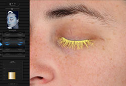
The proof-of-concept research, published today [May 29, 2018] in Experimental Eye Research, reports how stem cells (human corneal stromal cells) from a healthy donor cornea were mixed together with alginate and collagen to create a solution that could be printed, a ‘bio-ink’.

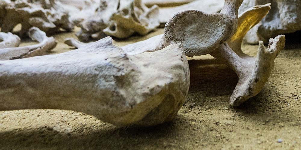
Here are the proud researchers with their cornea,

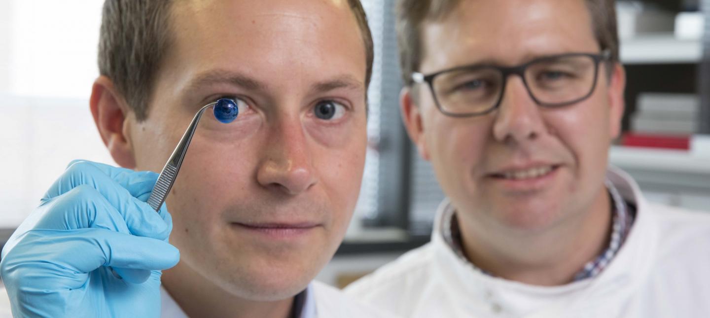
Caption: Dr. Steve Swioklo and Professor Che Connon with a dyed cornea. Credit: Newcastle University, UK

A May 30,2018 Newcastle University press release (also on EurekAlert but published on May 29, 2018), which originated the news item, adds more details,

Using a simple low-cost 3D bio-printer, the bio-ink was successfully extruded in concentric circles to form the shape of a human cornea. It took less than 10 minutes to print.

The stem cells were then shown to culture – or grow.

Che Connon, Professor of Tissue Engineering at Newcastle University, who led the work, said: “Many teams across the world have been chasing the ideal bio-ink to make this process feasible.

“Our unique gel – a combination of alginate and collagen – keeps the stem cells alive whilst producing a material which is stiff enough to hold its shape but soft enough to be squeezed out the nozzle of a 3D printer.

“This builds upon our previous work in which we kept cells alive for weeks at room temperature within a similar hydrogel. Now we have a ready to use bio-ink containing stem cells allowing users to start printing tissues without having to worry about growing the cells separately.”

The scientists, including first author and PhD student Ms Abigail Isaacson from the Institute of Genetic Medicine, Newcastle University, also demonstrated that they could build a cornea to match a patient’s unique specifications.

The dimensions of the printed tissue were originally taken from an actual cornea. By scanning a patient’s eye, they could use the data to rapidly print a cornea which matched the size and shape.

Professor Connon added: “Our 3D printed corneas will now have to undergo further testing and it will be several years before we could be in the position where we are using them for transplants.

“However, what we have shown is that it is feasible to print corneas using coordinates taken from a patient eye and that this approach has potential to combat the world-wide shortage.”

Here’s a link to and a citation for the paper,

3D bioprinting of a corneal stroma equivalent by Abigail Isaacson, Stephen Swioklo, Che J. Connon. Experimental Eye Research Volume 173, August 2018, Pages 188–193 and 2018 May 14 pii: S0014-4835(18)30212-4. doi: 10.1016/j.exer.2018.05.010. [Epub ahead of print]

This paper is behind a paywall.

A 3D printed copy of your brain

I love the title for this May 30, 2018 Wyss Institute for Biologically Inspired Engineering news release: Creating piece of mind by Lindsay Brownell (also on EurekAlert),

What if you could hold a physical model of your own brain in your hands, accurate down to its every unique fold? That’s just a normal part of life for Steven Keating, Ph.D., who had a baseball-sized tumor removed from his brain at age 26 while he was a graduate student in the MIT Media Lab’s Mediated Matter group. Curious to see what his brain actually looked like before the tumor was removed, and with the goal of better understanding his diagnosis and treatment options, Keating collected his medical data and began 3D printing his MRI [magnetic resonance imaging] and CT [computed tomography] scans, but was frustrated that existing methods were prohibitively time-intensive, cumbersome, and failed to accurately reveal important features of interest. Keating reached out to some of his group’s collaborators, including members of the Wyss Institute at Harvard University, who were exploring a new method for 3D printing biological samples.

“It never occurred to us to use this approach for human anatomy until Steve came to us and said, ‘Guys, here’s my data, what can we do?” says Ahmed Hosny, who was a Research Fellow with at the Wyss Institute at the time and is now a machine learning engineer at the Dana-Farber Cancer Institute. The result of that impromptu collaboration – which grew to involve James Weaver, Ph.D., Senior Research Scientist at the Wyss Institute; Neri Oxman, [emphasis mine] Ph.D., Director of the MIT Media Lab’s Mediated Matter group and Associate Professor of Media Arts and Sciences; and a team of researchers and physicians at several other academic and medical centers in the US and Germany – is a new technique that allows images from MRI, CT, and other medical scans to be easily and quickly converted into physical models with unprecedented detail. The research is reported in 3D Printing and Additive Manufacturing.

“I nearly jumped out of my chair when I saw what this technology is able to do,” says Beth Ripley, M.D. Ph.D., an Assistant Professor of Radiology at the University of Washington and clinical radiologist at the Seattle VA, and co-author of the paper. “It creates exquisitely detailed 3D-printed medical models with a fraction of the manual labor currently required, making 3D printing more accessible to the medical field as a tool for research and diagnosis.”

Imaging technologies like MRI and CT scans produce high-resolution images as a series of “slices” that reveal the details of structures inside the human body, making them an invaluable resource for evaluating and diagnosing medical conditions. Most 3D printers build physical models in a layer-by-layer process, so feeding them layers of medical images to create a solid structure is an obvious synergy between the two technologies.

However, there is a problem: MRI and CT scans produce images with so much detail that the object(s) of interest need to be isolated from surrounding tissue and converted into surface meshes in order to be printed. This is achieved via either a very time-intensive process called “segmentation” where a radiologist manually traces the desired object on every single image slice (sometimes hundreds of images for a single sample), or an automatic “thresholding” process in which a computer program quickly converts areas that contain grayscale pixels into either solid black or solid white pixels, based on a shade of gray that is chosen to be the threshold between black and white. However, medical imaging data sets often contain objects that are irregularly shaped and lack clear, well-defined borders; as a result, auto-thresholding (or even manual segmentation) often over- or under-exaggerates the size of a feature of interest and washes out critical detail.

The new method described by the paper’s authors gives medical professionals the best of both worlds, offering a fast and highly accurate method for converting complex images into a format that can be easily 3D printed. The key lies in printing with dithered bitmaps, a digital file format in which each pixel of a grayscale image is converted into a series of black and white pixels, and the density of the black pixels is what defines the different shades of gray rather than the pixels themselves varying in color.

Similar to the way images in black-and-white newsprint use varying sizes of black ink dots to convey shading, the more black pixels that are present in a given area, the darker it appears. By simplifying all pixels from various shades of gray into a mixture of black or white pixels, dithered bitmaps allow a 3D printer to print complex medical images using two different materials that preserve all the subtle variations of the original data with much greater accuracy and speed.

The team of researchers used bitmap-based 3D printing to create models of Keating’s brain and tumor that faithfully preserved all of the gradations of detail present in the raw MRI data down to a resolution that is on par with what the human eye can distinguish from about 9-10 inches away. Using this same approach, they were also able to print a variable stiffness model of a human heart valve using different materials for the valve tissue versus the mineral plaques that had formed within the valve, resulting in a model that exhibited mechanical property gradients and provided new insights into the actual effects of the plaques on valve function.

“Our approach not only allows for high levels of detail to be preserved and printed into medical models, but it also saves a tremendous amount of time and money,” says Weaver, who is the corresponding author of the paper. “Manually segmenting a CT scan of a healthy human foot, with all its internal bone structure, bone marrow, tendons, muscles, soft tissue, and skin, for example, can take more than 30 hours, even by a trained professional – we were able to do it in less than an hour.”

The researchers hope that their method will help make 3D printing a more viable tool for routine exams and diagnoses, patient education, and understanding the human body. “Right now, it’s just too expensive for hospitals to employ a team of specialists to go in and hand-segment image data sets for 3D printing, except in extremely high-risk or high-profile cases. We’re hoping to change that,” says Hosny.

In order for that to happen, some entrenched elements of the medical field need to change as well. Most patients’ data are compressed to save space on hospital servers, so it’s often difficult to get the raw MRI or CT scan files needed for high-resolution 3D printing. Additionally, the team’s research was facilitated through a joint collaboration with leading 3D printer manufacturer Stratasys, which allowed access to their 3D printer’s intrinsic bitmap printing capabilities. New software packages also still need to be developed to better leverage these capabilities and make them more accessible to medical professionals.

Despite these hurdles, the researchers are confident that their achievements present a significant value to the medical community. “I imagine that sometime within the next 5 years, the day could come when any patient that goes into a doctor’s office for a routine or non-routine CT or MRI scan will be able to get a 3D-printed model of their patient-specific data within a few days,” says Weaver.

Keating, who has become a passionate advocate of efforts to enable patients to access their own medical data, still 3D prints his MRI scans to see how his skull is healing post-surgery and check on his brain to make sure his tumor isn’t coming back. “The ability to understand what’s happening inside of you, to actually hold it in your hands and see the effects of treatment, is incredibly empowering,” he says.

“Curiosity is one of the biggest drivers of innovation and change for the greater good, especially when it involves exploring questions across disciplines and institutions. The Wyss Institute is proud to be a space where this kind of cross-field innovation can flourish,” says Wyss Institute Founding Director Donald Ingber, M.D., Ph.D., who is also the Judah Folkman Professor of Vascular Biology at Harvard Medical School (HMS) and the Vascular Biology Program at Boston Children’s Hospital, as well as Professor of Bioengineering at Harvard’s John A. Paulson School of Engineering and Applied Sciences (SEAS).

Here’s an image illustrating the work,

Caption: This 3D-printed model of Steven Keating’s skull and brain clearly shows his brain tumor and other fine details thanks to the new data processing method pioneered by the study’s authors. Credit: Wyss Institute at Harvard University

Here’s a link to and a citation for the paper,

From Improved Diagnostics to Presurgical Planning: High-Resolution Functionally Graded Multimaterial 3D Printing of Biomedical Tomographic Data Sets by Ahmed Hosny , Steven J. Keating, Joshua D. Dilley, Beth Ripley, Tatiana Kelil, Steve Pieper, Dominik Kolb, Christoph Bader, Anne-Marie Pobloth, Molly Griffin, Reza Nezafat, Georg Duda, Ennio A. Chiocca, James R.. Stone, James S. Michaelson, Mason N. Dean, Neri Oxman, and James C. Weaver. 3D Printing and Additive Manufacturing http://doi.org/10.1089/3dp.2017.0140 Online Ahead of Print:May 29, 2018

This paper appears to be open access.

A tangential Brad Pitt connection

It’s a bit of Hollywood gossip. There was some speculation in April 2018 that Brad Pitt was dating Dr. Neri Oxman highlighted in the Wyss Institute news release. Here’s a sample of an April 13, 2018 posting on Laineygossip (Note: A link has been removed),

It took him a long time to date, but he is now,” the insider tells PEOPLE. “He likes women who challenge him in every way, especially in the intellect department. Brad has seen how happy and different Amal has made his friend (George Clooney). It has given him something to think about.”

While a Pitt source has maintained he and Oxman are “just friends,” they’ve met up a few times since the fall and the insider notes Pitt has been flying frequently to the East Coast. He dropped by one of Oxman’s classes last fall and was spotted at MIT again a few weeks ago.

Pitt and Oxman got to know each other through an architecture project at MIT, where she works as a professor of media arts and sciences at the school’s Media Lab. Pitt has always been interested in architecture and founded the Make It Right Foundation, which builds affordable and environmentally friendly homes in New Orleans for people in need.

“One of the things Brad has said all along is that he wants to do more architecture and design work,” another source says. “He loves this, has found the furniture design and New Orleans developing work fulfilling, and knows he has a talent for it.”

It’s only been a week since Page Six first broke the news that Brad and Dr Oxman have been spending time together.

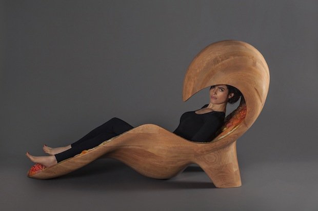
I’m fascinated by Oxman’s (and her colleagues’) furniture. Rose Brook writes about one particular Oxman piece in her March 27, 2014 posting for TCT magazine (Note: Links have been removed),

MIT Professor and 3D printing forerunner Neri Oxman has unveiled her striking acoustic chaise longue, which was made using Stratasys 3D printing technology.

Oxman collaborated with Professor W Craig Carter and Composer and fellow MIT Professor Tod Machover to explore material properties and their spatial arrangement to form the acoustic piece.

Christened Gemini, the two-part chaise was produced using a Stratasys Objet500 Connex3 multi-colour, multi-material 3D printer as well as traditional furniture-making techniques and it will be on display at the Vocal Vibrations exhibition at Le Laboratoire in Paris from March 28th 2014.

An Architect, Designer and Professor of Media, Arts and Science at MIT, Oxman’s creation aims to convey the relationship of twins in the womb through material properties and their arrangement. It was made using both subtractive and additive manufacturing and is part of Oxman’s ongoing exploration of what Stratasys’ ground-breaking multi-colour, multi-material 3D printer can do.

Brook goes on to explain how the chaise was made and the inspiration that led to it. Finally, it’s interesting to note that Oxman was working with Stratasys in 2014 and that this 2018 brain project is being developed in a joint collaboration with Statasys.

That’s it for 3D printing today.

Nanofibrous fish skins for wrinkle-free skin (New Zealand’s biggest seafood company moves into skincare)

I am utterly enchanted by this venture employing fish skins and nanotechnology-based processes for a new line of skin care products and, they hope, medical applications,


For those who like text (from a May 21, 2018 Sanford media advisory),

Nanofibre magic turns fish skins into wrinkle busting skin care

Sanford partners with kiwi nanotech experts to help develop a wrinkle-busting skincare product made from Hoki skins.

New Zealand’s biggest and oldest seafood company is moving into the future of skincare and medicine by becoming supporting partner to West Auckland nanofibre producer Revolution Fibres, which is launching a potentially game-changing nanotech face mask.

The actiVLayr face masks use collagen extracted from fish skins as a base ingredient which is then combined with elements such as fruit extracts and hyaluronic acid to make a 100 percent natural and sustainably sourced product.

They have achieved stunning results in third party tests which show that the nanofiber masks can reduce wrinkles by up to 31.5%.*

Revolution Fibres CEO Iain Hosie says it is no exaggeration to say the masks could be revolutionary.

“The wayactiVLayr is produced, and the unique application method of placing it onto wet skin like a mask, means ingredients are absorbed quickly and efficiently into the skin to maximise the repair and protection of the skin.”

Sanford is delighted to support the work that Revolution Fibres is doing by supplying hoki fish skins. Hoki is a sustainably caught fish and its skin has some unique properties.

Sanford’s General Manager of Innovation, Andrew Stanley, says these properties make it ideal for the actiVLayr technology. “Hoki skins are rich in collagen, which is an essential part of our bodies. But their marine collagen is unique – it has a very low melt point, so when placed on the skin, it can dissolve completely and be absorbed in a way that collagen f rom other animals cannot.”

Sanford’s Chief Customer Officer, Andre Gargiulo, says working with the team at Revolution Fibres is a natural fit, because both company’s think about innovation and sustainability in the same way.

“We hope actiVLayr gets the global attention it deserves, and we’re delighted that our sustainably caught Hoki is part of this fantastic New Zealand product. It’s exactly what we’re all about at Sanford – making the most of the precious resources from the sea, working in a sustainable way and getting the most value out of the goodness we harvest from nature.”

Sanford’s Business Development Manager Adrian Grey says the focus on sustainability and value creation are so important for the seafood company.

“Previously we have been making use of these hoki skins, which is great, but they were being used only for fish meal or pet food products. Being able to supply and support a high tech company that is going to earn increased export revenue for New Zealand is just fantastic. And the product created is completely natural, harvested from a globally certified sustainable fishery.”

Sanford provides the hoki skins and then turns these skins into pure collagen using the science and skills of the team at Plant and Food in Nelson [New Zealand for those of us who associate Nelson with British Columbia]. Revolution Fibres transforms the Sanford product into nanofibre using a technique called electrospinning of which Revolution Fibres are the New Zealand pioneers.

During the electrospinning process natural ingredients known as “bioactives” (such as kiwifruit and grapes) and hyaluronic acid (an ingredient to help the skin retain moisture) are bonded to the nanofibres to create sheets of actiVLayr. When it is exposed to wet skin the nanofibres dissolve rapidly and release the bioactives deep into the skin.

The product is being launched at the China Beauty Fair in Shanghai on May 22 [2018] and will go on sale in China this month followed by Hong Kong and New Zealand later in the year.   Revolution Fibres CEO Iain Hosie says there is big demand for unique delivery systems of natural skin and beauty products such as actiVLayr in Asia, which was the key reason to launch the product in China. But his view of the future is even bigger.

“There are endless uses for actiVLayr and the one we’re most proud of is in the medical area with the ability for drug compounds or medicines to be added to the actiVLayr formula. It will enable a controlled dose to be delivered to a patient with skin lesions, burns or acne.”

Revolution Fibres is presenting at Techweek NZ as part of The Fourth Revolution event on May 25 [2018] in Christchurch which introduces high tech engineers who are building a better place.

*Testing conducted by Easy Care using VISIA Complexion Analysis

The media advisory also includes some ‘fascinating ‘facts’,

1kg of hoki skin produces 400 square meters of nanofibre material

Nanofibres are 1/500th the width of a human hair

Revolution Fibres is the only nanofibre producer in the world to meet aerospace industry standards with its AS9100d quality assurance certification

The marine collagen found in hoki skins is unique because of its relatively low melt point, meaning it can dissolve at a lower temperature which makes it perfect for human use

Revolution Fibres is based in West Auckland and employs 12 people, of which 4 have P hDs in science related to nanotechnology. There are also a number of employees with strong engineering backgrounds to complement the company’s Research & Development expertise

Sanford is New Zealand’s oldest and biggest seafood company. It was founded by Albert Sanford in Auckland in 1904

New Zealand’s hoki fishery is certified as sustainable by the London-based Marine Stewardship Council, which audits fisheries all over the world

You can find Sanford here and Revolution Fibres here.

For some perspective on the business side of things, there’s a May 21, 2018 article by Nikki Mandow for newsroom.co.nz,

Revolution Fibres first started talking about the possibility of a collagen nanofibre made from hoki almost a decade ago, as part of a project with Plant & Food’s Seafood Research Centre in Nelson, Hosie [Revolution Fibres CEO Iain Hosie] said, and the company got serious about making a product in 2013.

Previously, the hoki waste skins were used for fish meal and pet food, said Sanford business development manager Adrian Grey.

“Being able to supply and support a high tech company that is going to earn increased export revenue for New Zealand is just fantastic.”

Revolution Fibres also manufactures nanofibres for a number of other uses. These include anti-dust mite pillow coverings, anti-pollution protective face masks, filters for pumps for HRV’s home ventilation systems, and reinforcing material for carbon fibre for fishing rods. The latter product is made from recycled fishing nets collected from South America.

He [Revolution Fibres CEO Iain Hosie] said the company could be profitable, but instead has chosen to continue to invest heavily in research and development.

About 75 percent of revenue comes from selling proprietary products, but increasingly Hosie said the company is working on “co-innovation” projects, where Revolution Fibres manufactures bespoke materials for outside companies.

Revolution Fibres completed its first external funding round last year, raising $1.5 million from the US, and it has just completed another round worth approximately $1million. Hosie, one of the founders, still holds around 20 percent of the company.

He said he hopes to keep the intellectual property in New Zealand, although manufacturing of some products is likely to move closer to their markets – China and the US potentially. However, he said actiVLayr manufacture will remain in New Zealand, because that’s where the raw hoki comes from.

I wonder if we’ll see this product in Canada.

One other thing,  I was curious about this ” … the nanofiber masks can reduce wrinkles by up to 31.5%”  and Visia Complexion Analysis, which is a product from Canfield Scientific, a company specializing in imaging.  Here’s some of what Visia can do (from the Visia product page),

Percentile Scores

Percentile Scores

VISIA’s patented comparison to norms analysis uses the world’s largest skin feature database to grade your patient’s skin relative to others of the same age and skin type. Measure spots, wrinkles, texture, pores, UV spots, brown spots, red areas, and porphyrins.

Meaningful Comparisons

Meaningful Comparisons

Compare results side by side for any combination of views, features or time points, including graphs and numerical data. Zoom and pan images in tandem for clear and easy comparisons.

And, there’s my personal favourite (although it has nothing to do with the topic of this posting0,

Eyelash Analysis

Eyelash Analysis

Evaluates the results of lash improvement treatments with numerical assessments and graphic visualizations.

For anyone who wondered about why the press release has both ‘nanofibre’ and ‘nanofiber’, It’s the difference between US and UK spelling. Perhaps the complexion analysis information came from a US company or one that uses US spellings.

‘Smart’ fabric that’s bony

Researchers at Australia’s University of New South of Wales (UNSW) have devised a means of ‘weaving’ a material that mimics *bone tissue, periosteum according to a Jan. 11, 2017 news item on ScienceDaily,

For the first time, UNSW [University of New South Wales] biomedical engineers have woven a ‘smart’ fabric that mimics the sophisticated and complex properties of one nature’s ingenious materials, the bone tissue periosteum.

Having achieved proof of concept, the researchers are now ready to produce fabric prototypes for a range of advanced functional materials that could transform the medical, safety and transport sectors. Patents for the innovation are pending in Australia, the United States and Europe.

Potential future applications range from protective suits that stiffen under high impact for skiers, racing-car drivers and astronauts, through to ‘intelligent’ compression bandages for deep-vein thrombosis that respond to the wearer’s movement and safer steel-belt radial tyres.

A Jan. 11, 2017 UNSW press release on EurekAlert, which originated the news item, expands on the theme,

Many animal and plant tissues exhibit ‘smart’ and adaptive properties. One such material is the periosteum, a soft tissue sleeve that envelops most bony surfaces in the body. The complex arrangement of collagen, elastin and other structural proteins gives periosteum amazing resilience and provides bones with added strength under high impact loads.

Until now, a lack of scalable ‘bottom-up’ approaches by researchers has stymied their ability to use smart tissues to create advanced functional materials.

UNSW’s Paul Trainor Chair of Biomedical Engineering, Professor Melissa Knothe Tate, said her team had for the first time mapped the complex tissue architectures of the periosteum, visualised them in 3D on a computer, scaled up the key components and produced prototypes using weaving loom technology.

“The result is a series of textile swatch prototypes that mimic periosteum’s smart stress-strain properties. We have also demonstrated the feasibility of using this technique to test other fibres to produce a whole range of new textiles,” Professor Knothe Tate said.

In order to understand the functional capacity of the periosteum, the team used an incredibly high fidelity imaging system to investigate and map its architecture.

“We then tested the feasibility of rendering periosteum’s natural tissue weaves using computer-aided design software,” Professor Knothe Tate said.

The computer modelling allowed the researchers to scale up nature’s architectural patterns to weave periosteum-inspired, multidimensional fabrics using a state-of-the-art computer-controlled jacquard loom. The loom is known as the original rudimentary computer, first unveiled in 1801.

“The challenge with using collagen and elastin is their fibres, that are too small to fit into the loom. So we used elastic material that mimics elastin and silk that mimics collagen,” Professor Knothe Tate said.

In a first test of the scaled-up tissue weaving concept, a series of textile swatch prototypes were woven, using specific combinations of collagen and elastin in a twill pattern designed to mirror periosteum’s weave. Mechanical testing of the swatches showed they exhibited similar properties found in periosteum’s natural collagen and elastin weave.

First author and biomedical engineering PhD candidate, Joanna Ng, said the technique had significant implications for the development of next-generation advanced materials and mechanically functional textiles.

While the materials produced by the jacquard loom have potential manufacturing applications – one tyremaker believes a titanium weave could spawn a new generation of thinner, stronger and safer steel-belt radials – the UNSW team is ultimately focused on the machine’s human potential.

“Our longer term goal is to weave biological tissues – essentially human body parts – in the lab to replace and repair our failing joints that reflect the biology, architecture and mechanical properties of the periosteum,” Ms Ng said.

An NHMRC development grant received in November [2016] will allow the team to take its research to the next phase. The researchers will work with the Cleveland Clinic and the University of Sydney’s Professor Tony Weiss to develop and commercialise prototype bone implants for pre-clinical research, using the ‘smart’ technology, within three years.

In searching for more information about this work, I found a Winter 2015 article (PDF; pp. 8-11) by Amy Coopes and Steve Offner for UNSW Magazine about Knothe Tate and her work (Note: In Australia, winter would be what we in the Northern Hemisphere consider summer),

Tucked away in a small room in UNSW’s Graduate School of Biomedical Engineering sits a 19th century–era weaver’s wooden loom. Operated by punch cards and hooks, the machine was the first rudimentary computer when it was unveiled in 1801. While on the surface it looks like a standard Jacquard loom, it has been enhanced with motherboards integrated into each of the loom’s five hook modules and connected to a computer. This state-of-the-art technology means complex algorithms control each of the 5,000 feed-in fibres with incredible precision.

That capacity means the loom can weave with an extraordinary variety of substances, from glass and titanium to rayon and silk, a development that has attracted industry attention around the world.

The interest lies in the natural advantage woven materials have over other manufactured substances. Instead of manipulating material to create new shades or hues as in traditional weaving, the fabrics’ mechanical properties can be modulated, to be stiff at one end, for example, and more flexible at the other.

“Instead of a pattern of colours we get a pattern of mechanical properties,” says Melissa Knothe Tate, UNSW’s Paul Trainor Chair of Biomedical Engineering. “Think of a rope; it’s uniquely good in tension and in bending. Weaving is naturally strong in that way.”


The interface of mechanics and physiology is the focus of Knothe Tate’s work. In March [2015], she travelled to the United States to present another aspect of her work at a meeting of the international Orthopedic Research Society in Las Vegas. That project – which has been dubbed “Google Maps for the body” – explores the interaction between cells and their environment in osteoporosis and other degenerative musculoskeletal conditions such as osteoarthritis.

Using previously top-secret semiconductor technology developed by optics giant Zeiss, and the same approach used by Google Maps to locate users with pinpoint accuracy, Knothe Tate and her team have created “zoomable” anatomical maps from the scale of a human joint down to a single cell.

She has also spearheaded a groundbreaking partnership that includes the Cleveland Clinic, and Brown and Stanford universities to help crunch terabytes of data gathered from human hip studies – all processed with the Google technology. Analysis that once took 25 years can now be done in a matter of weeks, bringing researchers ever closer to a set of laws that govern biological behaviour. [p. 9]

I gather she was recruited from the US to work at the University of New South Wales and this article was to highlight why they recruited her and to promote the university’s biomedical engineering department, which she chairs.

Getting back to 2017, here’s a link to and citation for the paper,

Scale-up of nature’s tissue weaving algorithms to engineer advanced functional materials by Joanna L. Ng, Lillian E. Knothe, Renee M. Whan, Ulf Knothe & Melissa L. Knothe Tate. Scientific Reports 7, Article number: 40396 (2017) doi:10.1038/srep40396 Published online: 11 January 2017

This paper is open access.

One final comment, that’s a lot of people (three out of five) with the last name Knothe in the author’s list for the paper.

*’the bone tissue’ changed to ‘bone tissue’ on July 17,2017.

Fish skin for wound healing

A Feb. 11, 2015 news item on Nanowerk features Chinese research on tilapia fish skin and possible applications for wound healing (Note: A link has been removed),

With a low price tag and mild flavor, tilapia has become a staple dinnertime fish for many Americans. Now it could have another use: helping to heal our wounds. In the journal ACS Applied Materials & Interfaces (“Development of Biomimetic Tilapia Collagen Nanofibers for Skin Regeneration through Inducing Keratinocytes Differentiation and Collagen Synthesis of Dermal Fibroblasts”), scientists have shown that a protein found in this fish can promote skin repair in rats without an immune reaction, suggesting possible future use for human patients.

A Feb. 11, 2015 American Chemical Society (ACS) news release, which originated the news item, provides a few more details about the work,

Jiao Sun, Xiumei Mo and colleagues explain that applying collagen — a major structural protein in animals — to wounds can help encourage skin to heal faster.  But when the protein dressing comes from mammals such as cows and pigs, it has the potential to transmit conditions such as foot-and-mouth disease. Searching for an alternative source of collagen, scientists recently turned to the ocean. Sun’s team wanted to test fish collagen’s potential as a more benign wound treatment.

The researchers developed nanofibers from tilapia collagen and used them to cover skin wounds on rats. The rats with the nanofiber dressing healed faster than those without it. In addition, lab tests on cells suggested that the fish collagen was not likely to cause an immune reaction. The researchers conclude that it could be a good candidate to develop for clinical use.

Here’s a link to and a citation for the paper,

Development of Biomimetic Tilapia Collagen Nanofibers for Skin Regeneration through Inducing Keratinocytes Differentiation and Collagen Synthesis of Dermal Fibroblasts by Tian Zhou, Nanping Wang, Yang Xue, Tingting Ding, Xin Liu, Xiumei Mo, and Jiao Sun. ACS Appl. Mater. Interfaces, 2015, 7 (5), pp 3253–3262 DOI: 10.1021/am507990m Publication Date (Web): January 19, 2015

Copyright © 2015 American Chemical Society

This article is behind a paywall.