Tag Archives: dental implants

Smart dental implant resists bacterial growth and generates own electricity

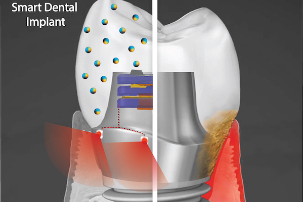
A “smart” dental implant could improve upon current devices by employing biofilm-resisting nanoparticles and a light powered by biomechanical forces to promote health of the surrounding gum tissue. (Image: Courtesy of Albert Kim)

A September 9, 2021 news item on ScienceDaily announces research into ‘smart’ dental implants,

More than 3 million people in America have dental implants, used to replace a tooth lost to decay, gum disease, or injury. Implants represent a leap of progress over dentures or bridges, fitting much more securely and designed to last 20 years or more.

But often implants fall short of that expectation, instead needing replacement in five to 10 years due to local inflammation or gum disease, necessitating a repeat of a costly and invasive procedure for patients.

“We wanted to address this issue, and so we came up with an innovative new implant,” says Geelsu Hwang, an assistant professor in the University of Pennsylvania School of Dental Medicine, who has a background in engineering that he brings to his research on oral health issues.

The novel implant would implement two key technologies, Hwang says. One is a nanoparticle-infused material that resists bacterial colonization. And the second is an embedded light source to conduct phototherapy, powered by the natural motions of the mouth, such as chewing or toothbrushing. In a paper in the journal ACS Applied Materials & Interfaces and a 2020 paper in the journal Advanced Healthcare Materials, Hwang and colleagues lay out their platform, which could one day be integrated not only into dental implants but other technologies, such as joint replacements, as well.

A September 9, 2021 University of Pennsylvania news release (also on EurekAlert), which originated the news item, provides more technical details about the proposed technology,

“Phototherapy can address a diverse set of health issues,” says Hwang. “But once a biomaterial is implanted, it’s not practical to replace or recharge a battery. We are using a piezoelectric material, which can generate electrical power from natural oral motions to supply a light that can conduct phototherapy, and we find that it can successfully protect gingival tissue from bacterial challenge.”

In the paper, the material the researchers explored was barium titanate (BTO), which has piezoelectric properties that are leveraged in applications such as capacitators and transistors, but has not yet been explored as a foundation for anti-infectious implantable biomaterials. To test its potential as the foundation for a dental implant, the team first used discs embedded with nanoparticles of BTO and exposed them to Streptococcus mutans, a primary component of the bacterial biofilm responsible for tooth decay commonly known as dental plaque. They found that the discs resisted biofilm formation in a dose-dependent manner. Discs with higher concentrations of BTO were better at preventing biofilms from binding.

While earlier studies had suggested that BTO might kill bacteria outright using reactive oxygen species generated by light-catalyzed or electric polarization reactions, Hwang and colleagues did not find this to be the case due to the short-lived efficacy and off-target effects of these approaches. Instead, the material generates enhanced negative surface charge that repels the negatively charged cell walls of bacteria. It’s likely that this repulsion effect would be long-lasting, the researchers say.

“We wanted an implant material that could resist bacterial growth for a long time because bacterial challenges are not a one-time threat,” Hwang says.

The power-generating property of the material was sustained and in tests over time the material did not leach. It also demonstrated a level of mechanical strength comparable to other materials used in dental applications.

Finally, the material did not harm normal gingival tissue in the researchers’ experiments, supporting the idea that this could be used without ill effect in the mouth.

The technology is a finalist in the Science Center’s research accelerator program, the QED Proof-of-Concept program. As one of 12 finalists, Hwang and colleagues will receive guidance from experts in commercialization. If the project advances to be one of three finalists, the group has the potential to receive up to $200,000 in funding.

In future work, the team hopes to continue to refine the “smart” dental implant system, testing new material types and perhaps even using assymetric properties on each side of the implant components, one that encourages tissue integration on the side facing the gums and one that resists bacterial formation on the side facing the rest of the mouth.

“We hope to further develop the implant system and eventually see it commercialized so it can be used in the dental field,” Hwang says.

Here’s a link to and a citation for the paper,

Bimodal Nanocomposite Platform with Antibiofilm and Self-Powering Functionalities for Biomedical Applications by Atul Dhall, Sayemul Islam, Moonchul Park, Yu Zhang, Albert Kim, and Geelsu Hwang. ACS Appl. Mater. Interfaces 2021, 13, 34, 40379–40391 DOI: https://doi.org/10.1021/acsami.1c11791 Publication Date:August 18, 2021 Copyright © 2021 American Chemical Society

This paper is behind a paywall.

The work from 2020, mentioned in the news release, laid groundwork for the latest paper.

Human Oral Motion-Powered Smart Dental Implant (SDI) for In Situ Ambulatory Photo-biomodulation Therapy by Moonchul Park, Sayemul Islam, Hye-Eun Kim, Jonathan Korosto, Markus B. Blatz, Geelsu Hwang, and Albert Kim. Adv. Healthcare Mater. 2020, 9, 2000658 DOI: 10.1002/adhm.202000658 First published: 01 July 2020 © 2020 WILEY-VCH Verlag GmbH & Co. KGaA, WeinheimHuman

This paper is behind a paywall.

Killer graphene spikes to kill bacteria on medical implants

Implants of all kinds (hip replacements, knee replacements, etc.) seem to be on the rise and along with that an increasing number of infections. A Swedish research team announces a technology that could make implants safer in an April 16, 2018 news item on Nanowerk,

A tiny layer of graphene flakes becomes a deadly weapon and kills bacteria, stopping infections during procedures such as implant surgery. This is the findings of new research from Chalmers University of Technology, Sweden, recently published in the scientific journal Advanced Materials Interfaces (“Vertically Aligned Graphene Coating is Bactericidal and Prevents the Formation of Bacterial Biofilms”).

An April 16, 2018 Chalmers University of Technology press release (also on EurekAlert), which originated the news item, provides more detail about the scope of the problem and the proposed solution (Note: A link has been removed),

Operations for surgical implants, such as hip and knee replacements or dental implants, have increased in recent years. However, in such procedures, there is always a risk of bacterial infection. In the worst case scenario, this can cause the implant to not attach to the skeleton, meaning it must be removed.

Bacteria travel around in fluids, such as blood, looking for a surface to cling on to. Once in place, they start to grow and propagate, forming a protective layer, known as a biofilm.

A research team at Chalmers has now shown that a layer of vertical graphene flakes forms a protective surface that makes it impossible for bacteria to attach. Instead, bacteria are sliced apart by the sharp graphene flakes and killed. Coating implants with a layer of graphene flakes can therefore help protect the patient against infection, eliminate the need for antibiotic treatment, and reduce the risk of implant rejection. The osseointegration – the process by which the bone structure grow to attach the implant – is not disturbed. In fact, the graphene has been shown to benefit the bone cells.

Chalmers University is a leader in the area of graphene research, but the biological applications did not begin to materialise until a few years ago. The researchers saw conflicting results in earlier studies. Some showed that graphene damaged the bacteria, others that they were not affected.

“We discovered that the key parameter is to orient the graphene vertically. If it is horizontal, the bacteria are not harmed” says Ivan Mijakovic, Professor at the Department of Biology and Biological Engineering.

The sharp flakes do not damage human cells. The reason is simple: one bacterium is one micrometer – one thousandth of a millimeter – in diameter, while a human cell is 25 micrometers. So, what constitutes a deadly knife attack for a bacterium, is therefore only a tiny scratch for a human cell.

“Graphene has high potential for health applications. But more research is needed before we can claim it is entirely safe. Among other things, we know that graphene does not degrade easily” says Jie Sun, Associate Professor at the Department of Micro Technology and Nanoscience.

Good bacteria are also killed by the graphene. But that’s not a problem, as the effect is localised and the balance of microflora in the body remains undisturbed.

“We want to prevent bacteria from creating an infection. Otherwise, you may need antibiotics, which could disrupt the balance of normal bacteria and also enhance the risk of antimicrobial resistance by pathogens” says Santosh Pandit, postdoc at Biology and Biological Engineering.

Vertical flakes of graphene are not a new invention, having existed for a few years. But the Chalmers research teams are the first to use the vertical graphene in this way. The next step for the research team will be to test the graphene flakes further, by coating implant surfaces and studying the effect on animal cells.

Chalmers cooperated with Wellspect Healthcare, a company which makes catheters and other medical instruments, in this research. They will now continue with a second study.

Here’s a link to and a citation for the paper,

Vertically Aligned Graphene Coating is Bactericidal and Prevents the Formation of Bacterial Biofilms by Santosh Pandit, Zhejian Cao, Venkata R. S. S. Mokkapati, Emanuele Celauro, Avgust Yurgens, Martin Lovmar, Fredrik Westerlund, Jie Sun, Ivan Mijakovic. Advanced Materials Interfaces Volume5, Issue7 April 9, 2018 Pages 1701331 [sic] https://doi.org/10.1002/admi.201701331 First published [online]: 2 February 2018

This paper is behind a paywall.

Finally, here’s a ‘killer spikes’ video made available by Chalmers University of Technology,

Nanocoating to reduce dental implant failures

Scientists at Plymouth University (UK) have developed a nanocoating that could reduce the number of dental implant failures. From a March 24, 2017 news item on Nanowerk (Note: A link has been removed),

According to the American Academy of Implant Dentistry (AAID), 15 million Americans have crown or bridge replacements and three million have dental implants — with this latter number rising by 500,000 a year. The AAID estimates that the value of the American and European market for dental implants will rise to $4.2 billion by 2022.

Dental implants are a successful form of treatment for patients, yet according to a study published in 2005, five to 10 per cent of all dental implants fail.

The reasons for this failure are several-fold – mechanical problems, poor connection to the bones in which they are implanted, infection or rejection. When failure occurs the dental implant must be removed.

The main reason for dental implant failure is peri-implantitis. This is the destructive inflammatory process affecting the soft and hard tissues surrounding dental implants. This occurs when pathogenic microbes in the mouth and oral cavity develop into biofilms, which protects them and encourages growth. Peri-implantitis is caused when the biofilms develop on dental implants.

A research team comprising scientists from the School of Biological Sciences, Peninsula Schools of Medicine and Dentistry and the School of Engineering at the University of Plymouth, have joined forces to develop and evaluate the effectiveness of a new nanocoating for dental implants to reduce the risk of peri-implantitis.

The results of their work are published in the journal Nanotoxicology (“Antibacterial activity and biofilm inhibition by surface modified titanium alloy medical implants following application of silver, titanium dioxide and hydroxyapatite nanocoatings”).

A March 27, 2017 Plymouth University press release, which originated the news item, gives more details about the research,

In the study, the research team created a new approach using a combination of silver, titanium oxide and hydroxyapatite nanocoatings.

The application of the combination to the surface of titanium alloy implants successfully inhibited bacterial growth and reduced the formation of bacterial biofilm on the surface of the implants by 97.5 per cent.

Not only did the combination result in the effective eradication of infection, it created a surface with anti-biofilm properties which supported successful integration into surrounding bone and accelerated bone healing.

Professor Christopher Tredwin, Head of Plymouth University Peninsula School of Dentistry, commented:

“In this cross-Faculty study we have identified the means to protect dental implants against the most common cause of their failure. The potential of our work for increased patient comfort and satisfaction, and reduced costs, is great and we look forward to translating our findings into clinical practice.”

The University of Plymouth was the first university in the UK to secure Research Council Funding in Nanoscience and this project is the latest in a long line of projects investigating nanotechnology and human health.

Nanoscience activity at the University of Plymouth is led by Professor Richard Handy, who has represented the UK on matters relating to the Environmental Safety and Human Health of Nanomaterials at the Organisation for Economic Cooperation and Development (OECD). He commented:

“As yet there are no nano-specific guidelines in dental or medical implant legislation and we are, with colleagues elsewhere, guiding the way in this area. The EU recognises that medical devices and implants must: perform as expected for its intended use, and be better than similar items in the market; be safe for the intended use or safer than an existing item, and; be biocompatible or have negligible toxicity.”

He added:

“Our work has been about proving these criteria which we have done in vitro. The next step would be to demonstrate the effectiveness of our discovery, perhaps with animal models and then human volunteers.”

Dr Alexandros Besinis, Lecturer in Mechanical Engineering at the School of Engineering, University of Plymouth, led the research team. He commented:

“Current strategies to render the surface of dental implants antibacterial with the aim to prevent infection and peri-implantitis development, include application of antimicrobial coatings loaded with antibiotics or chlorhexidine. However, such approaches are usually effective only in the short-term, and the use of chlorhexidine has also been reported to be toxic to human cells. The significance of our new study is that we have successfully applied a dual-layered silver-hydroxyapatite nanocoating to titanium alloy medical implants which helps to overcome these risks.”

Dr Besinis has been an Honorary Teaching Fellow at the Peninsula School of Dentistry since 2011 and has recently joined the School of Engineering. His research interests focus on advanced engineering materials and the use of nanotechnology to build novel biomaterials and medical implants with improved mechanical, physical and antibacterial properties.

Here’s a link to and a citation for the paper,

Antibacterial activity and biofilm inhibition by surface modified titanium alloy medical implants following application of silver, titanium dioxide and hydroxyapatite nanocoatings by A. Besinis, S. D. Hadi, H. R. Le, C. Tredwin & R. D. Handy.  Nanotoxicology Volume 11, 2017 – Issue 3  Pages 327-338  http://dx.doi.org/10.1080/17435390.2017.1299890 Published online: 17 Mar 2017

This paper appears to be open access.

Inspiration from the sea for titanium implants (mussels) and adhesive panels for flexible sensors (octopuses/octopi/octopodes)

I have two sea-inspired news bits both of which concern adhesion.

Mussels and titanium implants

A July 8, 2016 news item on ScienceDaily features some mussel-inspired research from Japan into how to make better titanium implants,

Titanium is used medically in applications such as artificial joints and dental implants. While it is strong and is not harmful to tissues, the metal lacks some of the beneficial biological properties of natural tissues such as bones and natural teeth. Now, based on insights from mussels–which are able to attach themselves very tightly to even metallic surfaces due to special proteins found in their byssal threads–scientists from RIKEN have successfully attached a biologically active molecule to a titanium surface, paving the way for implants that can be more biologically beneficial.

A July 11, 2016 RIKEN press release (also on EurekAlert but dated July 8, 2016), which originated the news item, provides more information,

The work began from earlier discoveries that mussels can attach to smooth surfaces so effectively thanks to a protein, L-DOPA, which is known to be able to bind very strongly to smooth surfaces such as rocks, ceramics, or metals (…). Interestingly, the same protein functions in humans as a precursor to dopamine, and is used as a treatment for Parkinson’s disease.

According to Chen Zhang of the RIKEN Nano Medical Engineering Laboratory, the first author of the paper published in Angewandte Chemie, “We thought it would be interesting to try to use various techniques to attach a biologically active protein—in our case we chose insulin-like growth factor-1, a promoter of cell proliferation—to a titanium surface like those used in implants” (…).

Using a combination of recombinant DNA technology and treatment with tyrosinase, they were able to create a hybrid protein that contained active parts of both the growth factor and L-DOPA. Tests showed that the proteins were able to fold normally, and further experiments in cell cultures demonstrated that the IGF-1 was still functioning normally. Thanks to the incorporation of the L-DOPA, the team was able to confirm that the proteins bound strongly to the titanium surface, and remained attached even when the metal was washed with phosphate-buffered saline, a water-based solution. Zhang says, “This is similar to the powerful properties of mussel adhesive, which can remain fixed to metallic materials even underwater.”

According to Yoshihiro Ito, Team Leader of the Emergent Bioengineering Research Team of the RIKEN Center for Emergent Matter Science, “We are very excited by this finding, because the modification process is a universal one that could be used with other proteins. It could allow us to prepare new cell-growth enhancing materials, with potential applications in cell culture systems and regenerative medicine. And it is particularly interesting that this is an example of biomimetics, where nature can teach us new ways to do things. The mussel has given us insights that could be used to allow us to live healthier lives.”

The work was done by RIKEN researchers in collaboration with Professor Peibiao Zhang of the Changchun Institute of Applied Chemistry, Chinese Academy of Sciences, and Professor Yi Wang of the School of Pharmaceutical Sciences, Jilin University. The work was partially supported by the Japan Society for the Promotion of Science KAKENHI (Grant Number 15H01810 and 22220009), CAS-JSPS joint fund (GJHZ1519), and RIKEN MOST joint project.

Here’s a link to and a citation for the paper,

A Bioorthogonal Approach for the Preparation of a Titanium-Binding Insulin-like Growth-Factor-1 Derivative by using Tyrosinase by Chen Zhang, Hideyuki Miyatake, Yu Wang, Takehiko Inaba, Yi Wang, Peibiao Zhang, and Prof. Yoshihiro Ito. Angewandte Chemie International Edition DOI: 10.1002/anie.201603155 Version of Record online: 6 JUL 2016

© 2016 WILEY-VCH Verlag GmbH & Co. KGaA, Weinheim

This paper is behind a paywall.

Octopuses/octopi/octopodes and adhesive panels

Before launching into the science part of this news bit, here’s some grammar (from the Octopus Wikipedia entry; Note: Links have been removed),

The standard pluralized form of “octopus” in the English language is “octopuses” /ˈɒktəpʊsɪz/,[10] although the Ancient Greek plural “octopodes” /ɒkˈtɒpədiːz/, has also been used historically.[9] The alternative plural “octopi” — which misguidedly assumes it is a Latin “-us”-word — is considered grammatically incorrect.[11][12][13][14] It is nevertheless used enough to make it notable, and was formally acknowledged by the descriptivist Merriam-Webster 11th Collegiate Dictionary and Webster’s New World College Dictionary. The Oxford English Dictionary (2008 Draft Revision)[15] lists “octopuses”, “octopi”, and “octopodes”, in that order, labelling “octopodes” as rare and noting that “octopi” derives from the apprehension that octōpus comes from Latin.[16] In contrast, New Oxford American Dictionary (3rd Edition 2010) lists “octopuses” as the only acceptable pluralization, with a usage note indicating “octopodes” as being still occasionally used but “octopi” as being incorrect.[17]

Now the news. A July 12, 2016 news item on Nanowerk highlights some research into adhesives and octopuses,

With increased study of bio-adhesives, a significant effort has been made in search for novel adhesives that will combine reversibility, repeated usage, stronger bonds and faster bonding time, non-toxic, and more importantly be effective in wet and other extreme conditions.

A team of Korean scientists-made up of scientists from Korea Institute of Science and Technology (KIST) and UNIST has recently found a way to make building flexible pressure sensors easier–by mimicking the suction cups on octopus’s tentacles.

A July 5, 2016 UNIST (Ulsan National Institute of Science and Technology) press release, which originated the news item, provides more information,

According to the research team, “Although flexible pressure sensors might give future prosthetics and robots a better sense of touch, building them requires a lot of laborious transferring of nano- and microribbons of inorganic semiconductor materials onto polymer sheets.”

In search of an easier way to process this transfer printing, Prof. Hyunhyub Ko (School of Energy and Chemical Engineering, UNIST) and his colleagues turned to the octopus suction cups for inspiration.

An octopus uses its tentacles to move to a new location and uses suction cups underneath each tentacle to grab onto something. Each suction cup contains a cavity whose pressure is controlled by surrounding muscles. These can be made thinner or thicker on demand, increasing or decreasing air pressure inside the cup, allowing for sucking and releasing as desired.

By mimicking muscle actuation to control cavity-pressure-induced adhesion of octopus suckers, Prof. Ko and his team engineered octopus-inspired smart adhesive pads. They used the rubbery material polydimethylsiloxane (PDMS) to create an array of microscale suckers, which included pores that are coated with a thermally responsive polymer to create sucker-like walls.

The team discovered that the best way to replicate organic nature of muscle contractions would be through applied heat. Indeed, at room temperature, the walls of each pit sit in an ‘open’ state, but when the mat is heated to 32°C, the walls contract, creating suction, therby allowing the entire mate to adhere to a material (mimicking the suction function of an octopus). The adhesive strength also spiked from .32 kilopascals to 94 kilopascals at high temperature.

The team reports that the mat worked as envisioned—they made some indium gallium arsenide transistors that sat on a flexible substrate and also used it to move some nanomaterials to a different type of flexible material.

Prof. Ko and his team expect that their smart adhesive pads can be used as the substrate for wearable health sensors, such as Band-Aids or sensors that stick to the skin at normal body temperatures but fall off when rinsed under cold water.

Here’s a link to and a citation for the paper,

Octopus-Inspired Smart Adhesive Pads for Transfer Printing of Semiconducting Nanomembranes by Hochan Lee, Doo-Seung Um, Youngsu Lee, Seongdong Lim, Hyung-jun Kim,  and Hyunhyub Ko. Advanced Materials DOI: 10.1002/adma.201601407 Version of Record online: 20 JUN 2016

© 2016 WILEY-VCH Verlag GmbH & Co. KGaA, Weinheim

This paper is behind a paywall.

Preventing deep bone infections with antibiotic-laced polymer layers in implants

I know someone who suffered a deep bone infection after some dental work. Devastatingly, she lost bone material as a consequence and it took years, more than one surgery, and multiple sessions in a hyperbaric chamber to recover, more or less.

While my friend’s infection was due to a dental procedure, the work at the University of Sheffield’s (UK) School of Clinical Dentistry, if successful, will help eliminate incidents of deep bone infection from one potential source, implants. From a May 28, 2015 news item on Azonano,

Leading scientists at the University of Sheffield have discovered nanotechnology could hold the key to preventing deep bone infections, after developing a treatment which prevents bacteria and other harmful microorganisms growing.

The pioneering research, led by the University of Sheffield’s School of Clinical Dentistry, showed applying small quantities of antibiotic to the surface of medical devices, from small dental implants to hip replacements, could protect patients from serious infection.

A May 27, 2015 University of Sheffield press release, which originated the news item, provides more information but few details about how this work is nanotechnology-enabled,

Scientists used revolutionary nanotechnology to work on small polymer layers inside implants which measure between 1 and 100 nanometers (nm) – a human hair is approximately 100,000 nm wide.

Lead researcher Paul Hatton, Professor of Biomaterials Sciences at the University of Sheffield, said: “Microorganisms can attach themselves to implants or replacements during surgery and once they grab onto a non-living surface they are notoriously difficult to treat which causes a lot of problems and discomfort for the patient.

“By making the actual surface of the hip replacement or dental implant inhospitable to these harmful microorganisms, the risk of deep bone infection is substantially reduced.

“Our research shows that applying small quantities of antibiotic to a surface between the polymer layers which make up each device could prevent not only the initial infection but secondary infection – it is like getting between the layers of an onion skin.”

Bone infection affects thousands of patients every year and results in a substantial cost to the NHS.

Treating the surface of medical devices would have a greater impact on patients considered at high risk of infection such as trauma victims from road traffic collisions or combat operations, and those who have had previous bone infections.

Professor Hatton added: “Deep bone infections associated with medical devices are increasing in number, especially among the elderly.

“As well as improving the quality of life, this new application for nanotechnology could save health providers such as the NHS millions of pounds every year.”

Here’s a link to and a citation for the paper,

Functionalised nanoscale coatings using layer-by-layer assembly for imparting antibacterial properties to polylactide-co-glycolide surfaces by Piergiorgio Gentile, ,Maria E. Frongia, Mar Cardellach, Cheryl A. Miller, Graham P. Stafford, Graham J. Leggettc, & Paul V. Hatton. Acta Biomaterialia Volume 21, 15 July 2015, Pages 35–43 doi: 10.1016/j.actbio.2015.04.009

This paper is behind a paywall.

Dental implants with a surface that affects genetic cellular expression

Intra-Lock International is trumpeting in triumph in the wake  of a study noting their OSSEAN-surfaced dental implants promote better bone-healing than an alternative used for comparison. From the June 10, 2014 news item on Azonano,

As reported in the internationally renowned scientific journal, Bone [in press for Aug. 2014], a research team from New York University [NYU] has confirmed what scientific developers at Intra-Lock® International, Inc. have known for several years: the fractal, nano-rough OSSEAN® surface developed for their dental implants actually changes the cellular genetic expression – or the fate of stem cells – at the nano-level, which in turn induces faster healing of implants.

A June 9, 2014 Intra-Lock news release, which originated the new item, describes what usually occurs when an implant is first situated in the tissue (cellular confusion) and how the OSSEAN surface affects the ‘confusion’,

Typically, when an implant is surgically placed, there is a period of cellular “confusion” and chaos around the implant, and usually a little bone resorbs before being formed again. The implant is then at risk from the moment it is inserted through the time when the bone is healed around it – a time period Giorno [Thierry M. Giorno, DDS, director of research and development, and CEO of Intra-Lock®, International] refers to as “the window of negative opportunity.”

However, the NYU researchers found that bone cells immediately start clustering around the OSSEAN implants and begin accelerated healing, with little confusion whatsoever.

This occurs primarily due to the biomimetic structure of the OSSEAN surface, designed and classified as nanorough and fractalii. Mimicking nature at the nano-level, the OSSEAN surface repeats a similar structural pattern to that of natural bone over and over, essentially “tricking” the body into accepting the implant as a natural substance and igniting the healing process far sooner than would occur with an artificial substance, which is smooth at the nano-level and without natural-seeming pattern repetition.

Typically, with an implant of any sort, whether it’s a dental implant in your jaw or a titanium rod in your leg, several weeks will pass before the bone begins to grow around it. During this time lapse, known as the “catabolic phase,” there can be great risk and instability with the implant.

Naturally, compressing the healing time and accelerating the degree of osseointegration – the merging of implant and bone – are highly desirable outcomes, and implants with an OSSEAN can provide a faster healing process, which thereby reduces patient discomfort and provides a higher potential for successful long-term results with the implant.

“If you’ve ever had dental implants, you can appreciate the outcomes the OSSEAN surface provides,” said Giorno. “The healing process has changed forever, and future patients with an OSSEAN surface implant can look forward to reduced complications, overall.”

Looking further into the future, Giorno said, “I believe the effects of OSSEAN can potentially revolutionize the implant industry beyond dentistry and into all types of orthopedics where patients must wait for their bodies to accept a foreign substance. With OSSEAN, the wait is over.”

Here’s a link to and a citation for the paper,

Nanometer-scale features on micrometer-scale surface texturing: A bone histological, gene expression, and nanomechanical study by Paulo G. Coelho, Tadahiro Takayama, Daniel Yoo, Ryo Jimboemail, Sanjay Karunagaran, Nick Tovar, Malvin N. Janal, and Seiichi Yamano. Bone, Issue 65, Aug. 2014. Bone (2014) DOI: http://dx.doi.org/10.1016/j.bone.2014.05.004 Published Online: May 07, 2014

This article is behind a paywall. You can find out more about Intra-Lock and OSSEAN here.