Tag Archives: diagnostics

Diagnosing diseases by using nanomembranes to isolate biomarkers in tears

How are they planning to make people cry on command or use a swab on your eyeball? In general, I like the idea of using tears instead of other bodily secretions but it’s the practicalities that have me questioning how this kind of diagnostic test could be implemented. In any event, here’s more from a July 20, 2022 news item on phys.org,

Going to the doctor might make you want to cry, and according to a new study, doctors could someday put those tears to good use. In ACS Nano, researchers report a nanomembrane system that harvests and purifies tiny blobs called exosomes from tears, allowing researchers to quickly analyze them for disease biomarkers. Dubbed iTEARS, the platform could enable more efficient and less invasive molecular diagnoses for many diseases and conditions, without relying solely on symptoms.

A July 20, 2022 American Chemical Society (ACS) news release (also on EurekAlert), which originated the news item, explains the work in more detail,

Diagnosing diseases often hinges on assessing a patient’s symptoms, which can be unobservable at early stages, or unreliably reported. Identifying molecular clues in samples from patients, such as specific proteins or genes from vesicular structures called exosomes, could improve the accuracy of diagnoses. However, current methods for isolating exosomes from these samples require long, complicated processing steps or large sample volumes. Tears are well-suited for sample collection because the fluid can be collected quickly and non-invasively, though only tiny amounts can be harvested at a time. So, Luke Lee, Fei Liu and colleagues wondered if a nanomembrane system, which they originally developed for isolating exosomes from urine and plasma, could allow them to quickly obtain these vesicles from tears and then analyze them for disease biomarkers.

The team modified their original system to handle the low volume of tears. The new system, called “Incorporated Tear Exosomes Analysis via Rapid-isolation System” (iTEARS), separated out exosomes in just 5 minutes by filtering tear solutions over nanoporous membranes with an oscillating pressure flow to reduce clogging. Proteins from the exosomes could be tagged with fluorescent probes while they were still on the device and then transferred to other instruments for further analysis. Nucleic acids were also extracted from the exosomes and analyzed. The researchers successfully distinguished between healthy controls and patients with various types of dry eye disease based on a proteomic assessment of extracted proteins. Similarly, iTEARS enabled researchers to observe differences in microRNAs between patients with diabetic retinopathy and those that didn’t have the eye condition, suggesting that the system could help track disease progression. The team says that this work could lead to a more sensitive, faster and less invasive molecular diagnosis of various diseases — using only tears.

Here’s a link to and a citation for the paper,

Discovering the Secret of Diseases by Incorporated Tear Exosomes Analysis via Rapid-Isolation System: iTEARS by Liang Hu, Ting Zhang, Huixiang Ma, Youjin Pan, Siyao Wang, Xiaoling Liu, Xiaodan Dai, Yuyang Zheng, Luke P. Lee, and Fei Liu. ACS Nano 2022, XXXX, XXX, XXX-XXX DOI: https://doi.org/10.1021/acsnano.2c02531 Publication Date:July 20, 2022 © 2022 American Chemical Society

This paper appears to be open access.
How do medical nanoparticles biodegrade?

With all the excitement about nanoscale carriers for drugs that will seek out disease processes in the body while avoiding the healthy processes, there hasn’t been much talk about what happens once those carriers have fulfilled their mission. Research from the Children’s Hospital of Philadelphia (CHOP) changes that situation (from an April 28, 2014 news item on ScienceDaily),

Nanoparticles have been heralded as a potential “disruptive technology” in biomedicine, a versatile platform that could supplant conventional technologies, both as drug delivery vehicles and diagnostic tools.

First, however, researchers must demonstrate the properly timed disintegration of these extremely small structures, a process essential for their performance and their ability to be safely cleared out of a patient’s body after their job is done. A new study presents a unique method to directly measure nanoparticle degradation in real time within biological environments.

“Nanoparticles are made with very diverse designs and properties, but all of them need to be eventually eliminated from the body after they complete their task,” said cardiology researcher Michael Chorny, Ph.D., of The Children’s Hospital of Philadelphia (CHOP). “We offer a new method to analyze and characterize nanoparticle disassembly, as a necessary step in translating nanoparticles into clinical use.”

An April 28, 2014 CHOP news release on EurekAlert, which originated the news item, provides information about the particles and about their research process,

The CHOP team has long investigated biodegradable nanoparticles for medical applications. With diameters ranging from a few tens to a few hundreds of nanometers, these particles are 10 to 1000 times smaller than red blood cells (a nanometer is one millionth of a millimeter). One major challenge is to continuously monitor the fate of nanoparticles in model biological settings and in living cells without disrupting cell functions.

“Accurately measuring nanoparticle disassembly in real time directly in media of interest, such as the interior of a living cell or other types of complex biological milieu, is challenging. Our goal here was to develop such a noninvasive method providing unbiased results,” said Chorny. “These results will help researchers to customize nanoparticle formulations for specific therapeutic and diagnostic applications.”

The study team used a physical phenomenon called Förster resonance energy transfer, or FRET, as a sort of molecular ruler to measure the distance between the components of their particles.

For this, the researchers labelled their formulations with fluorescent probes exhibiting the radiationless transfer of energy, i.e., FRET, when located within the same particle. This process results in a special pattern of fluorescence, a “fingerprint” of physically intact particles, which gradually disappears as particle disassembly proceeds. This change in the nanoparticle fluorescent properties can be monitored directly without separating the particles from their environment, allowing for undistorted, continuous measurements of their integrity.

“The molecules must be very close together, just several nanometers apart, for the energy transfer to take place,” said Chorny. “The changes in the fluorescence patterns sensitively reflect the kinetics of nanoparticle disassembly. Based on these results, we can improve the particle design in order to make them safer and more effective.”

The rate of disassembly is highly relevant to specific potential applications. For instance, some nanoparticles might carry a drug intended for quick action, while others should keep the drug protected and released in a controlled fashion over time. Tailoring formulation properties for these tasks may require carefully adjusting the time frame of the nanoparticle disassembly. This is where this technique can become a valuable tool, greatly facilitating the optimization process

The researchers also share some of their results (from the news release),

In the current study, the scientists analyzed how nanoparticles disintegrated both in liquid and semi-liquid media, and in vascular cells simulating the fate of particles used to deliver therapy to injured blood vessels. “We found that disassembly is likely to occur more rapidly early in the vessel healing process and slow down later. This may have implications for the design of nanoparticles intended for targeted drug, gene or cell therapy of vascular disease,” said Chorny.

Chorny and colleagues have long studied using nanoparticles formulated as carriers delivering antiproliferative drugs and biotherapeutics to blood vessels subject to dangerous restenosis (re-blockage). Many of these studies, in the Cardiology Research laboratory of CHOP co-author Robert J. Levy, M.D., use external magnetic fields to guide iron oxide-impregnated nanoparticles to metallic arterial stents, narrow scaffolds implanted within blood vessels.

The current research, said Chorny, while immediately relevant to restenosis therapy and magnetically guided delivery, has much broader potential applications. “Nanoparticles could be formulated with contrast agents for diagnostic imaging, or could deliver anticancer drugs to a tumor,” he said. “Our measuring tool can help researchers to develop and optimize their nanomedicine formulations for a range of medical uses.”

Here’s a link to and a citation for the paper,

Real-time analysis of composite magnetic nanoparticle disassembly in vascular cells and biomimetic media by Jillian E. Tengood, Ivan S. Alferiev, Kehan Zhang, Ilia Fishbein, Robert J. Levy, and Michael Chorny.  Proceedings of the National Academy of Sciences, published ahead of print, March 3, 2014, in March 18, 2014 print issue. 27, 2014. http://doi.org/10.1073/pnas.1324104111

This paper is behind a paywall.

Nanopore instruments, femtomolar concentrations, Ireland, and New Zealand

It was the word femtomolar that did it for me. While I have somehow managed to conceptualize the nanoscale, the other scales (pico, femto, atto, zetto, and yocto) continue to  elude me. If my experience with the ‘nanoscale ‘ is any guide, the only solution will be to find as much information as I can on these other ones and immerse myself in them. With that said, here’s more from the July 19, 2012 Izon press release,

Researchers at the Lee Bionanosciences Laboratory at UCD [University College Dublin] School of Chemistry and Chemical Biology in Dublin have demonstrated the detection and measurement of biological analytes down to femtomolar concentration levels using an off the shelf qNano instrument. This ultra low level biodetection capability has implications for biomedical research and clinical development as trace amounts of a biological substance in a sample can now be detected amd quantfied using standard commercially available equipment.

Platt [Dr Mark Platt] and colleagues’ [Professor Gil Lee and Dr Geoff Willmott] method for femtomolar-level detection uses magnetic particle systems and can be applied to any biological particle or protein for which specific aptamers or antibodies exist. Resistive pulse sensing, the underlying technology of the qNano [Izon product], was used to monitor individual and aggregated rod-shaped nanoparticles as they move through tunable pores in elastomeric membranes.

Dr Platt says, “The strength of using the qNano is the ability to interrogate individual particles through a nanopore. This allowed us to establish a very sensitive measurement of concentration because we could detect the interactions occurring down to individual particle level.

”The unique and technically innovative approach of the authors was to detect a molecule’s presence by a process that results in end on end or side by side aggregation of rod shaped nickel-gold particles. The rods were designed so that the aptamers could be attached to one end only.

“By comparing particles of similar dimensions we demonstrated that the resistive pulse signal is fundamentally different for rod and sphere-shaped particles, and for rod shaped particles of different lengths. We could exploit these differences in a new agglutina¬tion assay to achieve these low detection levels,” says Dr Platt.

In the agglutination assay particles with a particular aspect ratio can be distinguished by two measurements: the measured drop in current as particles traverse the pore (∆ip), which reveals the particle’s size; and the full width at half maximum (FWHM) duration of the resistive pulse, which relates to the particle’s speed and length. Limits of detection down to femtomolar levels were thus able to be demonstrated.

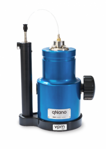
I’m a little unclear as to what qNano actually is. I did find this description on the qNano product page,

qNano uses unique nanopore-based detection to enable the physical properties of a wide range of particle types to be measured with unsurpassed accuracy.

Detailed Multi-Parameter Analysis.

Particle-by-particle measurement through qNano enables detailed determination of:

Nanopore-based detection allows thousands of particles to be measured individually, providing far greater detail and accuracy than light-based techniques.

Applications & Particle Types

A wide range of biological and synthetic particle types, spanning 50 nm – 10 μm, can be measured, across a broad range of research fields.

qNano Package

qNano is sold as a full system ready for use including the base instrument, variable pressure module, fluid cell and a starter kit of nanopores, buffer solution and standard particle sets.

Here’s what the product looks like,

qNano (from the Izon website)

As for what this all might mean to those of us who exist at the macroscale (from the Izon press release),

Izon Science will continue to work with Dr Platt at Loughborough, and with University College Dublin and various customers to develop a series of diagnostic kits that can be used with the qNano to identify and measure biomolecules, viruses, and microvesicles.“This is a real milestone for Izon’s technology as being able to measure biomolecules down to these extremely low levels opens up new bio-analysis options for researchers. 10 femtomolar was achieved, which is the equivalent dilution to 1 gram in 3.3 billion litres, or 1 gram in 1300 Olympic sized swimming pools,” says Hans van der Voorn, Executive Chairman of Izon Science.

For those interested in finding out about nanopores, these were mentioned in my July 18, 2012 posting while aptamers were discussed in my interview (Oct. 25, 2011 posting) with Dr. Maria DeRosa who researches them in her Carleton University laboratory (Ottawa, Canada).

Portable x-ray machine

It’s all about the adhesive tape according to the researchers at Tribogenics. Yes, they can create x-rays by unrolling scotch tape in a vacuum. Neal Ungerleider’s Dec. 8, 2011 article for Fast Company,

Tribogenics’ products rely on a counterintuitive discovery: X-rays are generated when unrolling Scotch tape in a vacuum. In a Nature article, UCLA researchers Carlos Camara, Juan Escobar, Jonathan Hird, and Seth Putterman detailed how Scotch tape can generate surprisingly large amounts of X-rays thanks to visible radiation generated by static electricity between two contacting surfaces. The research encountered challenges thanks to the fact that Scotch tape and generic brand adhesive tapes generated slightly different energy signatures; the composition of Scotch tape adhesive is a closely guarded 3M trade secret. …

Fox [Dale Fox, Tribogenics’ Chief Scientist] told Fast Company that “every other X-ray source in the world uses a high-voltage transformer connected to a vacuum tube. In contrast, we’ve harnessed the power of the immense voltages in static electricity to create tiny, low-cost, battery-operated X-ray sources for the first time in history. It’s like the jump the electronics industry took when it moved from vacuum tubes to transistors.” According to Fox, Tribogenics has already developed X-ray energy sources the size of a USB memory stick. While Tribogenics representatives declined to discuss pricing for upcoming products, the firm “very comfortably” promised that the cost would be less than 10% than that of any existing X-ray technology.

This technology can be traced back to DARPA (Defense Advanced Research Projects Agency) in 2007 when the agency funded the company’s first research, according to the company website. There have been other military funds as well, the US Army Telemedicine and Advanced Research Center in 2010.

The company describes itself this way (from the home page),

Tribogenics patented technology enables portable, compact x-ray solutions for applications in precious metal, mining, military, medical imaging, security and other industries. By miniaturizing X-ray sources and eliminating the need for high voltage, we can create products and solutions unattainable using existing X-ray technology. Tribogenics revolutionary X-ray solution emerged from DARPA and TATRC-funded initiatives at UCLA and was developed by prominent scientists.

Ungerleider notes that the company has not launched any commercial products yet but this one sure looks interesting,

… ultra-portable X-ray machines show the greatest potential for becoming a disruptive medical technology. Tribogenics’ methods have revolutionary ramifications for catheterized radiation therapy, which currently poses significant radiation risks for patients, doctors, and nurses. According to Fox, the company’s products eliminate the need for radioactive isotopes.

If you are interested in this technology, I would suggest reading Ungerleider’s article for additional details.