Tag Archives: dragonflies

Spiky materials that can pop bacteria?

Bacteria interacting with four different topographies Courtesy: Imperial College London

A February 9, 2022 news item on phys.org describes some bioinspired research that could help cut down on the use of disinfectants,

Researchers have created intricately patterned materials that mimic antimicrobial, adhesive and drag reducing properties found in natural surfaces.

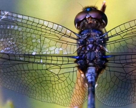
The team from Imperial College London found inspiration in the wavy and spiky surfaces found in insects, including on cicada and dragonfly wings, which ward off bacteria.

They hope the new materials could be used to create self-disinfecting surfaces and offer an alternative to chemically functionalized surfaces and cleaners, which can promote the growth of antibiotic-resistant bacteria.

A February 9, 2022 Imperial College London (ICL) press release by Caroline Brogan, which originated the news item, describes the work in more technical detail,

The tiny waves, which overlap at defined angles to create spikes and ripples, could also help to reduce drag on marine transport by mimicking shark skin, and to enhance the vibrancy of color without needing pigment, by mimicking insects.

Senior author Professor Joao Cabral, of Imperial’s Department of Chemical Engineering, said, “It’s inspiring to see in miniscule detail how the wings and skins of animals help them master their environments. Animals evolved wavy surfaces to kill bacteria, enhance color, and reduce drag while moving through water. We’re borrowing these natural tricks for the very same purposes, using a trick reminiscent of a Fourier wave superposition.”
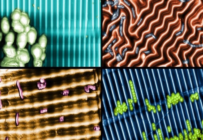
Spiky structures

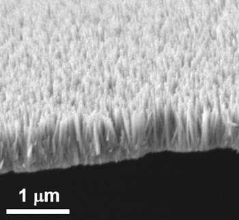
Researchers created the new materials by stretching and compressing a thin, soft, sustainable plastic resembling clingfilm to create three-dimensional nano- and microscale wavy patterns, compatible with sustainable and biodegradable polymers. 

The spiky structure was inspired by the way insects and fish have evolved to interact with their environments. The corrugated ripple effect is seen in the wings of cicadas and dragonflies, whose surfaces are made of tiny spikes which pop bacterial cells to keep the insects clean.  

The structure could also be applied to ships to reduce drag and boost efficiency – an application inspired by shark skin, which contains nanoscale horizontal ridges to reduce friction and drag.

Another application is in producing vibrant colours like those seen in the wings of morpho blue butterflies, whose cells are arranged to reflect and bend light into a brilliant blue without using pigment. Known as structural colour, other examples include the blue in peacock feathers, the shells of iridescent beetles, and blue human eyes.

Scaling up waves

To conduct the research, which is published in Physical Review Letters, the researchers studied specimens of cicadas and dragonflies from the Natural History Museum, and sedimentary deposits and rock formations documented by Trinity College Dublin.

They discovered that they could recreate these naturally occurring surface waves by stretching and then relaxing thin polymer skins in precise directions at the nanoscale.

While complex patterns can be fabricated by lithography and other methods, for instance in silicon microchip production, these are generally prohibitively expensive to use over large areas. This new technique, on the other hand, is ready to be scaled up relatively inexpensively if confirmed to be effective and robust. 

Potential applications include self-disinfecting surfaces in hospitals, schools, public transport, and food manufacturing. They could even help keep medical implants clean, which is important as these can host networks of bacterial matter known as biofilms that are notoriously difficult to kill. 

Naturally occurring wave patterns are also seen in the wrinkling of the human brain and fingertips as well as the ripples in sand beds. First author Dr Luca Pellegrino from the Department of Chemical Engineering, said: “The idea is compelling because it is simple: by mimicking the surface waves found in nature, we can create a palette of patterns with important applications. Through this work we can also learn more about the possible origins of these natural forms – a field called morphogenesis.” 

he next focus for the team is to test the effectiveness and robustness of the material in real-world settings, like on bus surfaces. The researchers hope it can contribute to solutions to surface cleanliness that are not reliant on chemical cleaners. To this end, they have been awarded a €5.4million EU HORIZON grant with collaborators ranging from geneticists at KU Leuven to a bus manufacturer to develop sustainable and robust antimicrobial surfaces for high traffic contexts. 

Here’s a link (the press release also has a link) to and a citation for the paper,

Ripple Patterns Spontaneously Emerge through Sequential Wrinkling Interference in Polymer Bilayers by Luca Pellegrino, Annabelle Tan, and João T. Cabral. Phys. Rev. Lett. 128, 058001 Vol. 128, Issue 5 — 4 February 2022 Published online 2 February 2022

This paper is behind a paywall.

This work reminds me of Sharklet, a company that was going to produce materials that mimicked the structure of sharkskin. Apparently, sharks have nanostructures on their skin which prevents bacteria and more from finding a home there.

An electronics-free, soft robotic dragonfly

From the description on YouTube,

With the ability to sense changes in pH, temperature and oil, this completely soft, electronics-free robot dubbed “DraBot” could be the prototype for future environmental sentinels. …

Music: Joneve by Mello C from the Free Music Archive

A favourite motif in the Art Nouveau movement (more about that later in the post), dragonflies or a facsimile thereof feature in March 25, 2021 Duke University news release (also on EurekAlert) by Ken Kingery,

Engineers at Duke University have developed an electronics-free, entirely soft robot shaped like a dragonfly that can skim across water and react to environmental conditions such as pH, temperature or the presence of oil. The proof-of-principle demonstration could be the precursor to more advanced, autonomous, long-range environmental sentinels for monitoring a wide range of potential telltale signs of problems.

The soft robot is described online March 25 [2021] in the journal Advanced Intelligent Systems.

Soft robots are a growing trend in the industry due to their versatility. Soft parts can handle delicate objects such as biological tissues that metal or ceramic components would damage. Soft bodies can help robots float or squeeze into tight spaces where rigid frames would get stuck.

The expanding field was on the mind of Shyni Varghese, professor of biomedical engineering, mechanical engineering and materials science, and orthopaedic surgery at Duke, when inspiration struck.

“I got an email from Shyni from the airport saying she had an idea for a soft robot that uses a self-healing hydrogel that her group has invented in the past to react and move autonomously,” said Vardhman Kumar, a PhD student in Varghese’s laboratory and first author of the paper. “But that was the extent of the email, and I didn’t hear from her again for days. So the idea sort of sat in limbo for a little while until I had enough free time to pursue it, and Shyni said to go for it.”

In 2012, Varghese and her laboratory created a self-healing hydrogel that reacts to changes in pH in a matter of seconds. Whether it be a crack in the hydrogel or two adjoining pieces “painted” with it, a change in acidity causes the hydrogel to form new bonds, which are completely reversible when the pH returns to its original levels.

Varghese’s hastily written idea was to find a way to use this hydrogel on a soft robot that could travel across water and indicate places where the pH changes. Along with a few other innovations to signal changes in its surroundings, she figured her lab could design such a robot as a sort of autonomous environmental sensor.

With the help of Ung Hyun Ko, a postdoctoral fellow also in Varghese’s laboratory, Kumar began designing a soft robot based on a fly. After several iterations, the pair settled on the shape of a dragonfly engineered with a network of interior microchannels that allow it to be controlled with air pressure.

They created the body–about 2.25 inches long with a 1.4-inch wingspan–by pouring silicon into an aluminum mold and baking it. The team used soft lithography to create interior channels and connected with flexible silicon tubing.

DraBot was born.

“Getting DraBot to respond to air pressure controls over long distances using only self-actuators without any electronics was difficult,” said Ko. “That was definitely the most challenging part.”

DraBot works by controlling the air pressure coming into its wings. Microchannels carry the air into the front wings, where it escapes through a series of holes pointed directly into the back wings. If both back wings are down, the airflow is blocked, and DraBot goes nowhere. But if both wings are up, DraBot goes forward.

To add an element of control, the team also designed balloon actuators under each of the back wings close to DraBot’s body. When inflated, the balloons cause the wings to curl upward. By changing which wings are up or down, the researchers tell DraBot where to go.

“We were happy when we were able to control DraBot, but it’s based on living things,” said Kumar. “And living things don’t just move around on their own, they react to their environment.”

That’s where self-healing hydrogel comes in. By painting one set of wings with the hydrogel, the researchers were able to make DraBot responsive to changes in the surrounding water’s pH. If the water becomes acidic, one side’s front wing fuses with the back wing. Instead of traveling in a straight line as instructed, the imbalance causes the robot to spin in a circle. Once the pH returns to a normal level, the hydrogel “un-heals,” the fused wings separate, and DraBot once again becomes fully responsive to commands.

To beef up its environmental awareness, the researchers also leveraged the sponges under the wings and doped the wings with temperature-responsive materials. When DraBot skims over water with oil floating on the surface, the sponges will soak it up and change color to the corresponding color of oil. And when the water becomes overly warm, DraBot’s wings change from red to yellow.

The researchers believe these types of measurements could play an important part in an environmental robotic sensor in the future. Responsiveness to pH can detect freshwater acidification, which is a serious environmental problem affecting several geologically-sensitive regions. The ability to soak up oils makes such long-distance skimming robots an ideal candidate for early detection of oil spills. Changing colors due to temperatures could help spot signs of red tide and the bleaching of coral reefs, which leads to decline in the population of aquatic life.

The team also sees many ways that they could improve on their proof-of-concept. Wireless cameras or solid-state sensors could enhance the capabilities of DraBot. And creating a form of onboard propellant would help similar bots break free of their tubing.

“Instead of using air pressure to control the wings, I could envision using some sort of synthetic biology that generates energy,” said Varghese. “That’s a totally different field than I work in, so we’ll have to have a conversation with some potential collaborators to see what’s possible. But that’s part of the fun of working on an interdisciplinary project like this.”

Here’s a link to and a citation for the paper,

Microengineered Materials with Self‐Healing Features for Soft Robotics by Vardhman Kumar, Ung Hyun Ko, Yilong Zhou, Jiaul Hoque, Gaurav Arya, Shyni Varghese. Advanced Intelligent Systems DOI: https://doi.org/10.1002/aisy.202100005 First published: 25 March 2021

This paper is open access.

The earlier reference to Art Nouveau gives me an excuse to introduce this March 7, 2020 (?) essay by Bex Simon (artist blacksmith) on her eponymous website.

Dragonflies, in particular, are a very poplar subject matter in the Art Nouveau movement. Art Nouveau, with its wonderful flowing lines and hidden fantasies, is full of symbolism.  The movement was a response to the profound social changes and industrialization of every day life and the style of the moment was, in part, inspired by Japanese art.

Simon features examples of Art Nouveau dragonfly art along with examples of her own take on the subject. She also has this,

[downloaded from https://www.bexsimon.com/dragonflies-and-butterflies-in-art-nouveau/]

This is a closeup of a real dragonfly as seen on Simon’s website. If you have an interest, reading her March 7, 2020 (?) essay and gazing at the images won’t take much time.

Killing bacteria on contact with dragonfly-inspired nanocoating

Scientists in Singapore were inspired by dragonflies and cicadas according to a March 28, 2018 news item on Nanowerk (Note: A link has been removed),

Studies have shown that the wings of dragonflies and cicadas prevent bacterial growth due to their natural structure. The surfaces of their wings are covered in nanopillars making them look like a bed of nails. When bacteria come into contact with these surfaces, their cell membranes get ripped apart immediately and they are killed. This inspired researchers from the Institute of Bioengineering and Nanotechnology (IBN) of A*STAR to invent an anti-bacterial nano coating for disinfecting frequently touched surfaces such as door handles, tables and lift buttons.

This technology will prove particularly useful in creating bacteria-free surfaces in places like hospitals and clinics, where sterilization is important to help control the spread of infections. Their new research was recently published in the journal Small (“ZnO Nanopillar Coated Surfaces with Substrate-Dependent Superbactericidal Property”)

Image 1: Zinc oxide nanopillars that looked like a bed of nails can kill a broad range of germs when used as a coating on frequently-touched surfaces. Courtesy: A*STAR

A March 28, 2018 Agency for Science Technology and Research (A*STAR) press release, which originated the news item, describes the work further,

80% of common infections are spread by hands, according to the B.C. [province of Canada] Centre for Disease Control1. Disinfecting commonly touched surfaces helps to reduce the spread of harmful germs by our hands, but would require manual and repeated disinfection because germs grow rapidly. Current disinfectants may also contain chemicals like triclosan which are not recognized as safe and effective 2, and may lead to bacterial resistance and environmental contamination if used extensively.

“There is an urgent need for a better way to disinfect surfaces without causing bacterial resistance or harm to the environment. This will help us to prevent the transmission of infectious diseases from contact with surfaces,” said IBN Executive Director Professor Jackie Y. Ying.

To tackle this problem, a team of researchers led by IBN Group Leader Dr Yugen Zhang created a novel nano coating that can spontaneously kill bacteria upon contact. Inspired by studies on dragonflies and cicadas, the IBN scientists grew nanopilllars of zinc oxide, a compound known for its anti-bacterial and non-toxic properties. The zinc oxide nanopillars can kill a broad range of germs like E. coli and S. aureus that are commonly transmitted from surface contact.

Tests on ceramic, glass, titanium and zinc surfaces showed that the coating effectively killed up to 99.9% of germs found on the surfaces. As the bacteria are killed mechanically rather than chemically, the use of the nano coating would not contribute to environmental pollution. Also, the bacteria will not be able to develop resistance as they are completely destroyed when their cell walls are pierced by the nanopillars upon contact.

Further studies revealed that the nano coating demonstrated the best bacteria killing power when it is applied on zinc surfaces, compared with other surfaces. This is because the zinc oxide nanopillars catalyzed the release of superoxides (or reactive oxygen species), which could even kill nearby free floating bacteria that were not in direct contact with the surface. This super bacteria killing power from the combination of nanopillars and zinc broadens the scope of applications of the coating beyond hard surfaces.

Subsequently, the researchers studied the effect of placing a piece of zinc that had been coated with zinc oxide nanopillars into water containing E. coli. All the bacteria were killed, suggesting that this material could potentially be used for water purification.

Dr Zhang said, “Our nano coating is designed to disinfect surfaces in a novel yet practical way. This study demonstrated that our coating can effectively kill germs on different types of surfaces, and also in water. We were also able to achieve super bacteria killing power when the coating was used on zinc surfaces because of its dual mechanism of action. We hope to use this technology to create bacteria-free surfaces in a safe, inexpensive and effective manner, especially in places where germs tend to accumulate.”

IBN has recently received a grant from the National Research Foundation, Prime Minister’s Office, Singapore, under its Competitive Research Programme to further develop this coating technology in collaboration with Tan Tock Seng Hospital for commercial application over the next 5 years.

1 B.C. Centre for Disease Control

2 U.S. Food & Drug Administration

(I wasn’t expecting to see a reference to my home province [BC Centre for Disease Control].) Back to the usual, here’s a link to and a citation for the paper,

ZnO Nanopillar Coated Surfaces with Substrate‐Dependent Superbactericidal Property by Guangshun Yi, Yuan Yuan, Xiukai Li, Yugen Zhang. Small https://doi.org/10.1002/smll.201703159 First published: 22 February 2018

This paper is behind a paywall.

One final comment, this research reminds me of research into simulating shark skin because that too has bacteria-killing nanostructures. My latest about the sharkskin research is a Sept, 18, 2014 posting.

Artists classified the animal kingdom?

Where taxonomy and biology are concerned, my knowledge begins and end with Carl Linnaeus, the Swedish scientist who ushered in modern taxonomy. It was with some surprise that I find out artists also helped develop the field. From a June 21, 2016 news item on ScienceDaily,

In the sixteenth and seventeenth centuries artists were fascinated by how the animal kingdom was classified. They were in some instances ahead of natural historians.

This is one of the findings of art historian Marrigje Rikken. She will defend her PhD on 23 June [2016] on animal images in visual art. In recent years she has studied how images of animals between 1550 and 1630 became an art genre in themselves. ‘The close relationship between science and art at that time was remarkable,’ Rikken comments. ‘Artists tried to bring some order to the animal kingdom, just as biologists did.’

A June 21, 2016 Universiteit Leiden (Leiden University, Netherlands) press release, which originated the news item, expands on the theme,

In some cases the artists were ahead of their times. They became interested in insects, for example, before they attracted the attention of natural historians. It was artist Joris Hoefnagel who in 1575 made the first miniatures featuring beetles, butterflies and dragonflies, indicating how they were related to one another. In his four albums Hoefnagel divided the animal species according to the elements of fire, water, air and earth, but within these classifications he grouped animals on the basis of shared characteristics.

Courtesy: Universiteit Leiden

Beetles, butterflies, and dragonflies by Joris Hoefnagel. Courtesy: Universiteit Leiden

The press release goes on,

Other illustrators, print-makers and painters tried to bring some cohesion to the animal kingdom.  Some of them used an alphabetical system but artist Marcus Gheeraerts  published a print as early as 1583 [visible below, Ed.] in which grouped even-toed ungulates together. The giraffe and sheep – both visible on Gheeraerts’ print – belong to this species of animals. This doesn’t apply to all Gheeraerts’ animals. The mythical unicorn, which was featured by Gheeraerts, no longer appears in contemporary biology books.

Wealthy courtiers

According to Rikken, the so-called menageries played an important role historically in how animals were represented. These forerunners of today’s zoos were popular in the sixteenth and seventeenth centuries particularly among wealthy rulers and courtiers. Unfamiliar exotic animals regularly arrived that were immediately committed to paper by artists. Rikken: ‘The toucan, for example, was immortalised in 1615 by Jan Brueghel the Elder, court painter in Brussels.’  [See the main image, Ed.].’

In the flesh

Rikken also discovered that the number of animals featured in a work gradually increased. ‘Artists from the 1570s generally included one or just a few animals per work. With the arrival of print series a decade later, each illustration tended to include more and more animals. This trend reached its peak in the lavish paintings produced around 1600.’ These paintings are also much more varied than the drawings and prints. Illustrators and print-makers often blindly copied one another’s motifs, even showing the animals in an identical pose. Artists had no hesitation in including the same animal in different positions. Rikken: ‘This allowed them to show that they had observed the animal in the flesh.’

Even-toed ungulates by Marcus Gheeraerts. Courtesy: Leiden Universiteit

Even-toed ungulates by Marcus Gheeraerts. Courtesy: Leiden Universiteit

Yet more proof or, at least, a very strong suggestion that art and science are tightly linked.

Dragonfly and locust rubber

There’s a protein in some insects such as dragonflies, mosquitoes (!) and locusts which is superior to synthetic rubber according to a July 30, 2013 news release from the American Chemical Society (ACS) [also on EurekAlert],

Kristi Kiick and colleagues explain that scientists discovered resilin a half-century ago in the wing hinges of locusts and elastic tendons of dragonflies. The extraordinary natural protein tops the best synthetic rubbers. Resilin can stretch to three times its original length, for instance, and then spring back to its initial shape without losing its elasticity, despite repeated stretching and relaxing cycles. That’s a crucial trait for insects that must flap or jump millions of times over their lifetimes. Scientists first synthesized resilin in 2005 and have been striving to harness its properties in medicine.

Kiick’s team describes how their own research and experiments by other scientists are making major strides toward practical applications of resilin. Scientists have modified resilin with gold nanoparticles for possible use in diagnostics, engineered mosquito-based resin to act like human cartilage and developed a hybrid material for cardiovascular applications. “This increasing amount of knowledge gained from studies on natural resilin and resilin-like polypeptides continues to inspire new designs and applications of recombinant resilin-based biopolymers in biomedical and biotechnological applications,” the scientists state.

Illustrating 'insect rubber' [downloaded from http://pubs.acs.org/doi/full/10.1021/mz4002194]

Illustrating ‘insect rubber’ [downloaded from http://pubs.acs.org/doi/full/10.1021/mz4002194]

Here’s a link to and a citation for the researchers’ biomimicry paper published by ACS Macro Letters,

Resilin-Based Materials for Biomedical Applications by Linqing Li and Kristi L. Kiick. ACS Macro Lett., 2013, 2, pp 635–640 DOI: 10.1021/mz4002194 Publication Date (Web): July 11, 2013
Copyright © 2013 American Chemical Society

This paper is open access.

Seeing things from a bug’s perspective—a new type of digital camera

The new digital cameras exploit large arrays of tiny focusing lenses and miniaturized detectors in hemispherical layouts, just like eyes found in arthropods

The new digital cameras exploit large arrays of tiny focusing lenses and miniaturized detectors in hemispherical layouts, just like eyes found in arthropods

A May 1, 2013 news item on Nanowerk provides some details about a new ‘bug-eyed’ digital camera,

An interdisciplinary team of researchers has created the first digital cameras with designs that mimic those of ocular systems found in dragonflies, bees, praying mantises and other insects. This class of technology offers exceptionally wide-angle fields of view, with low aberrations, high acuity to motion, and nearly infinite depth of field.

Taking cues from Mother Nature, the cameras exploit large arrays of tiny focusing lenses and miniaturized detectors in hemispherical layouts, just like eyes found in arthropods. The devices combine soft, rubbery optics with high performance silicon electronics and detectors, using ideas first established in research on skin and brain monitoring systems by John A. Rogers, a Swanlund Chair Professor at the University of Illinois at Urbana-Champaign, and his collaborators.

The May 1, 2013 University of Illinois news release by John Kubetz, which originated the news item, describes the special properties of an insect eye and how the camera mimics those properties,

Eyes in arthropods use compound designs, in which arrays of smaller eyes act together to provide image perception. Each small eye, known as an ommatidium, consists of a corneal lens, a crystalline cone, and a light sensitive organ at the base. The entire system is configured to provide exceptional properties in imaging, many of which lie beyond the reach of existing man-made cameras.

The researchers developed new ideas in materials and fabrication strategies allowing construction of artificial ommatidia in large, interconnected arrays in hemispherical layouts. Building such systems represents a daunting task, as all established camera technologies rely on bulk glass lenses and detectors constructed on the planar surfaces of silicon wafers which cannot be bent or flexed, much less formed into a hemispherical shape.

“A critical feature of our fly’s eye cameras is that they incorporate integrated microlenses, photodetectors, and electronics on hemispherically curved surfaces,” said Jianliang Xiao, an assistant professor of mechanical engineering at University of Colorado Boulder and coauthor of the study. “To realize this outcome, we used soft, rubbery optics bonded to detectors/electronics in mesh layouts that can be stretched and deformed, reversibly and without damage.”

On a more technical note, from the news release,

The fabrication starts with electronics, detectors and lens arrays formed on flat surfaces using advanced techniques adapted from the semiconductor industry, said Xiao [Jianliang Xiao, an assistant professor of mechanical engineering at University of Colorado Boulder and coauthor of the study], who began working on the project as a postdoctoral researcher in Rogers’ lab at Illinois. The lens sheet—made from a polymer material similar to a contact lens—and the electronics/detectors are then aligned and bonded together. Pneumatic pressure deforms the resulting system into the desired hemispherical shape, in a process much like blowing up a balloon, but with precision engineering control.

The individual electronic detectors and microlenses are coupled together to avoid any relative motion during this deformation process. Here, the spaces between these artificial ommatidia can stretch to allow transformation in geometry from planar to hemispherical. The electrical interconnections are thin, and narrow, in filamentary serpentine shapes; they deform as tiny springs during the stretching process.

According to the researchers, each microlens produces a small image of an object with a form dictated by the parameters of the lens and the viewing angle. An individual detector responds only if a portion of the image formed by the associated microlens overlaps the active area. The detectors stimulated in this way produce a sampled image of the object that can then be reconstructed using models of the optics.

Katherine Bourzac in her May 1, 2013 article for Nature magazine provides some additional insight and a perspective (intentional wordplay) from a researcher who has an idea of how he might like to integrate this new type of camera into his own work,

Insect eyes are made up of hundreds or even thousands of light-sensing structures called ommatidia. Each contains a lens and a cone that funnels light to a photosensitive organ. The long, thin ommatidia are bunched together to form the hemispherical eye, with each ommatidium pointing in a slightly different direction. This structure gives bugs a wide field of view, with objects in the periphery just as clear as those in the centre of the visual field, and high motion sensitivity. It also allows a large depth of field — objects are in focus whether they’re nearby or at a distance.

“The whole thing [the new digital camera] is stretchy and thin, and we blow it up like a balloon” so that it curves like a compound eye, says Rogers. The current prototype produces black-and-white images only, but Rogers says a colour version could be made with the same design.

With the basic designs in place, Rogers says, his team can now increase the resolution of the camera by incorporating more ommatidia. “We’d like to do a dragonfly, with 20,000 ommatidia,” he says, which will require some miniaturization of the components.

Alexander Borst, who builds miniature flying robots at the Max Planck Institute of Neurobiology in Martinsried, Germany, says that he is eager to integrate the camera into his machines. Insects’ wide field of vision helps them to monitor and stabilize their position during flight; robots with artificial compound eyes might be better fliers, he says.

For interested parties, here’s a link to and a citation for the research paper,

Digital cameras with designs inspired by the arthropod eye by Young Min Song, Yizhu Xie, Viktor Malyarchuk, Jianliang Xiao, Inhwa Jung, Ki-Joong Choi, Zhuangjian Liu, Hyunsung Park, Chaofeng Lu, Rak-Hwan Kim, Rui Li, Kenneth B. Crozier, Yonggang Huang, & John A. Rogers.
Nature 497, 95–99 (02 May 2013) doi:10.1038/nature12083 Published online 01 May 2013

This article is behind a paywall.

I last mentioned John A. Rogers and the University of Illinois in a Feb. 28, 2013 posting about a bendable, stretchable lithium-ion battery.

Dragonflies: beautiful and smart according to Adelaide University (Australia) researchers

[downloaded from http://en.wikipedia.org/wiki/File:Tiffany_dragonfly_hg.jpg] Attribution: pendant Dragonfly - replica from the lamp by Louis Comfort Tiffany (50 cm diameter, 20 cm hight, about 400 glass pieces), Own work, Hannes Grobe 19:33, 20 June 2007 (UTC) Permission Own work, share alike, attribution required (Creative Commons CC-BY-SA-2.5)

[downloaded from http://en.wikipedia.org/wiki/File:Tiffany_dragonfly_hg.jpg] Attribution: pendant Dragonfly – replica from the lamp by Louis Comfort Tiffany (50 cm diameter, 20 cm hight, about 400 glass pieces), Own work, Hannes Grobe 19:33, 20 June 2007 (UTC) Permission Own work, share alike, attribution required (Creative Commons CC-BY-SA-2.5)

Long a subject of inspiration for artists, dragonflies have now been observed to exhibit signs of selective intelligence similar to human selective intelligence. From the Dec. 20, 2012 news release on EurekAlert,

In a discovery that may prove important for cognitive science, our understanding of nature and applications for robot vision, researchers at the University of Adelaide have found evidence that the dragonfly is capable of higher-level thought processes when hunting its prey.

The discovery, to be published online today in the journal Current Biology [link to article which behind a paywall], is the first evidence that an invertebrate animal has brain cells for selective attention, which has so far only been demonstrated in primates.

Here’s how the researchers made the observation (from the EurekAlert news release),

Using a tiny glass probe with a tip that is only 60 nanometers wide – 1500 times smaller than the width of a human hair – the researchers have discovered neuron activity in the dragonfly’s brain that enables this selective attention.

They found that when presented with more than one visual target, the dragonfly brain cell ‘locks on’ to one target and behaves as if the other targets don’t exist.

“Selective attention is fundamental to humans’ ability to select and respond to one sensory stimulus in the presence of distractions,” Dr Wiederman [Dr. Steven Wiederman, University of Adelaide] says.

Wiederman’s research partner suggests this observation has the potential for a number of widespread applications,

“Recent studies reveal similar mechanisms at work in the primate brain, but you might expect it there. We weren’t expecting to find something so sophisticated in lowly insects from a group that’s been around for 325 million years.

“We believe our work will appeal to neuroscientists and engineers alike. For example, it could be used as a model system for robotic vision. Because the insect brain is simple and accessible, future work may allow us to fully understand the underlying network of neurons and copy it into intelligent robots,” he [Associate Professor David O’Carroll, University of Adelaide] says.

You can find more information including pictures and a video in the Dec. 21, 2012 University of Adelaide news release.