Tag Archives: Elisa Riedo

Hot nano-chisel for creating artificial bones?

If ‘chisel’ made you think of sculpting, you are correct. The researchers are alluding to the process of sculpting in their research.
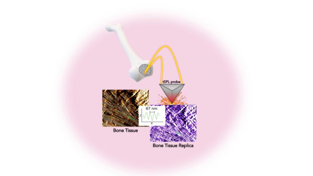
Researchers were able to replicate — with sub-15 nm resolution — bone tissue structure in a biocompatible material using thermal scanning probe lithography. This method opens up unprecedented possibilities for pioneering new stem cell studies and biomedical applications. Courtesy: New York University Tandon School of Engineering

From a February 9, 2021 news item on phys.org (Note: Links have been removed),

A holy grail for orthopedic research is a method for not only creating artificial bone tissue that precisely matches the real thing, but does so in such microscopic detail that it includes tiny structures potentially important for stem cell differentiation, which is key to bone regeneration.

Researchers at the NYU [New York University] Tandon School of Engineering and New York Stem Cell Foundation Research Institute (NYSF) have taken a major step by creating the exact replica of a bone using a system that pairs biothermal imaging with a heated “nano-chisel.” In a study, “Cost and Time Effective Lithography of Reusable Millimeter Size Bone Tissue Replicas with Sub-15 nm Feature Size on a Biocompatible Polymer,” which appears in the journal Advanced Functional Materials, the investigators detail a system allowing them to sculpt, in a biocompatible material, the exact structure of the bone tissue, with features smaller than the size of a single protein—a billion times smaller than a meter. This platform, called, bio-thermal scanning probe lithography (bio-tSPL), takes a “photograph” of the bone tissue, and then uses the photograph to produce a bona-fide replica of it.

The team, led by Elisa Riedo, professor of chemical and biomolecular engineering at NYU Tandon, and Giuseppe Maria de Peppo, a Ralph Lauren Senior Principal Investigator at the NYSF, demonstrated that it is possible to scale up bio-tSPL to produce bone replicas on a size meaningful for biomedical studies and applications, at an affordable cost. These bone replicas support the growth of bone cells derived from a patient’s own stem cells, creating the possibility of pioneering new stem cell applications with broad research and therapeutic potential. This technology could revolutionize drug discovery and result in the development of better orthopedic implants and devices.

A February 8, 2021 NYU Tandon School of Engineering news release (also on EurekAlert but published February 9, 2021), which originated the news item, explains the work in further detail,

In the human body, cells live in specific environments that control their behavior and support tissue regeneration via provision of morphological and chemical signals at the molecular scale. In particular, bone stem cells are embedded in a matrix of fibers — aggregates of collagen molecules, bone proteins, and minerals. The bone hierarchical structure consists of an assembly of micro- and nano- structures, whose complexity has hindered their replication by standard fabrication methods so far.

“tSPL is a powerful nanofabrication method that my lab pioneered a few years ago, and it is at present implemented by using a commercially available instrument, the NanoFrazor,” said Riedo. “However, until today, limitations in terms of throughput and biocompatibility of the materials have prevented its use in biological research. We are very excited to have broken these barriers and to have led tSPL into the realm of biomedical applications.”

Its time- and cost-effectiveness, as well as the cell compatibility and reusability of the bone replicas, make bio-tSPL an affordable platform for the production of surfaces that perfectly reproduce any biological tissue with unprecedented precision.

“I am excited about the precision achieved using bio-tSPL. Bone-mimetic surfaces, such as the one reproduced in this study, create unique possibilities for understanding cell biology and modeling bone diseases, and for developing more advanced drug screening platforms,” said de Peppo. “As a tissue engineer, I am especially excited that this new platform could also help us create more effective orthopedic implants to treat skeletal and maxillofacial defects resulting from injury or disease.”

Here’s a link to and a citation for the paper,

Cost and Time Effective Lithography of Reusable Millimeter Size Bone Tissue Replicas With Sub‐15 nm Feature Size on A Biocompatible Polymer by Xiangyu Liu, Alessandra Zanut, Martina Sladkova‐Faure, Liyuan Xie, Marcus Weck, Xiaorui Zheng, Elisa Riedo, Giuseppe Maria de Peppo. Advanced Functional Materials DOI: https://doi.org/10.1002/adfm.202008662 First published: 05 February 2021

This paper is behind a paywall.
Bulletproof graphene

A December 18, 2017 news item on Nanowerk announces research that demonstrates graphene can be harder than diamonds (Note: A link has been removed),

Imagine a material as flexible and lightweight as foil that becomes stiff and hard enough to stop a bullet on impact. In a newly published paper in Nature Nanotechnology (“Ultrahard carbon film from epitaxial two-layer graphene”), researchers across The City University of New York (CUNY) describe a process for creating diamene: flexible, layered sheets of graphene that temporarily become harder than diamond and impenetrable upon impact.

Scientists at the Advanced Science Research Center (ASRC) at the Graduate Center, CUNY, worked to theorize and test how two layers of graphene — each one-atom thick — could be made to transform into a diamond-like material upon impact at room temperature. The team also found the moment of conversion resulted in a sudden reduction of electric current, suggesting diamene could have interesting electronic and spintronic properties. The new findings will likely have applications in developing wear-resistant protective coatings and ultra-light bullet-proof films.

A December 18, 2017 CUNY news release, which originated the news item, provides a little more detail,

“This is the thinnest film with the stiffness and hardness of diamond ever created,” said Elisa Riedo, professor of physics at the ASRC and the project’s lead researcher. “Previously, when we tested graphite or a single atomic layer of graphene, we would apply pressure and feel a very soft film. But when the graphite film was exactly two-layers thick, all of a sudden we realized that the material under pressure was becoming extremely hard and as stiff, or stiffer, than bulk diamond.”

Angelo Bongiorno, associate professor of chemistry at CUNY College of Staten Island and part of the research team, developed the theory for creating diamene. He and his colleagues used atomistic computer simulations to model potential outcomes when pressurizing two honeycomb layers of graphene aligned in different configurations. Riedo and other team members then used an atomic force microscope to apply localized pressure to two-layer graphene on silicon carbide substrates and found perfect agreement with the calculations. Experiments and theory both show that this graphite-diamond transition does not occur for more than two layers or for a single graphene layer.

“Graphite and diamonds are both made entirely of carbon, but the atoms are arranged differently in each material, giving them distinct properties such as hardness, flexibility and electrical conduction,” Bongiorno said. “Our new technique allows us to manipulate graphite so that it can take on the beneficial properties of a diamond under specific conditions.”

The research team’s successful work opens up possibilities for investigating graphite-to-diamond phase transition in two-dimensional materials, according to the paper. Future research could explore methods for stabilizing the transition and allow for further applications for the resulting materials.

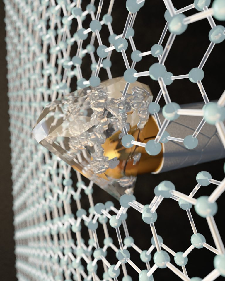
There’s an artist’s representation of a bullet’s impact on graphene,

By applying pressure at the nanoscale with an indenter to two layers of graphene, each one-atom thick, CUNY researchers transformed the honeycombed graphene into a diamond-like material at room temperature. Photo credit: Ella Maru Studio Courtesy: CUNY

Here’s a link to and a citation for the paper,

Ultrahard carbon film from epitaxial two-layer graphene by Yang Gao, Tengfei Cao, Filippo Cellini, Claire Berger, Walter A. de Heer, Erio Tosatti, Elisa Riedo, & Angelo Bongiorno. Nature Nanotechnology (2017) doi:10.1038/s41565-017-0023-9 Published online: 18 December 2017

This paper is behind a paywall.

Mini Lisa made possible by ThermoChemical NanoLithography

One of the world’s most recognizable images has undergone a makeover of sorts. According to an Aug. 6, 2013 news item on Azonano, researchers Georgia institute of Technology (Georgia Tech) in the US, have created a mini Mona Lisa,

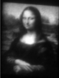
The world’s most famous painting has now been created on the world’s smallest canvas. Researchers at the Georgia Institute of Technology have “painted” the Mona Lisa on a substrate surface approximately 30 microns in width – or one-third the width of a human hair.

The team’s creation, the “Mini Lisa,” demonstrates a technique that could potentially be used to achieve nanomanufacturing of devices because the team was able to vary the surface concentration of molecules on such short-length scales.

The Aug. 5, 2013 Georgia Tech news release, which originated the news item, provides more technical details,

The image was created with an atomic force microscope and a process called ThermoChemical NanoLithography (TCNL). Going pixel by pixel, the Georgia Tech team positioned a heated cantilever at the substrate surface to create a series of confined nanoscale chemical reactions. By varying only the heat at each location, Ph.D. Candidate Keith Carroll controlled the number of new molecules that were created. The greater the heat, the greater the local concentration. More heat produced the lighter shades of gray, as seen on the Mini Lisa’s forehead and hands. Less heat produced the darker shades in her dress and hair seen when the molecular canvas is visualized using fluorescent dye. Each pixel is spaced by 125 nanometers.

“By tuning the temperature, our team manipulated chemical reactions to yield variations in the molecular concentrations on the nanoscale,” said Jennifer Curtis, an associate professor in the School of Physics and the study’s lead author. “The spatial confinement of these reactions provides the precision required to generate complex chemical images like the Mini Lisa.”

Production of chemical concentration gradients and variations on the sub-micrometer scale are difficult to achieve with other techniques, despite a wide range of applications the process could allow. The Georgia Tech TCNL research collaboration, which includes associate professor Elisa Riedo and Regents Professor Seth Marder, produced chemical gradients of amine groups, but expects that the process could be extended for use with other materials.

“We envision TCNL will be capable of patterning gradients of other physical or chemical properties, such as conductivity of graphene,” Curtis said. “This technique should enable a wide range of previously inaccessible experiments and applications in fields as diverse as nanoelectronics, optoelectronics and bioengineering.”

Another advantage, according to Curtis, is that atomic force microscopes are fairly common and the thermal control is relatively straightforward, making the approach accessible to both academic and industrial laboratories.  To facilitate their vision of nano-manufacturing devices with TCNL, the Georgia Tech team has recently integrated nanoarrays of five thermal cantilevers to accelerate the pace of production. Because the technique provides high spatial resolutions at a speed faster than other existing methods, even with a single cantilever, Curtis is hopeful that TCNL will provide the option of nanoscale printing integrated with the fabrication of large quantities of surfaces or everyday materials whose dimensions are more than one billion times larger than the TCNL features themselves.

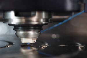
Here’s an image of the AFM and the cantilever used in the TCNL process to create the ‘Mini Lisa’,

Atomic force microscope (AFM) modified with a thermal cantilever. The AFM scanner allows for precise positioning on the nanoscale while the thermal cantilever induces local nanoscale chemical reactions. Courtesy Georgia Tech

Atomic force microscope (AFM) modified with a thermal cantilever. The AFM scanner allows for precise positioning on the nanoscale while the thermal cantilever induces local nanoscale chemical reactions. Courtesy Georgia Tech

Finally, the “Mini Lisa’,

Georgia Tech researchers have created the "Mini Lisa" on a substrate surface approximately 30 microns in width. The image demonstrates a technique that could potentially be used to achieve nano-manufacturing of devices because the team was able to vary the surface concentration of molecules on such short length scales. Courtesy Georgia Tech

Georgia Tech researchers have created the “Mini Lisa” on a substrate surface approximately 30 microns in width. The image demonstrates a technique that could potentially be used to achieve nano-manufacturing of devices because the team was able to vary the surface concentration of molecules on such short length scales. Courtesy Georgia Tech

For those who can’t get enough of the ‘Mini Lisa’ or TCNL, here’s a link to and a citation for the research team’s published paper,

Fabricating Nanoscale Chemical Gradients with ThermoChemical NanoLithography by Keith M. Carroll, Anthony J. Giordano, Debin Wang, Vamsi K. Kodali, Jan Scrimgeour, William P. King, Seth R. Marder, Elisa Riedo, and Jennifer E. Curtis. Langmuir, 2013, 29 (27), pp 8675–8682 DOI: 10.1021/la400996w Publication Date (Web): June 10, 2013
Copyright © 2013 American Chemical Society

This article is behind a paywall.