Tag Archives: European Synchrotron Radiation Facility (ESRF)

Seeing a single nanoparticle catalyst at work

https://www.desy.de/e409/e116959/e119238/media/9833/alloy_kat_np_close-up.jpg
Carbon monoxide oxidises to carbon dioxide on the surface of the nanoparticle. Credit: Science Communication Lab for DESY

An October 1, 2021 news item on ScienceDaily announces research enabling scientists to observe a single nanoparticle at work,

A DESY-led research team has been using high-intensity X-rays to observe a single catalyst nanoparticle at work. The experiment has revealed for the first time how the chemical composition of the surface of an individual nanoparticle changes under reaction conditions, making it more active. The team led by DESY’s Andreas Stierle is presenting its findings in the journal Science Advances. This study marks an important step towards a better understanding of real, industrial catalytic materials.

An October 1, 2021 Deutsches Elektronen-Synchrotron (DESY) press release (also on EurekAlert), which originated the news item, explains why this research is important and provides more technical details,

Catalysts are materials that promote chemical reactions without being consumed themselves. Today, catalysts are used in numerous industrial processes, from fertiliser production to manufacturing plastics. Because of this, catalysts are of huge economic importance. A very well-known example is the catalytic converter installed in the exhaust systems of cars. These contain precious metals such as platinum, rhodium and palladium, which allow highly toxic carbon monoxide (CO) to be converted into carbon dioxide (CO2) and reduce the amount of harmful nitrogen oxides (NOx).

“In spite of their widespread use and great importance, we are still ignorant of many important details of just how the various catalysts work,” explains Stierle, head of the DESY NanoLab. “That’s why we have long wanted to study real catalysts while in operation.” This is not easy, because in order to make the active surface as large as possible, catalysts are typically used in the form of tiny nanoparticles, and the changes that affect their activity occur on their surface.

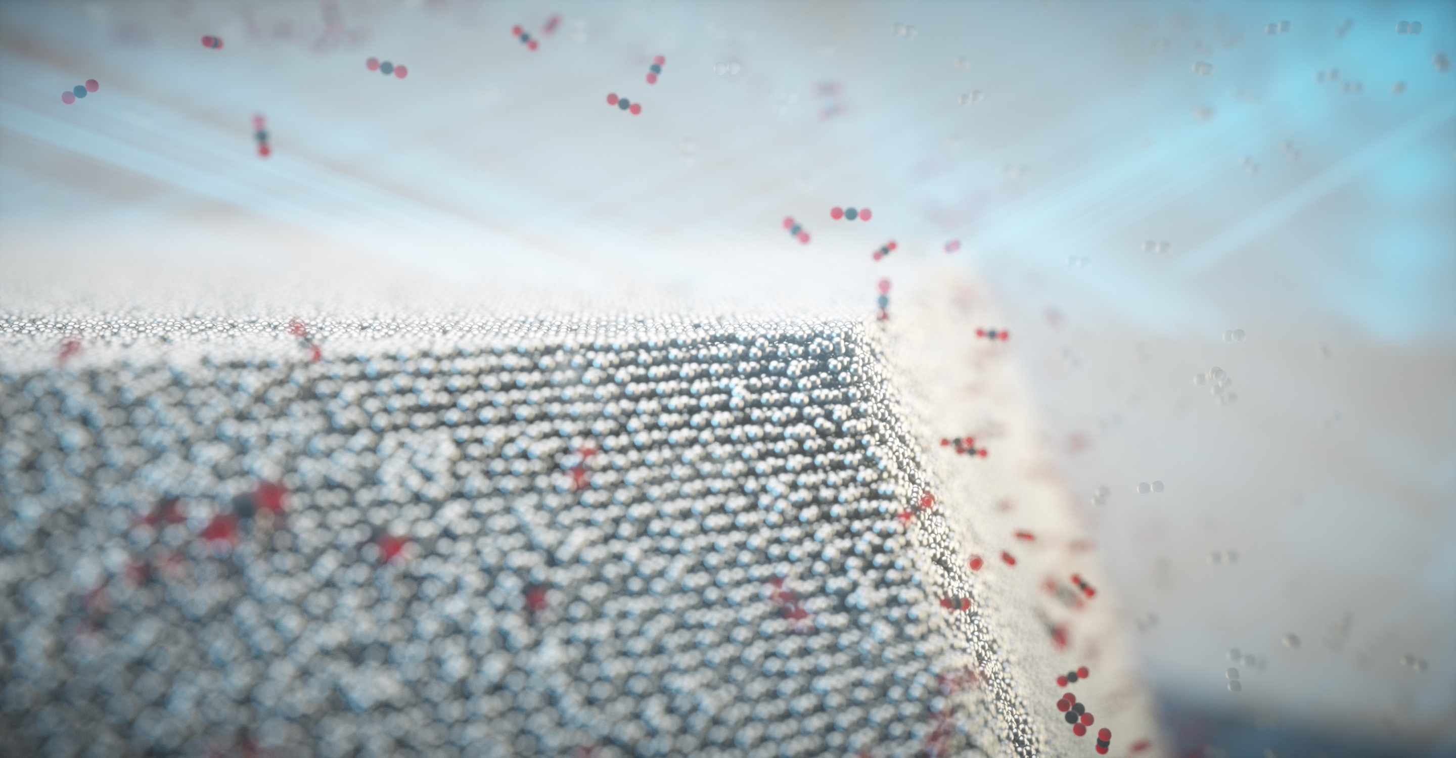
Surface strain relates to chemical composition

In the framework of the EU project Nanoscience Foundries and Fine Analysis (NFFA), the team from DESY NanoLab has developed a technique for labelling individual nanoparticles and thereby identifying them in a sample. “For the study, we grew nanoparticles of a platinum-rhodium alloy on a substrate in the lab and labelled one specific particle,” says co-author Thomas Keller from DESY NanoLab and in charge of the project at DESY. “The diameter of the labelled particle is around 100 nanometres, and it is similar to the particles used in a car’s catalytic converter.” A nanometre is a millionth of a millimetre.

Using X-rays from the European Synchrotron Radiation Facility ESRF in Grenoble, France, the team was not only able to create a detailed image of the nanoparticle; it also measured the mechanical strain within its surface. “The surface strain is related to the surface composition, in particular the ratio of platinum to rhodium atoms,” explains co-author Philipp Plessow from the Karlsruhe Institute of Technology (KIT), whose group computed strain as a function of surface composition. By comparing the observed and computed facet-dependent strain, conclusions can be drawn concerning the chemical composition at the particle surface. The different surfaces of a nanoparticle are called facets, just like the facets of a cut gemstone.

When the nanoparticle is grown, its surface consists mainly of platinum atoms, as this configuration is energetically favoured. However, the scientists studied the shape of the particle and its surface strain under different conditions, including the operating conditions of an automotive catalytic converter. To do this, they heated the particle to around 430 degrees Celsius and allowed carbon monoxide and oxygen molecules to pass over it. “Under these reaction conditions, the rhodium inside the particle becomes mobile and migrates to the surface because it interacts more strongly with oxygen than the platinum,” explains Plessow. This is also predicted by theory.

“As a result, the surface strain and the shape of the particle change,” reports co-author Ivan Vartaniants, from DESY, whose team converted the X-ray diffraction data into three-dimensional spatial images. “A facet-dependent rhodium enrichment takes place, whereby additional corners and edges are formed.” The chemical composition of the surface, and the shape and size of the particles have a significant effect on their function and efficiency. However, scientists are only just beginning to understand exactly how these are connected and how to control the structure and composition of the nanoparticles. The X-rays allow researchers to detect changes of as little as 0.1 in a thousand in the strain, which in this experiment corresponds to a precision of about 0.0003 nanometres (0.3 picometres).

Crucial step towards analysing industrial catalyst materials

“We can now, for the first time, observe the details of the structural changes in such catalyst nanoparticles while in operation,” says Stierle, Lead Scientist at DESY and professor for nanoscience at the University of Hamburg. “This is a major step forward and is helping us to understand an entire class of reactions that make use of alloy nanoparticles.” Scientists at KIT and DESY now want to explore this systematically at the new Collaborative Research Centre 1441, funded by the German Research Foundation (DFG) and entitled “Tracking the Active Sites in Heterogeneous Catalysis for Emission Control (TrackAct)”.

“Our investigation is an important step towards analysing industrial catalytic materials,” Stierle points out. Until now, scientists have had to grow model systems in the laboratory in order to conduct such investigations. “In this study, we have gone to the limit of what can be done. With DESY’s planned X-ray microscope PETRA IV, we will be able to look at ten times smaller individual particles in real catalysts, and under reaction conditions.”
 
DESY is one of the world’s leading particle accelerator centres and investigates the structure and function of matter – from the interaction of tiny elementary particles and the behaviour of novel nanomaterials and vital biomolecules to the great mysteries of the universe. The particle accelerators and detectors that DESY develops and builds at its locations in Hamburg and Zeuthen are unique research tools. They generate the most intense X-ray radiation in the world, accelerate particles to record energies and open up new windows onto the universe. DESY is a member of the Helmholtz Association, Germany’s largest scientific association, and receives its funding from the German Federal Ministry of Education and Research (BMBF) (90 per cent) and the German federal states of Hamburg and Brandenburg (10 per cent).

Here’s a link to and a citation for the paper,

Single alloy nanoparticle x-ray imaging during a catalytic reaction by Young Yong Kim, Thomas F. Keller, Tiago J. Goncalves, Manuel Abuin, Henning Runge, Luca Gelisio, Jerome Carnis, Vedran Vonk, Philipp N. Plessow, Ivan A. Vartaniants, Andreas Stierle. Science Advances • 1 Oct 2021 • Vol 7, Issue 40 • DOI: 10.1126/sciadv.abh0757

This paper is open access.

Mother-of-pearl self-assembles from disorder into perfection

Courtesy: Mother-of-pearl Courtesy: Technische Universitaet (TU) Dresden

Mother-of-pearl (also known as nacre) research has been featured here a few times (links at the end of this post). This time it touches on self-assembly, which is the source of much interest and, on occasion, much concern in the field of nanotechnology.

In any case, the latest mother-of-pearl work comes from the Technische Universität (TU) Dresden (Technical University of Dresden), located in Germany. From a January 4, 2021 news item on phys.org,

In a new study published in Nature Physics, researchers from the B CUBE—Center for Molecular Bioengineering at TU Dresden and European Synchrotron Radiation Facility (ESRF) in Grenoble [Grance] describe, for the first time, that structural defects in self-assembling nacre attract and cancel each other out, eventually leading to a perfect periodic structure.

A January 4, 2021 Technische Universität (TU) Dresden press release (also on EurekAlert), which originated the news item, explains the reason for the ongoing interest in mother-of-pearl and reveals an unexpected turn in the research,

Mollusks build shells to protect their soft tissues from predators. Nacre, also known as the mother of pearl, has an intricate, highly regular structure that makes it an incredibly strong material. Depending on the species, nacres can reach tens of centimeters in length. No matter the size, each nacre is built from materials deposited by a multitude of single cells at multiple different locations at the same time. How exactly this highly periodic and uniform structure emerges from the initial disorder was unknown until now.

Nacre formation starts uncoordinated with the cells depositing the material simultaneously at different locations. Not surprisingly, the early nacre structure is not very regular. At this point, it is full of defects. “In the very beginning, the layered mineral-organic tissue is full of structural faults that propagate through a number of layers like a helix. In fact, they look like a spiral staircase, having either right-handed or left-handed orientation,” says Dr. Igor Zlotnikov, research group leader at the B CUBE – Center for Molecular Bioengineering at TU Dresden. “The role of these defects in forming such a periodic tissue has never been established. On the other hand, the mature nacre is defect-free, with a regular, uniform structure. How could perfection emerge from such disorder?”

The researchers from the Zlotnikov group collaborated with the European Synchrotron Radiation Facility (ESRF) in Grenoble to take a very detailed look at the internal structure of the early and mature nacre. Using synchrotron-based holographic X-ray nano-tomography the researchers could capture the growth of nacre over time. “Nacre is an extremely fine structure, having organic features below 50 nm in size. Beamline ID16A at the ESRF provided us with an unprecedented capability to visualize nacre in three-dimensions,” explains Dr. Zlotnikov. “The combination of electron dense and highly periodical inorganic platelets with delicate and slender organic interfaces makes nacre a challenging structure to image. Cryogenic imaging helped us to obtain the resolving power we needed,” explains Dr. ‘Alexandra] Pacureanu from the X-ray Nanoprobe group at the ESRF.

The analysis of data was quite a challenge. The researchers developed a segmentation algorithm using neural networks and trained it to separate different layers of nacre. In this way, they were able to follow what happens to the structural defects as nacre grows.

The behavior of structural defects in a growing nacre was surprising. Defects of opposite screw direction were attracted to each other from vast distances. The right-handed and left-handed defects moved through the structure, until they met, and cancelled each other out. These events led to a tissue-wide synchronization. Over time, it allowed the structure to develop into a perfectly regular and defect-free.

Periodic structures similar to nacre are produced by many different animal species. The researchers think that the newly discovered mechanism could drive not only the formation of nacre but also other biogenic structures.

Here’s a link to and a citation for the paper,

Dynamics of topological defects and structural synchronization in a forming periodic tissue by Maksim Beliaev, Dana Zöllner, Alexandra Pacureanu, Paul Zaslansky & Igor Zlotnikov. Nature Physics (2021) First published online: 17 September 2020 Published: 04 January 2021

This paper is behind a paywall.

As promised here are the links for One tough mother, imitating mother-of-pearl for stronger ceramics (a March 14, 2014 posting) and Clues as to how mother of pearl is made (a December 15, 2015 posting).

Nanoparticles from tattoo inks circulate through your body

English: Tattoo of Hand of Fatima,. Model: Casini. Date: 4 July 2017, 18:13:41. Source : Own work. Author: Stephencdickson.

For those who like their news in video format, there’s this Canadian Broadcasting Corporation (CBC) news item broadcast on Sep. 11, 2017 (after the commercials),

For those who like text and more detail, scientists at the European Synchrotron Radiation Facility (ESRF) have produced a study of the (at the nanoparticle scale) inks in tattoos. From a Sept. 12, 2017 news item on phys.org,

The elements that make up the ink in tattoos travel inside the body in micro and nanoparticle forms and reach the lymph nodes, according to a study published in Scientific Reports on 12 September [2017] by scientists from Germany and the ESRF, the European Synchrotron, Grenoble (France). It is the first time researchers have found analytical evidence of the transport of organic and inorganic pigments and toxic element impurities as well as in depth characterization of the pigments ex vivo in tattooed tissues. Two ESRF beamlines were crucial in this breakthrough.

A Sept. 12, 2017 ESRF press release (also on EurkeAlert), which originated the news item, explains further,

The reality is that little is known about the potential impurities in the colour mixture applied to the skin. Most tattoo inks contain organic pigments, but also include preservatives and contaminants like nickel, chromium, manganese or cobalt. Besides carbon black, the second most common ingredient used in tattoo inks is titanium dioxide (TiO2), a white pigment usually applied to create certain shades when mixed with colorants. Delayed healing, along with skin elevation and itching, are often associated with white tattoos, and by consequence with the use of TiO2. TiO2 is also commonly used in food additives, sun screens and paints. Scientists from the ESRF, the German Federal Institute for Risk Assessment, Ludwig-Maximilians University, and the Physikalisch-Technische Bundesanstalt have managed to get a very clear picture on the location of titanium dioxide once it gets in the tissue. This work was done on the ESRF beamlines ID21 and ID16B.

drawing tattookinetics.jpg

Translocation of tattoo particles from skin to lymph nodes. Upon injection of tattoo inks, particles can be either passively transported via blood and lymph fluids or phagocytized by immune cells and subsequently deposited in regional lymph nodes. After healing, particles are present in the dermis and in the sinusoids of the draining lymph nodes. Credits: C. Seim.

The hazards that potentially derive from tattoos were, until now, only investigated by chemical analysis of the inks and their degradation products in vitro. “We already knew that pigments from tattoos would travel to the lymph nodes because of visual evidence: the lymph nodes become tinted with the colour of the tattoo. It is the response of the body to clean the site of entrance of the tattoo. What we didn’t know is that they do it in a nano form, which implies that they may not have the same behaviour as the particles at a micro level. And that is the problem: we don’t know how nanoparticles react”, explains Bernhard Hesse, one of the two first authors of the study (together with Ines Schreiver, from the German Federal Institute for Risk Assessment) and ESRF visiting scientist.

titaniumdistribution.jpg

Particle mapping and size distribution of different tattoo pigment elements.  a, d) Ti and the Br containing pigment phthalocyanine green 36 are located next to each other. b, e) Log scale mappings of Ti, Br and Fe in the same areas as displayed in a) and d) reveal primary particle sizes of different pigment species. c, f) Magnifications of the indicated areas in b) and e), respectively. Credits: C. Seim.

X-ray fluorescence measurements on ID21 allowed the team to locate titanium dioxide at the micro and nano range in the skin and the lymphatic environment. They found a broad range of particles with up to several micrometres in size in human skin, but only smaller (nano) particles transported to the lymph nodes. This can lead to the chronic enlargement of the lymph nodes and lifelong exposure. Scientists also used the technique of Fourier transform infrared spectroscopy to assess biomolecular changes in the tissues in the proximity of the tattoo particles.

ESRF16_Tattoo-1low.jpg

Ines Schreiver doing experiments on ID16B with Julie Villanova. Credits: B. Hesse.

Altogether the scientists report strong evidence for both migration and long-term deposition of toxic elements and tattoo pigments as well as for conformational alterations of biomolecules that are sometimes linked to cutaneous adversities upon tattooing.

Then next step for the team is to inspect further samples of patients with adverse effects in their tattoos in order to find links with chemical and structural properties of the pigments used to create these tattoos.

Here’s a link to and  a citation for the paper,

Synchrotron-based ν-XRF mapping and μ-FTIR microscopy enable to look into the fate and effects of tattoo pigments in human skin by Ines Schreiver, Bernhard Hesse, Christian Seim, Hiram Castillo-Michel, Julie Villanova, Peter Laux, Nadine Dreiack, Randolf Penning, Remi Tucoulou, Marine Cotte, & Andreas Luch. Scientific Reports 7, Article number: 11395 (2017) doi:10.1038/s41598-017-11721-z Published online: 12 September 2017

This paper is open access.

X-raying fungus on paper to conserve memory

Civilization is based on memory. Our libraries and archives serve as memories of how things are made, why we use certain materials rather than others, how the human body is put together, what the weather patterns have been, etc. For centuries we have preserved our memories on paper. While this has many advantages, there are some drawbacks including fungus infestations.

A July 21, 2015 news item on ScienceDaily describes how a technique used to x-ray rocks has provided insights into paper and its fungal infestations,

Believe it or not: X-ray works a lot better on rocks than on paper. This has been a problem for conservators trying to save historical books and letters from the ravages of time and fungi. They frankly did not know what they were up against once the telltale signs of vandals such as Dothidales or Pleosporales started to spot the surface of their priceless documents

Now Diwaker Jha, an imaging specialist from Department of Chemistry, University of Copenhagen, has managed to adapt methods developed to investigate interiors of rocks to work on paper too, thus getting a first look at how fungus goes about infesting paper. …

A July 21, 2015 University of Copenhagen press release (also on EurekAlert), which originated the news item, expands on the theme,

This is good news for paper conservators and others who wish to study soft materials with X-ray tomography. “Rocks are easy because they are hard. The X-ray images show a very good contrast between the solid and the pores or channels, which are filled with low density materials such as air or fluids. In this case, however, paper and fungi, both are soft and carbon based, which makes them difficult to distinguish,” says Diwaker.

Diwaker Jha is a PhD student in the NanoGeoScience group, which is a part of the Nano-Science Center at Department of Chemistry. He investigates methods to improve imaging techniques used by chemists and physicists to investigate how fluids move in natural porous materials. At a recent conference, he was presenting an analysis method he developed for X-ray tomography data, for which he was awarded the Presidential Scholar Award by the Microscopy Society of America. And this sparked interest with a conservator in the audience.

Hanna Szczepanowska works as a research conservator with the Smithsonian Institution in the USA. She had been wondering how fungi interact with the paper. Does it sit on the surface, or does it burrow deeper? If they are surface dwellers, it should be easy to just brush them off, but no such luck, says Jha.

“As it turns out, microscopic fungi that infest paper grow very much the same way as mushrooms on a forest floor. However, unlike mushrooms, where the fruiting body emerges out of the soil to the surface, here the fruiting bodies can be embedded within the paper fibres, making it difficult to isolate them. This is not great news for conservators because the prevalent surface cleaning approaches are not adequate,” explains Diwaker Jha.

In working out a way to see into the paper, Jha investigated a 17th century letter on a handmade sheet and a 1920 engraving on machine-made paper. Compared with mushrooms, these fungi are thousands of times smaller, which required an advanced X-ray imaging technique available at the European Synchrotron Radiation Facility (ESRF), Grenoble, France. The technique is very similar to medical tomography (CT scanning) done at hospitals but in Grenoble the X-ray is produced by electrons accelerated to about the speed of light in an 844 meter long circular tube. A handy comparison: “If I were to use medical X-ray tomography to look at an Olympic village, I would be able to make out only the stadium. With the synchrotron based X-ray tomography, I would be able to distinguish individual blades of grass on the field..”

Diwaker hopes that conservators will be able to use the new insight to develop conservation strategies not just for paper artefacts but for combating biodegradation on a host of other types of cultural heritage materials. And that the developed methods can be extended to other studies related to soft matter.

Here’s a citation and a link for the paper,

Morphology and characterization of Dematiaceous fungi on a cellulose paper substrate using synchrotron X-ray microtomography, scanning electron microscopy and confocal laser scanning microscopy in the context of cultural heritage by H. M. Szczepanowska, D. Jha, and Th. G. Mathia. Anal. At. Spectrom. (Journal of Analystical Atomic Spetrometry), 2015,30, 651-657 DOI: 10.1039/C4JA00337C First published online 27 Nov 2014

This paper is behind a paywall. By the way, it is part of something the journal calls a themed collection:  Synchrotron radiation and neutrons in art and archaeology. Clicking on the ‘themed collection’ link will give you a view of the collection, i.e., titles, authors and brief abstracts.