This development could be looked at as a form of synthetic biology without the genetic engineering. From a July 1, 2016 news item on ScienceDaily,
Ideally, injectable or implantable medical devices should not only be small and electrically functional, they should be soft, like the body tissues with which they interact. Scientists from two UChicago labs set out to see if they could design a material with all three of those properties.
The material they came up with, published online June 27, 2016, in Nature Materials, forms the basis of an ingenious light-activated injectable device that could eventually be used to stimulate nerve cells and manipulate the behavior of muscles and organs.
“Most traditional materials for implants are very rigid and bulky, especially if you want to do electrical stimulation,” said Bozhi Tian, an assistant professor in chemistry whose lab collaborated with that of neuroscientist Francisco Bezanilla on the research.
The new material, in contrast, is soft and tiny — particles just a few micrometers in diameter (far less than the width of a human hair) that disperse easily in a saline solution so they can be injected. The particles also degrade naturally inside the body after a few months, so no surgery would be needed to remove them.
Each particle is built of two types of silicon that together form a structure full of nano-scale pores, like a tiny sponge. And like a sponge, it is squishy — a hundred to a thousand times less rigid than the familiar crystalline silicon used in transistors and solar cells. “It is comparable to the rigidity of the collagen fibers in our bodies,” said Yuanwen Jiang, Tian’s graduate student. “So we’re creating a material that matches the rigidity of real tissue.”
The material constitutes half of an electrical device that creates itself spontaneously when one of the silicon particles is injected into a cell culture, or, eventually, a human body. The particle attaches to a cell, making an interface with the cell’s plasma membrane. Those two elements together — cell membrane plus particle — form a unit that generates current when light is shined on the silicon particle.
“You don’t need to inject the entire device; you just need to inject one component,” João L. Carvalho-de-Souza , Bezanilla’s postdoc said. “This single particle connection with the cell membrane allows sufficient generation of current that could be used to stimulate the cell and change its activity. After you achieve your therapeutic goal, the material degrades naturally. And if you want to do therapy again, you do another injection.”
The scientists built the particles using a process they call nano-casting. They fabricate a silicon dioxide mold composed of tiny channels — “nano-wires” — about seven nanometers in diameter (less than 10,000 times smaller than the width of a human hair) connected by much smaller “micro-bridges.” Into the mold they inject silane gas, which fills the pores and channels and decomposes into silicon.
And this is where things get particularly cunning. The scientists exploit the fact the smaller an object is, the more the atoms on its surface dominate its reactions to what is around it. The micro-bridges are minute, so most of their atoms are on the surface. These interact with oxygen that is present in the silicon dioxide mold, creating micro-bridges made of oxidized silicon gleaned from materials at hand. The much larger nano-wires have proportionately fewer surface atoms, are much less interactive, and remain mostly pure silicon. [I have a note regarding ‘micro’ and ‘nano’ later in this posting.]
“This is the beauty of nanoscience,” Jiang said. “It allows you to engineer chemical compositions just by manipulating the size of things.”
Web-like nanostructure
Finally, the mold is dissolved. What remains is a web-like structure of silicon nano-wires connected by micro-bridges of oxidized silicon that can absorb water and help increase the structure’s softness. The pure silicon retains its ability to absorb light.
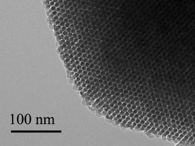
Transmission electron microscopy image shows an ordered nanowire array. The 100-nanometer scale bar is 1,000 times narrower than a hair. Courtesy of Tian Lab
The scientists have added the particles onto neurons in culture in the lab, shone light on the particles, and seen current flow into the neurons which activates the cells. The next step is to see what happens in living animals. They are particularly interested in stimulating nerves in the peripheral nervous system that connect to organs. These nerves are relatively close to the surface of the body, so near-infra-red wavelength light can reach them through the skin.
Tian imagines using the light-activated devices to engineer human tissue and create artificial organs to replace damaged ones. Currently, scientists can make engineered organs with the correct form but not the ideal function.
To get a lab-built organ to function properly, they will need to be able to manipulate individual cells in the engineered tissue. The injectable device would allow a scientist to do that, tweaking an individual cell using a tightly focused beam of light like a mechanic reaching into an engine and turning a single bolt. The possibility of doing this kind of synthetic biology without genetic engineering [emphasis mine] is enticing.
“No one wants their genetics to be altered,” Tian said. “It can be risky. There’s a need for a non-genetic system that can still manipulate cell behavior. This could be that kind of system.”
Tian’s graduate student Yuanwen Jiang did the material development and characterization on the project. The biological part of the collaboration was done in the lab of Francisco Bezanilla, the Lillian Eichelberger Cannon Professor of Biochemistry and Molecular Biology, by postdoc João L. Carvalho-de-Souza. They were, said Tian, the “heroes” of the work.
I was a little puzzled about the use of the word ‘micro’ in a context suggesting it was smaller than something measured at the nanoscale. Dr. Tian very kindly cleared up my confusion with this response in a July 4, 2016 email,
In fact, the definition of ‘micro’ and ’nano’ have been quite ambiguous in literature. For example, microporous materials (e.g., zeolite) usually refer to materials with pore sizes of less than 2 nm — this is defined based on IUPAC [International Union of Pure and Applied Chemistry] definition (http://goldbook.iupac.org/M03853.html). We used ‘micro-bridges’ because they come from the ‘micropores’ in the original template.
Thank you Dr. Tian for that very clear reply and Steve Koppes for forwarding my request to Dr. Tian!
Here’s a link to and a citation for the paper,
Heterogeneous silicon mesostructures for lipid-supported bioelectric interfaces by Yuanwen Jiang, João L. Carvalho-de-Souza, Raymond C. S. Wong, Zhiqiang Luo, Dieter Isheim, Xiaobing Zuo, Alan W. Nicholls, Il Woong Jung, Jiping Yue, Di-Jia Liu, Yucai Wang, Vincent De Andrade, Xianghui Xiao, Luizetta Navrazhnykh, Dara E. Weiss, Xiaoyang Wu, David N. Seidman, Francisco Bezanilla, & Bozhi Tian. Nature Materials (2016) doi:10.1038/nmat4673 Published online 27 June 2016
This paper is behind a paywall.
I gather animal testing will be the next step as they continue to develop this exciting technology. Good luck!
I have two items about implants and brains and an item about being able to exert remote control of the brain, all of which hint at a cyborg future for at least a few of us.
e-Dura, the spinal column, and the brain
The first item concerns some research, at the École Polytechnique de Lausanne (EPFL) which features flexible electronics. From a March 24, 2015 article by Ben Schiller for Fast Company (Note: Links have been removed),
Researchers at the Swiss Federal Institute of Technology, in Lausanne, have developed the e-Dura—a tiny skinlike device that attaches directly to damaged spinal cords. By sending out small electrical pulses, it stimulates the cord as if it were receiving signals from the brain, thus allowing movement.
“The purpose of the neuro-prosthesis is to excite the neurons that are on the spinal cord below the site of the injury and activate them, just like if they were receiving information from the brain,” says Stéphanie Lacour, a professor at the institute.
EPFL scientists have managed to get rats walking on their own again using a combination of electrical and chemical stimulation. But applying this method to humans would require multifunctional implants that could be installed for long periods of time on the spinal cord without causing any tissue damage. This is precisely what the teams of professors Stéphanie Lacour and Grégoire Courtine have developed. Their e-Dura implant is designed specifically for implantation on the surface of the brain or spinal cord. The small device closely imitates the mechanical properties of living tissue, and can simultaneously deliver electric impulses and pharmacological substances. The risks of rejection and/or damage to the spinal cord have been drastically reduced. An article about the implant will appear in early January [2015] in Science Magazine.
So-called “surface implants” have reached a roadblock; they cannot be applied long term to the spinal cord or brain, beneath the nervous system’s protective envelope, otherwise known as the “dura mater,” because when nerve tissues move or stretch, they rub against these rigid devices. After a while, this repeated friction causes inflammation, scar tissue buildup, and rejection.
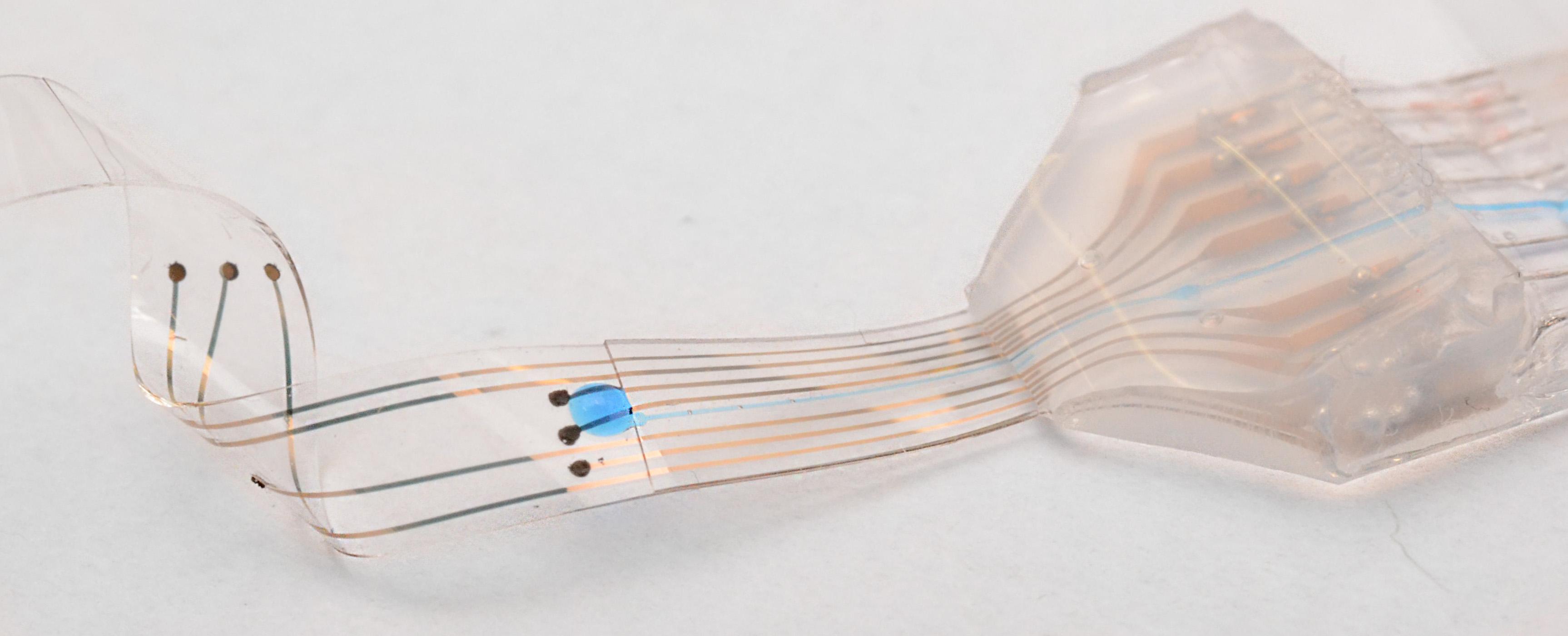
Here’s what the implant looks like,
Courtesy: EPFL
The press release describes how the implant is placed (Note: A link has been removed),
Flexible and stretchy, the implant developed at EPFL is placed beneath the dura mater, directly onto the spinal cord. Its elasticity and its potential for deformation are almost identical to the living tissue surrounding it. This reduces friction and inflammation to a minimum. When implanted into rats, the e-Dura prototype caused neither damage nor rejection, even after two months. More rigid traditional implants would have caused significant nerve tissue damage during this period of time.
The researchers tested the device prototype by applying their rehabilitation protocol — which combines electrical and chemical stimulation – to paralyzed rats. Not only did the implant prove its biocompatibility, but it also did its job perfectly, allowing the rats to regain the ability to walk on their own again after a few weeks of training.
“Our e-Dura implant can remain for a long period of time on the spinal cord or the cortex, precisely because it has the same mechanical properties as the dura mater itself. This opens up new therapeutic possibilities for patients suffering from neurological trauma or disorders, particularly individuals who have become paralyzed following spinal cord injury,” explains Lacour, co-author of the paper, and holder of EPFL’s Bertarelli Chair in Neuroprosthetic Technology.
The press release goes on to describe the engineering achievements,
Developing the e-Dura implant was quite a feat of engineering. As flexible and stretchable as living tissue, it nonetheless includes electronic elements that stimulate the spinal cord at the point of injury. The silicon substrate is covered with cracked gold electric conducting tracks that can be pulled and stretched. The electrodes are made of an innovative composite of silicon and platinum microbeads. They can be deformed in any direction, while still ensuring optimal electrical conductivity. Finally, a fluidic microchannel enables the delivery of pharmacological substances – neurotransmitters in this case – that will reanimate the nerve cells beneath the injured tissue.
The implant can also be used to monitor electrical impulses from the brain in real time. When they did this, the scientists were able to extract with precision the animal’s motor intention before it was translated into movement.
“It’s the first neuronal surface implant designed from the start for long-term application. In order to build it, we had to combine expertise from a considerable number of areas,” explains Courtine, co-author and holder of EPFL’s IRP Chair in Spinal Cord Repair. “These include materials science, electronics, neuroscience, medicine, and algorithm programming. I don’t think there are many places in the world where one finds the level of interdisciplinary cooperation that exists in our Center for Neuroprosthetics.”
For the time being, the e-Dura implant has been primarily tested in cases of spinal cord injury in paralyzed rats. But the potential for applying these surface implants is huge – for example in epilepsy, Parkinson’s disease and pain management. The scientists are planning to move towards clinical trials in humans, and to develop their prototype in preparation for commercialization.
EPFL has provided a video of researcher Stéphanie Lacour describing e-Dura and expressing hopes for its commercialization,
Here’s a link to and a citation for the paper,
Electronic dura mater for long-term multimodal neural interfaces by Ivan R. Minev, Pavel Musienko, Arthur Hirsch, Quentin Barraud, Nikolaus Wenger, Eduardo Martin Moraud, Jérôme Gandar, Marco Capogrosso, Tomislav Milekovic, Léonie Asboth, Rafael Fajardo Torres, Nicolas Vachicouras, Qihan Liu, Natalia Pavlova, Simone Duis, Alexandre Larmagnac, Janos Vörös, Silvestro Micera, Zhigang Suo, Grégoire Courtine, Stéphanie P. Lacour. Science 9 January 2015: Vol. 347 no. 6218 pp. 159-163 DOI: 10.1126/science.1260318
This paper is behind a paywall.
Carbon nanotube fibres could connect to the brain
Researchers at Rice University (Texas, US) are excited about the possibilities that carbon nanotube fibres offer in the field of implantable electronics for the brain. From a March 25, 2015 news item on Nanowerk,
Carbon nanotube fibers invented at Rice University may provide the best way to communicate directly with the brain.
The fibers have proven superior to metal electrodes for deep brain stimulation and to read signals from a neuronal network. Because they provide a two-way connection, they show promise for treating patients with neurological disorders while monitoring the real-time response of neural circuits in areas that control movement, mood and bodily functions.
New experiments at Rice demonstrated the biocompatible fibers are ideal candidates for small, safe electrodes that interact with the brain’s neuronal system, according to the researchers. They could replace much larger electrodes currently used in devices for deep brain stimulation therapies in Parkinson’s disease patients.
They may also advance technologies to restore sensory or motor functions and brain-machine interfaces as well as deep brain stimulation therapies for other neurological disorders, including dystonia and depression, the researchers wrote.
The fibers created by the Rice lab of chemist and chemical engineer Matteo Pasquali consist of bundles of long nanotubes originally intended for aerospace applications where strength, weight and conductivity are paramount.
The individual nanotubes measure only a few nanometers across, but when millions are bundled in a process called wet spinning, they become thread-like fibers about a quarter the width of a human hair.
“We developed these fibers as high-strength, high-conductivity materials,” Pasquali said. “Yet, once we had them in our hand, we realized that they had an unexpected property: They are really soft, much like a thread of silk. Their unique combination of strength, conductivity and softness makes them ideal for interfacing with the electrical function of the human body.”
The simultaneous arrival in 2012 of Caleb Kemere, a Rice assistant professor who brought expertise in animal models of Parkinson’s disease, and lead author Flavia Vitale, a research scientist in Pasquali’s lab with degrees in chemical and biomedical engineering, prompted the investigation.
“The brain is basically the consistency of pudding and doesn’t interact well with stiff metal electrodes,” Kemere said. “The dream is to have electrodes with the same consistency, and that’s why we’re really excited about these flexible carbon nanotube fibers and their long-term biocompatibility.”
Weeks-long tests on cells and then in rats with Parkinson’s symptoms proved the fibers are stable and as efficient as commercial platinum electrodes at only a fraction of the size. The soft fibers caused little inflammation, which helped maintain strong electrical connections to neurons by preventing the body’s defenses from scarring and encapsulating the site of the injury.
The highly conductive carbon nanotube fibers also show much more favorable impedance – the quality of the electrical connection — than state-of-the-art metal electrodes, making for better contact at lower voltages over long periods, Kemere said.
The working end of the fiber is the exposed tip, which is about the width of a neuron. The rest is encased with a three-micron layer of a flexible, biocompatible polymer with excellent insulating properties.
The challenge is in placing the tips. “That’s really just a matter of having a brain atlas, and during the experiment adjusting the electrodes very delicately and putting them into the right place,” said Kemere, whose lab studies ways to connect signal-processing systems and the brain’s memory and cognitive centers.
Doctors who implant deep brain stimulation devices start with a recording probe able to “listen” to neurons that emit characteristic signals depending on their functions, Kemere said. Once a surgeon finds the right spot, the probe is removed and the stimulating electrode gently inserted. Rice carbon nanotube fibers that send and receive signals would simplify implantation, Vitale said.
The fibers could lead to self-regulating therapeutic devices for Parkinson’s and other patients. Current devices include an implant that sends electrical signals to the brain to calm the tremors that afflict Parkinson’s patients.
“But our technology enables the ability to record while stimulating,” Vitale said. “Current electrodes can only stimulate tissue. They’re too big to detect any spiking activity, so basically the clinical devices send continuous pulses regardless of the response of the brain.”
Kemere foresees a closed-loop system that can read neuronal signals and adapt stimulation therapy in real time. He anticipates building a device with many electrodes that can be addressed individually to gain fine control over stimulation and monitoring from a small, implantable device.
“Interestingly, conductivity is not the most important electrical property of the nanotube fibers,” Pasquali said. “These fibers are intrinsically porous and extremely stable, which are both great advantages over metal electrodes for sensing electrochemical signals and maintaining performance over long periods of time.”
The paper is open access provided you register on the website.
Remote control for stimulation of the brain
Mo Costandi, neuroscientist and freelance science writer, has written a March 24, 2015 post for the Guardian science blog network focusing on neuronal remote control,
Two teams of scientists have developed new ways of stimulating neurons with nanoparticles, allowing them to activate brain cells remotely using light or magnetic fields. The new methods are quicker and far less invasive than other hi-tech methods available, so could be more suitable for potential new treatments for human diseases.
Researchers have various methods for manipulating brain cell activity, arguably the most powerful being optogenetics, which enables them to switch specific brain cells on or off with unprecedented precision, and simultaneously record their behaviour, using pulses of light.
This is very useful for probing neural circuits and behaviour, but involves first creating genetically engineered mice with light-sensitive neurons, and then inserting the optical fibres that deliver light into the brain, so there are major technical and ethical barriers to its use in humans.
Nanomedicine could get around this. Francisco Bezanilla of the University of Chicago and his colleagues knew that gold nanoparticles can absorb light and convert it into heat, and several years ago they discovered that infrared light can make neurons fire nervous impulses by heating up their cell membranes.
…
Polina Anikeeva’s team at the Massachusetts Institute of Technology adopted a slightly different approach, using spherical iron oxide particles that give off heat when exposed to an alternating magnetic field.
…
Although still in the experimental stages, research like this may eventually allow for wireless and minimally invasive deep brain stimulation of the human brain. Bezanilla’s group aim to apply their method to develop treatments for macular degeneration and other conditions that kill off light-sensitive cells in the retina. This would involve injecting nanoparticles into the eye so that they bind to other retinal cells, allowing natural light to excite them into firing impulses to the optic nerve.
Costandi’s article is intended for an audience that either understands the science or can deal with the uncertainty of not understanding absolutely everything. Provided you fall into either of those categories, the article is well written and it provides links and citations to the papers for both research teams being featured.
Taken together, the research at EPFL, Rice University, University of Chicago, and Massachusetts Institute of Technology provides a clue as to how much money and intellectual power is being directed at the brain.