Tag Archives: gold nanoparticles

Gold nanoparticles make a new promise: a non-invasive COVID-19 breathalyser

I believe that swab they stick up your nose to test for COVDI-19 is 10 inches long so it seems to me that discomfort or unpleasant are not the words that best describe the testing experience .

Hopefully, no one will have to find inadequate vocabulary for this new COVID-19 testing assuming that future trials are successful and they are able to put the technology into production. From an August 19, 2020 news item on Nanowerk,

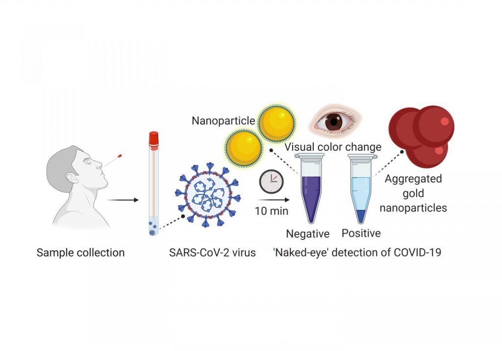
Few people who have undergone nasopharyngeal swabs for coronavirus testing would describe it as a pleasant experience. The procedure involves sticking a long swab up the nose to collect a sample from the back of the nose and throat, which is then analyzed for SARS-CoV-2 RNA [ribonucleic acid] by the reverse-transcription polymerase chain reaction (RT-PCR).

Now, researchers reporting in [American Chemical Society] ACS Nano (“Multiplexed Nanomaterial-Based Sensor Array for Detection of COVID-19 in Exhaled Breath”) have developed a prototype device that non-invasively detected COVID-19 in the exhaled breath of infected patients.

An August 19, 2020 ACS news release (also received via email and on EurekAlert), which originated the news item, provides more technical details,

In addition to being uncomfortable, the current gold standard for COVID-19 testing requires RT-PCR, a time-consuming laboratory procedure. Because of backlogs, obtaining a result can take several days. To reduce transmission and mortality rates, healthcare systems need quick, inexpensive and easy-to-use tests. Hossam Haick, Hu Liu, Yueyin Pan and colleagues wanted to develop a nanomaterial-based sensor that could detect COVID-19 in exhaled breath, similar to a breathalyzer test for alcohol intoxication. Previous studies have shown that viruses and the cells they infect emit volatile organic compounds (VOCs) that can be exhaled in the breath.

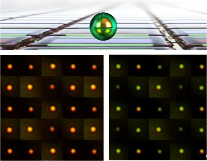
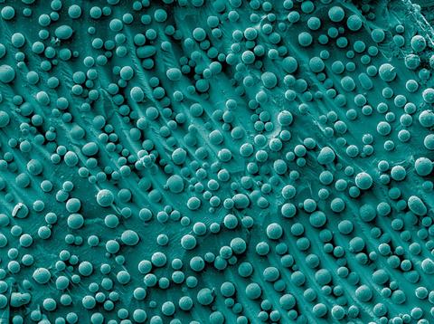
The researchers made an array of gold nanoparticles linked to molecules that are sensitive to various VOCs. When VOCs interact with the molecules on a nanoparticle, the electrical resistance changes. The researchers trained the sensor to detect COVID-19 by using machine learning to compare the pattern of electrical resistance signals obtained from the breath of 49 confirmed COVID-19 patients with those from 58 healthy controls and 33 non-COVID lung infection patients in Wuhan, China. Each study participant blew into the device for 2-3 seconds from a distance of 1¬-2 cm. Once machine learning identified a potential COVID-19 signature, the team tested the accuracy of the device on a subset of participants. In the test set, the device showed 76% accuracy in distinguishing COVID-19 cases from controls and 95% accuracy in discriminating COVID-19 cases from lung infections. The sensor could also distinguish, with 88% accuracy, between sick and recovered COVID-19 patients. Although the test needs to be validated in more patients, it could be useful for screening large populations to determine which individuals need further testing, the researchers say.

The authors acknowledge funding from the Technion-Israel Institute of Technology.

Here’s a link to and a citation for the paper,

Multiplexed Nanomaterial-Based Sensor Array for Detection of COVID-19 in Exhaled Breath by Benjie Shan, Yoav Y Broza, Wenjuan Li, Yong Wang, Sihan Wu, Zhengzheng Liu, Jiong Wang, Shuyu Gui, Lin Wang, Zhihong Zhang, Wei Liu, Shoubing Zhou, Wei Jin, Qianyu Zhang, Dandan Hu, Lin Lin, Qiujun Zhang, Wenyu Li, Jinquan Wang, Hu Liu, Yueyin Pan, and Hossam Haick. ACS Nano 2020, XXXX, XXX, XXX-XXX DOI: https://doi.org/10.1021/acsnano.0c05657 Publication Date:August 18, 2020 Copyright © 2020 American Chemical Society

This paper is behind a paywall.

Chameleon skin (nanomaterial made of gold nanoparticles) for robots

A June 17, 2020 news item on Nanowerk trumpets research into how robots might be able to sport chameleon-like skin one day,

A new film made of gold nanoparticles changes color in response to any type of movement. Its unprecedented qualities could allow robots to mimic chameleons and octopi — among other futuristic applications.

Unlike other materials that try to emulate nature’s color changers, this one can respond to any type of movement, like bending or twisting. Robots coated in it could enter spaces that might be dangerous or impossible for humans, and offer information just based on the way they look.

For example, a camouflaged robot could enter tough-to-access underwater crevices. If the robot changes color, biologists could learn about the pressures facing animals that live in these environments.

Although some other color-changing materials can also respond to motion, this one can be printed and programmed to display different, complex patterns that are difficult to replicate.

This video from the University of California at Riverside researchers shows the material in action (Note: It gets more interesting after the first 20 secs.),

A June 15, 2020 University of California at Riverside (UCR) news release (also on EurekAlert but published on June 17, 2020) by Jules Bernstein, which originated the news item, delves further,

Nanomaterials are simply materials that have been reduced to an extremely small scale — tens of nanometers in width and length, or, about the size of a virus. When materials like silver or gold become smaller, their colors will change depending on their size, shape, and the direction they face.

“In our case, we reduced gold to nano-sized rods. We knew that if we could make the rods point in a particular direction, we could control their color,” said chemistry professor Yadong Yin. “Facing one way, they might appear red. Move them 45 degrees, and they change to green.”

The problem facing the research team was how to take millions of gold nanorods floating in a liquid solution and get them all to point in the same direction to display a uniform color.

Their solution was to fuse smaller magnetic nanorods onto the larger gold ones. The two different-sized rods were encapsulated in a polymer shield, so that they would remain side by side. That way, the orientation of both rods could be controlled by magnets.

“Just like if you hold a magnet over a pile of needles, they all point in the same direction. That’s how we control the color,” Yin said.

Once the nanorods are dried into a thin film, their orientation is fixed in place and they no longer respond to magnets. “But, if the film is flexible, you can bend and rotate it, and will still see different colors as the orientation changes,” Yin said.

Other materials, like butterfly wings, are shiny and colorful at certain angles, and can also change color when viewed at other angles. However, those materials rely on precisely ordered microstructures, which are difficult and expensive to make for large areas. But this new film can be made to coat the surface of any sized object just as easily as applying spray paint on a house.

Though futuristic robots are an ultimate application of this film, it can be used in many other ways. UC Riverside chemist Zhiwei Li, the first author on this paper, explained that the film can be incorporated into checks or cash as an authentication feature. Under normal lighting, the film is gray, but when you put on sunglasses and look at it through polarized lenses, elaborate patterns can be seen. In addition, the color contrast of the film may change dramatically if you twist the film.

The applications, in fact, are only limited by the imagination. “Artists could use this technology to create fascinating paintings that are wildly different depending on the angle from which they are viewed,” Li said. “It would be wonderful to see how the science in our work could be combined with the beauty of art.”

Here’s a link to and a citation for the paper,

Coupling magnetic and plasmonic anisotropy in hybrid nanorods for mechanochromic responses by Zhiwei Li, Jianbo Jin, Fan Yang, Ningning Song & Yadong Yin. Nature Communications volume 11, Article number: 2883 (2020) DOI: https://doi.org/10.1038/s41467-020-16678-8 Published: 08 June 2020

This paper is open access.

Gold nanoparticles could help detect the presence of COVID-19 in ten minutes

If this works out, it would make testing for COVID-19 an infinitely easier task. From a May 29, 2020 news item on phys.org,

Scientists from the University of Maryland School of Medicine (UMSOM) developed an experimental diagnostic test for COVID-19 that can visually detect the presence of the virus in 10 minutes. It uses a simple assay containing plasmonic gold nanoparticles to detect a color change when the virus is present. The test does not require the use of any advanced laboratory techniques, such as those commonly used to amplify DNA, for analysis. The authors published their work last week [May 21, 2020] in the American Chemical Society’s nanotechnology journal ACS Nano.

“Based on our preliminary results, we believe this promising new test may detect RNA [ribonucleic acid] material from the virus as early as the first day of infection. Additional studies are needed, however, to confirm whether this is indeed the case,” said study leader Dipanjan Pan, PhD, Professor of Diagnostic Radiology and Nuclear Medicine and Pediatrics at the UMSOM.

Caption: A nasal swab containing a test sample is mixed with a simple lab test. It contains a liquid mixed with gold nanoparticles attached to a molecule that binds to the novel coronavirus. If the virus is present, the gold nanoparticles turns the solution a deep blue color (bottom of the tube) and a precipitation is noticed. If it is not present, the solution retains its original purple color. Credit: University of Maryland School of Medicine

A May 28, 2020 University of Maryland news release (also on EurekAlert), which originated the news item, provides more detail,

Once a nasal swab or saliva sample is obtained from a patient, the RNA is extracted from the sample via a simple process that takes about 10 minutes. The test uses a highly specific molecule attached to the gold nanoparticles to detect a particular protein. This protein is part of the genetic sequence that is unique to the novel coronavirus. When the biosensor binds to the virus’s gene sequence, the gold nanoparticles respond by turning the liquid reagent from purple to blue.

“The accuracy of any COVID-19 test is based on being able to reliably detect any virus. This means it does not give a false negative result if the virus actually is present, nor a false positive result if the virus is not present,” said Dr. Pan. “Many of the diagnostic tests currently on the market cannot detect the virus until several days after infection. For this reason, they have a significant rate of false negative results.”

Dr. Pan created a company called VitruVian Bio to develop the test for commercial application. He plans to have a pre-submission meeting with the U.S. Food and Drug Administration (FDA) within the next month to discuss requirements for getting an emergency use authorization for the test. New FDA policy allows for the marketing of COVID-19 tests without requiring them to go through the usual approval or clearance process. These tests do, however, need to meet certain validation testing requirements to ensure that they provide reliable results.

“This RNA-based test appears to be very promising in terms of detecting the virus. The innovative approach provides results without the need for a sophisticated laboratory facility,” said study co-author Matthew Frieman, PhD, Associate Professor of Microbiology and Immunology at UMSOM.

Although more clinical studies are warranted, this test could be far less expensive to produce and process than a standard COVID-19 lab test; it does not require laboratory equipment or trained personnel to run the test and analyze the results. If this new test meets FDA expectations, it could potentially be used in daycare centers, nursing homes, college campuses, and work places as a surveillance technique to monitor any resurgence of infections.

In Dr. Pan’s laboratory, research scientist Parikshit Moitra, PhD, and UMSOM research fellow Maha Alafeef conducted the studies along with research fellow Ketan Dighe from UMBC.

Dr. Pan holds a joint appointment with the College of Engineering at the University of Maryland Baltimore County and is also a faculty member of the Center for Blood Oxygen Transport and Hemostasis (CBOTH).

“This is another example of how our faculty is driving innovation to fulfill a vital need to expand the capacity of COVID-19 testing,” said Dean E. Albert Reece, MD, PhD, MBA, who is also Executive Vice President for Medical Affairs, UM Baltimore, and the John Z. and Akiko K. Bowers Distinguished Professor, University of Maryland School of Medicine. “Our nation will be relying on inexpensive, rapid tests that can be dispersed widely and used often until we have effective vaccines against this pandemic.”

Here’s a link to and a citation for the paper,

Selective Naked-Eye Detection of SARS-CoV-2 Mediated by N Gene Targeted Antisense Oligonucleotide Capped Plasmonic Nanoparticles by Parikshit Moitra, Maha Alafeef, Ketan Dighe, Matthew B. Frieman, and Dipanjan Pan. ACS Nano 2020, XXXX, XXX, XXX-XXX DOI: https://doi.org/10.1021/acsnano.0c03822 Publication Date:May 21, 2020 Copyright © 2020 American Chemical Society

This paper appears to be open access.

I tried to find Dr. Pan’s company, VitruVian Bio and found a business with an almost identical name, Vitruvian Biomedical, which does not include Dr. Pan on its management team list and this company’s focus is on Alzheimer’s Disease. Finally, there is no mention of the COVID-19 test anywhere on the Vitruvian Biomedical website.

An artificial tongue, gold, and maple syrup

I have always imagined the love of maple syrup to be a universal love. A friend who moved to Canada from somewhere else in the world disillusioned me on that subject. She claims to be unable to grasp why anyone would love maple syrup. Should you recognize yourself in those words you may not find this post all that interesting.

However, maple syrup lovers may find this May 5, 2020 news item on Nanowerk a bit disconcerting,

It’s said that maple syrup is Quebec’s liquid gold. Now scientists at Université de Montréal have found a way to use real gold — in the form of nanoparticles — to quickly find out how the syrup tastes.

The new method — a kind of artificial tongue — is validated in a study published in Analytical Methods (“High-throughput plasmonic tongue using an aggregation assay and nonspecific interactions: classification of taste profiles in maple syrup”), the journal of the Royal Society of Chemistry, in the United Kingdom.

The “tongue” is a colorimetric test that detects changes in colour to show how a sample of maple syrup tastes. The result is visible to the naked eye in a matter of seconds and is useful to producers.

“The artificial tongue is simpler than a human tongue: it can’t distinguish the complex flavour profiles that we can detect,” said UdeM chemistry professor Jean-François Masson, who led the study. “Our device works specifically to detect flavour differences in maple syrup as it’s being produced.”

A chemistry professor at Université de Montréal has developed a new test using gold nanoparticles to establish the flavour profile of maple syrup and help producers evaluate its quality. Courtesy: Université de Montréal

There is more information but the central question as to why anyone would want an artificial tongue for tasting maple syrup is never answered (presumably they want to speed up production and ensure more consistent classification) nor is there much in the way of technical detail in a May 5, 2019 Université de Montréal news release (also on EurekAlert),

1,818 samples tested

The artificial tongue was validated by analyzing 1,818 samples of maple syrup from different regions of Quebec. The syrups that were analyzed represented the various known aromatic profiles and colours of syrup, from golden to dark brown.

“We designed the ‘tongue’ at the request of the Québec Maple Syrup Producers to detect the presence of different flavour profiles,” explained Simon Forest, the study’s first author. “The tool takes into account the product’s olfactory and taste properties.”

Maple syrup has a molecular complexity similar to that of wine. Its taste is delicate, without bitterness, and it has a subtle aroma. During the production process, specialized human tasters are employed to judge which profile each batch fits into.

“The development of the artificial tongue is intended to support the colossal work that is being done in the field to do the first sorting of syrups quickly and classify them according to their qualities,” said Masson.

Red for the best, blue for the rest

The researchers compare the artificial tongue to a pH test for a swimming pool. You simply pour a few drops of syrup into the gold nanoparticle reagent and wait about 10 seconds.

If the result stays in the red spectrum, it has the characteristics of a premium quality syrup, the kind best loved by consumers and sold in grocery stores or exported.

If, on the other hand, the test turns blue, the syrup may have a flavour “defect”, which may be treated as an industrial syrup for use in processing.

“It doesn’t mean the syrup is not good for consumption or that it has a different sugar level,” Masson said of the “blue” type syrup, which the food industry uses as a natural sweetener in other products. “It just may not have the usual desired characteristics, and so can’t be sold directly in bottles to consumers.”

60 categories of taste

Caramelized, woody, green, smoked, salty, burnt — the taste of maple syrup has as many as 60 categories to fit into. Maple syrup is essentially a concentrated sugar solution of 66 per cent sucrose and 33 per cent water; the remaining one per cent of other compounds determines the taste.

Like wine, the taste of maple syrup changes according to a variety of factors, including the harvest period, the region, production and storage methods and, of course, the weather. Too much variation in temperature over a weekend, for instance, can greatly affect the taste profile of the product.

The artificial tongue developed at UdeM could someday be adapted for tasting wine or fruit juice, Masson said, as well as be useful in a number of other agrifood contexts.

Here’s a link to and a citation for the paper,

A high-throughput plasmonic tongue using an aggregation assay and nonspecific interactions: classification of taste profiles in maple syrup by Simon Forest, Trevor Théorêt, Julien Coutu, and Jean-Francois Masson. Anal. Methods, 2020, Advance Article DOI: https://doi.org/10.1039/C9AY01942A First published 05 May 2020

This paper is behind a paywall.

Are nano electronics as good as gold?

“As good as gold” was a behavioural goal when I was a child. It turns out, the same can be said of gold in electronic devices according to the headline for a March 26, 2020 news item on Nanowerk (Note: Links have been removed),

As electronics shrink to nanoscale, will they still be good as gold?

Deep inside computer chips, tiny wires made of gold and other conductive metals carry the electricity used to process data.

But as these interconnected circuits shrink to nanoscale, engineers worry that pressure, such as that caused by thermal expansion when current flows through these wires, might cause gold to behave more like a liquid than a solid, making nanoelectronics unreliable. That, in turn, could force chip designers to hunt for new materials to make these critical wires.

But according to a new paper in Physical Review Letters (“Nucleation of Dislocations in 3.9 nm Nanocrystals at High Pressure”), chip designers can rest easy. “Gold still behaves like a solid at these small scales,” says Stanford mechanical engineer Wendy Gu, who led a team that figured out how to pressurize gold particles just 4 nanometers in length — the smallest particles ever measured — to assess whether current flows might cause the metal’s atomic structure to collapse.

I have seen the issue about gold as a metal or liquid before but I can’t find it here (search engines, sigh). However, I found this somewhat related story from almost five years ago. In my April 14, 2015 posting (Gold atoms: sometimes they’re a metal and sometimes they’re a molecule), there was news that the number of gold atoms present means the difference between being a metal and being a molecule .This could have implications as circuit elements (which include some gold in their fabrication) shrink down past a certain point.

A March 24, 2020 Stanford University news release (also on Eurekalert but published on March 25, 2020) by Andrew Myers, which originated the news item, provides details about research designed to investigate a similar question, i.e, can we used gold as we shrink the scale?*,

To conduct the experiment, Gu’s team first had to devise a way put tiny gold particles under extreme pressure, while simultaneously measuring how much that pressure damaged gold’s atomic structure.

To solve the first problem, they turned to the field of high-pressure physics to borrow a device known as a diamond anvil cell. As the name implies, both hammer and anvil are diamonds that are used to compress the gold. As Gu explained, a nanoparticle of gold is built like a skyscraper with atoms forming a crystalline lattice of neat rows and columns. She knew that pressure from the anvil would dislodge some atoms from the crystal and create tiny defects in the gold.

The next challenge was to detect these defects in nanoscale gold. The scientists shined X-rays through the diamond onto the gold. Defects in the crystal caused the X-rays to reflect at different angles than they would on uncompressed gold. By measuring variations in the angles at which the X-rays bounced off the particles before and after pressure was applied, the team was able to tell whether the particles retained the deformations or reverted to their original state when pressure was lifted.

In practical terms, her findings mean that chipmakers can know with certainty that they’ll be able to design stable nanodevices using gold — a material they have known and trusted for decades — for years to come.

“For the foreseeable future, gold’s luster will not fade,” Gu says.

*The 2015 research measured the gold nanoclusters by the number of atoms within the cluster with the changes occurring at some where between 102 atoms and 144 atoms. This 2020 work measures the amount of gold by nanometers as in 3.9 nm gold nanocrystals . So, how many gold atoms in a nanometer? Cathy Murphy provides the answer and the way to calculate it for yourself in a July 26, 2016 posting on the Sustainable Nano blog ( a blog by the Center for Sustainable Nanotechnology),

Two years ago, I wrote a blog post called Two Ways to Make Nanoparticles, describing the difference between top-down and bottom-up methods for making nanoparticles. In the post I commented, “we can estimate, knowing how gold atoms pack into crystals, that there are about 2000 gold atoms in one 4 nm diameter gold nanoparticle.” Recently, a Sustainable Nano reader wrote in to ask about how this calculation is done. It’s a great question!

So, a 3.9 nm gold nanocrystal contains approximately 2000 gold atoms. (If you have time, do read Murphy’s description of how to determine the number of gold atoms in a gold nanoparticle.) So, this research does not answer the question posed by the 2015 research.

It may take years before researchers can devise tests for gold nanoclusters consisting of 102 atoms as opposed to nanoparticles consisting of 2000 atoms. In the meantime, here’s a link to and a citation for the latest on how gold reacts as we shrink the size of our electronics,

Nucleation of Dislocations in 3.9 nm Nanocrystals at High Pressure by Abhinav Parakh, Sangryun Lee, K. Anika Harkins, Mehrdad T. Kiani, David Doan, Martin Kunz, Andrew Doran, Lindsey A. Hanson, Seunghwa Ryu, and X. Wendy Gu. Phys. Rev. Lett. 124, 106104 DOI:https://doi.org/10.1103/PhysRevLett.124.106104 Published 13 March 2020 © 2020 American Physical Society

This paper is behind a paywall.

Gold nanoparticle loaded with CRISPR used to edit genes

CRISPR (clustered regularly interspaced short palindromic repeats) gene editing is usually paired with a virus (9, 12a, etc.) but this time scientists are using a gold nanoparticle. From a May 27, 2019 news item on Nanowerk (Note: Links have been removed),

Scientists at Fred Hutchinson Cancer Research Center took a step toward making gene therapy more practical by simplifying the way gene-editing instructions are delivered to cells. Using a gold nanoparticle instead of an inactivated virus, they safely delivered gene-editing tools in lab models of HIV and inherited blood disorders, as reported in Nature Materials (“Targeted homology-directed repair in blood stem and progenitor cells with CRISPR nanoformulations”).

A May 27, 2019 Fred Hutchinson Cancer Research Center news release (also on EurekAlert) by Jake Siegel, which originated the news item, expands on the theme, provides more detail,

It’s the first time that a gold nanoparticle loaded with CRISPR has been used to edit genes in a rare but powerful subset of blood stem cells, the source of all blood cells. The CRISPR-carrying gold nanoparticle led to successful gene editing in blood stem cells with no toxic effects.

“As gene therapies make their way through clinical trials and become available to patients, we need a more practical approach,” said senior author Dr. Jennifer Adair, an assistant member of the Clinical Research Division at Fred Hutch, adding that current methods of performing gene therapy are inaccessible to millions of people around the world. “I wanted to find something simpler, something that would passively deliver gene editing to blood stem cells.”

While CRISPR has made it faster and easier to precisely deliver genetic modifications to the genome, it still has challenges. Getting cells to accept CRISPR gene-editing tools involves a small electric shock that can damage and even kill the cells. And if precise gene edits are required, then additional molecules must be engineered to deliver them –adding cost and time.

Gold nanoparticles are a promising alternative because the surface of these tiny spheres (around 1 billionth the size of a grain of table salt) allows other molecules to easily stick to them and stay adhered.

“We engineered the gold nanoparticles to quickly cross the cell membrane, dodge cell organelles that seek to destroy them and go right to the cell nucleus to edit genes,” said Dr. Reza Shahbazi, a Fred Hutch postdoctoral researcher who has worked with gold nanoparticles for drug and gene delivery for seven years.

Shahbazi made the gold particles from laboratory-grade gold that is purified and comes as a liquid in a small lab bottle. He mixed the purified gold into a solution that causes the individual gold ions to form tiny particles, which the researchers then measured for size.

They found that a particular size – 19 nanometers wide – was the best for being big and sticky enough to add gene-editing materials to the surface of the particles, while still being small enough for cells to absorb them.

Packed onto the gold particles, the Fred Hutch team added these gene-editing components (diagram available [see below]):

A type of molecular guide called crRNA acts as a genetic GPS to show the CRISPR complex where in the genome to make the cut.

CRISPR nuclease protein, often called “genetic scissors,” makes the cut in the DNA. The CRISPR nuclease protein most often used is Cas9. But the Fred Hutch researchers also studied Cas12a (formerly called Cpf1) because Cas12a makes a staggered cut in DNA. The researchers hoped this would allow the cells to more efficiently repair the cut and while so doing embed the new genetic instructions into the cell. Another advantage of Cas12a over Cas9 is that it only requires one molecular guide, which is important because of space constraints on the nanoparticles. Cas9 requires two molecular guides.

Instructions for what genetic changes to make (“ssDNA”). The Fred Hutch team chose two inherited genetic changes that bestow protection from disease: CCR5, which protects against HIV, and gamma hemoglobin, which protects against blood disorders such as sickle cell disease and thalassemia.

A coating of a polyethylenimine swarms the surface of the particles to give them a more positive charge, which enables them to more readily be absorbed into cells. This is an improvement over another method of getting cells to take up gene editing tools, called electroporation, which involves lightly shocking the cells to get them to open and allow the genetic instructions to enter.

Then the researchers isolated blood stem cells with a protein marker on their surface called CD34. These CD34-positive cells contain the blood-making progenitor cells that give rise to the entire blood and immune system.

“These cells replenish blood in the body every day, making them a good candidate for one-time gene therapy because it will last a lifetime as the cells replace themselves,” Adair said.

Observing human blood stem cells in a lab dish, the researchers found that their fully loaded gold nanoparticles were taken up naturally by cells within six hours of being added and within 24 to 48 hours they could see gene editing happening. They observed that the Cas12a CRISPR protein partner was better at delivering very precise genetic edits to the cells than the more commonly used cas9 protein partner.

The gene-editing effect reached a peak eight weeks after the researchers injected the cells into mouse models; 22 weeks after injection the edited cells were still there. The Fred Hutch researchers also found edited cells in the bone marrow, spleen and thymus of the mouse models, a sign that the dividing blood cells in those organs could carry on the treatment without the mice having to be treated again.

“We believe we have a good candidate for two diseases — HIV and hemoglobinopathies — though we are also evaluating other disease targets where small genetic changes can have a big impact, as well as ways to make bigger genetic changes,” Adair said. “The next step is to increase how much gene editing happens in each cell, which is definitely doable. That will make it closer to being an effective therapy.”

In the study, the researchers report 10 to 20 percent of cells took on the gene edits, which is a promising start, but the researchers would like to aim for 50% or more of the cells being edited, which they believe will have a good chance of combatting these diseases.

###

Adair and Shahbazi are looking for commercial partners to develop the technology into therapies for people. They hope to begin clinical trials within a few years.

Here’s the diagram of a gold nanoparticle loaded with CRISPR,

Caption: Graphic of a fully loaded gold nanoparticle with CRISPR and other gene editing tools. Credit: Image courtesy of the Adair lab at Fred Hutch.

Here’s a link to and a citation for the paper,

Targeted homology-directed repair in blood stem and progenitor cells with CRISPR nanoformulations by Reza Shahbazi, Gabriella Sghia-Hughes, Jack L. Reid, Sara Kubek, Kevin G. Haworth, Olivier Humbert, Hans-Peter Kiem & Jennifer E. Adair. Nature Materials (2019) DOI https://doi.org/10.1038/s41563-019-0385-5Published 27 May 2019

This paper is behind a paywall.

Colo(u)r-changing building surfaces thanks to gold nanoparticles

Gold, at the nanoscale, has different properties than it has at the macroscale and research at the University of Cambridge has found a new way to exploit gold’s unique properties at the nanoscale according to a May 13, 2019 news item item on ScienceDaily,

The smallest pixels yet created — a million times smaller than those in smartphones, made by trapping particles of light under tiny rocks of gold — could be used for new types of large-scale flexible displays, big enough to cover entire buildings.

The colour pixels, developed by a team of scientists led by the University of Cambridge, are compatible with roll-to-roll fabrication on flexible plastic films, dramatically reducing their production cost. The results are reported in the journal Science Advances [May 10, 2019].

A May 10,2019 University of Cambridge press release (also on EurekAlert), which originated the news item, delves further into the research,

It has been a long-held dream to mimic the colour-changing skin of octopus or squid, allowing people or objects to disappear into the natural background, but making large-area flexible display screens is still prohibitively expensive because they are constructed from highly precise multiple layers.

At the centre of the pixels developed by the Cambridge scientists is a tiny particle of gold a few billionths of a metre across. The grain sits on top of a reflective surface, trapping light in the gap in between. Surrounding each grain is a thin sticky coating which changes chemically when electrically switched, causing the pixel to change colour across the spectrum.

The team of scientists, from different disciplines including physics, chemistry and manufacturing, made the pixels by coating vats of golden grains with an active polymer called polyaniline and then spraying them onto flexible mirror-coated plastic, to dramatically drive down production cost.

The pixels are the smallest yet created, a million times smaller than typical smartphone pixels. They can be seen in bright sunlight and because they do not need constant power to keep their set colour, have an energy performance that makes large areas feasible and sustainable. “We started by washing them over aluminized food packets, but then found aerosol spraying is faster,” said co-lead author Hyeon-Ho Jeong from Cambridge’s Cavendish Laboratory.

“These are not the normal tools of nanotechnology, but this sort of radical approach is needed to make sustainable technologies feasible,” said Professor Jeremy J Baumberg of the NanoPhotonics Centre at Cambridge’s Cavendish Laboratory, who led the research. “The strange physics of light on the nanoscale allows it to be switched, even if less than a tenth of the film is coated with our active pixels. That’s because the apparent size of each pixel for light is many times larger than their physical area when using these resonant gold architectures.”

The pixels could enable a host of new application possibilities such as building-sized display screens, architecture which can switch off solar heat load, active camouflage clothing and coatings, as well as tiny indicators for coming internet-of-things devices.
The team are currently working at improving the colour range and are looking for partners to develop the technology further.

The research is funded as part of a UK Engineering and Physical Sciences Research Council (EPSRC) investment in the Cambridge NanoPhotonics Centre, as well as the European Research Council (ERC) and the China Scholarship Council.

This image accompanies the press release,

Caption: eNPoMs formed from gold nanoparticles (Au NPs) encapsulated in a conductive polymer shell. Credit: NanoPhotonics Cambridge/Hyeon-Ho Jeong, Jialong Peng Credit: NanoPhotonics Cambridge/Hyeon-Ho Jeong, Jialong Peng

Here’s a link to and a citation for the paper,

Scalable electrochromic nanopixels using plasmonics by Jialong Peng, Hyeon-Ho Jeong, Qianqi Lin, Sean Cormier, Hsin-Ling Liang, Michael F. L. De Volder, Silvia Vignolini, and Jeremy J. Baumberg. Science Advances Vol. 5, no. 5, eaaw2205 DOI: 10.1126/sciadv.aaw2205 Published: 01 May 2019

This paper appears to be open access.

Non-viral ocular gene therapy with gold nanoparticles and femtosecond lasers

I love the stylistic choice the writer made (pay special attention to the second paragraph) when producing this November 19, 2018 Polytechnique Montréal news release (also on EurekAlert),

A scientific breakthrough by Professor Michel Meunier of Polytechnique Montréal and his collaborators offers hope for people with glaucoma, retinitis or macular degeneration.

In January 2009, the life of engineer Michel Meunier, a professor at Polytechnique Montréal, changed dramatically. Like others, he had observed that the extremely short pulse of a femtosecond laser (0.000000000000001 second) could make nanometre-sized holes appear in silicon when it was covered by gold nanoparticles. But this researcher, recognized internationally for his skills in laser and nanotechnology, decided to go a step further with what was then just a laboratory curiosity. He wondered if it was possible to go from silicon to living matter, from inorganic to organic. Could the gold nanoparticles and the femtosecond laser, this “light scalpel,” reproduce the same phenomenon with living cells?

A very pretty image illustrating the work,

Caption: Gold nanoparticles, which act like “nanolenses,” concentrate the energy produced by the extremely short pulse of a femtosecond laser to create a nanoscale incision on the surface of the eye’s retina cells. This technology, which preserves cell integrity, can be used to effectively inject drugs or genes into specific areas of the eye, offering new hope to people with glaucoma, retinitis or macular degeneration. Credit and Copyright: Polytechnique Montréal

The news release goes on to describe the technology in more detail,

Professor Meunier started working on cells in vitro in his Polytechnique laboratory. The challenge was to make a nanometric incision in the cells’ extracellular membrane without damaging it. Using gold nanoparticles that acted as “nanolenses,” Professor Meunier realized that it was possible to concentrate the light energy coming from the laser at a wavelength of 800 nanometres. Since there is very little energy absorption by the cells at this wavelength, their integrity is preserved. Mission accomplished!

Based on this finding, Professor Meunier decided to work on cells in vivo, cells that are part of a complex living cell structure, such as the eye for example.

The eye and the light scalpel

In April 2012, Professor Meunier met Przemyslaw Sapieha, an internationally renowned eye specialist, particularly recognized for his work on the retina. “Mike”, as he goes by, is a professor in the Department of Ophthalmology at Université de Montréal and a researcher at Centre intégré universitaire de santé et de services sociaux (CIUSSS) de l’Est-de-l’Île-de-Montréal. He immediately saw the potential of this new technology and everything that could be done in the eye if you could block the ripple effect that occurs following a trigger that leads to glaucoma or macular degeneration, for example, by injecting drugs, proteins or even genes.

Using a femtosecond laser to treat the eye–a highly specialized and fragile organ–is very complex, however. The eye is part of the central nervous system, and therefore many of the cells or families of cells that compose it are neurons. And when a neuron dies, it does not regenerate like other cells do. Mike Sapieha’s first task was therefore to ensure that a femtosecond laser could be used on one or several neurons without affecting them. This is what is referred to as “proof of concept.”

Proof of concept

Mike and Michel called on biochemistry researcher Ariel Wilson, an expert in eye structures and vision mechanisms, as well as Professor Santiago Costantino and his team from the Department of Ophthalmology at Université de Montréal and the CIUSSS de l’Est-de-l’Île-de-Montréal for their expertise in biophotonics. The team first decided to work on healthy cells, because they are better understood than sick cells. They injected gold nanoparticles combined with antibodies to target specific neuronal cells in the eye, and then waited for the nanoparticles to settle around the various neurons or families of neurons, such as the retina. Following the bright flash generated by the femtosecond laser, the expected phenomenon occurred: small holes appeared in the cells of the eye’s retina, making it possible to effectively inject drugs or genes in specific areas of the eye. It was another victory for Michel Meunier and his collaborators, with these conclusive results now opening the path to new treatments.

The key feature of the technology developed by the researchers from Polytechnique and CIUSSS de l’Est-de-l’Île-de-Montréal is its extreme precision. With the use of functionalized gold nanoparticles, the light scalpel makes it possible to precisely locate the family of cells where the doctor will have to intervene.

Having successfully demonstrated proof of concept, Professor Meunier and his team filed a patent application in the United States. This tremendous work was also the subject of a paper reviewed by an impressive reading committee and published in the renowned journal Nano Letters in October 2018.

While there is still a lot of research to be done–at least 10 years’ worth, first on animals and then on humans–this technology could make all the difference in an aging population suffering from eye deterioration for which there are still no effective long-term treatments. It also has the advantage of avoiding the use of viruses commonly employed in gene therapy. These researchers are looking at applications of this technology in all eye diseases, but more particularly in glaucoma, retinitis and macular degeneration.

This light scalpel is unprecedented.

Here’s a link to and a citation for the paper,

In Vivo Laser-Mediated Retinal Ganglion Cell Optoporation Using KV1.1 Conjugated Gold Nanoparticles by Ariel M. Wilson, Javier Mazzaferri, Éric Bergeron, Sergiy Patskovsky, Paule Marcoux-Valiquette, Santiago Costantino, Przemyslaw Sapieha, Michel Meunier. Nano Lett.201818116981-6988 DOI: https://doi.org/10.1021/acs.nanolett.8b02896 Publication Date: October 4, 2018  Copyright © 2018 American Chemical Society

This paper is behind a paywall.

Gold nanoparticles not always always biologically stable

It’s usually silver nanoparticles (with a nod to titanium dioxide as another problem nanoparticle) which star in scenarios regarding environmental concerns, especially with water. According to an Aug. 28, 2018 news item on Nanowerk, gold nanoparticles under certain conditions could also pose problems,

It turns out gold isn’t always the shining example of a biologically stable material that it’s assumed to be, according to environmental engineers at Duke’s Center for the Environmental Implications of NanoTechnology (CEINT).

In a nanoparticle form, the normally very stable, inert, noble metal actually gets dismantled by a microbe found on a Brazilian aquatic weed.

While the findings don’t provide dire warnings about any unknown toxic effects of gold, they do provide a warning to researchers on how it is used in certain experiments.

Here’s an image of one of the researchers standing in the test bed where they made their discovery (the caption will help to make sense of the reference to mesocosms in the news release, which follows,,

Mark Wiesner stands with rows of mesocosms—small, manmade structures containing different plants and microorganisms meant to represent a natural environment with experimental controls. Courtesy: Duke University

An August 28, 2018 Duke University news release (also on EurekAlert) by Ken Kingery, which originated the news item, provides more detail about gold nanoparticle instability,

CEINT researchers from Duke, Carnegie Mellon and the University of Kentucky were running an experiment to investigate how nanoparticles used as a commercial pesticide affect wetland environments in the presence of added nutrients. Although real-world habitats often receive doses of both pesticides and fertilizers, most studies on the environmental effects of such compounds only look at a single contaminant at a time.

For nine months, the researchers released low doses of nitrogen, phosphorus and copper hydroxide nanoparticles into wetland mesocosms [emphasis mine]– small, manmade structures containing different plants and microorganisms meant to represent a natural environment with experimental controls. The goal was to see where the nanoparticle pesticides ended up and how they affected the plant and animal life within the mesocosm.

The researchers also released low doses of gold nanoparticles as tracers, assuming the biologically inert nanoparticles would remain stable while migrating through the ecosystem. This would help the researchers interpret data on the pesticide particles that partly dissolve by showing them how a solid metal particle acts within the system.

But when the researchers went to analyze their results, they found that many of the gold nanoparticles had been oxidized and dissolved.

“We were taken completely by surprise,” said Mark Wiesner, the James B. Duke Professor and chair of civil and environmental engineering at Duke. “The nanoparticles that were supposed to be the most stable turned out to be the least stable of all.”

After further inspection, the researchers found the culprit — the microbiome growing on a common Brazilian waterweed called Egeria densa. Many bacteria secrete chemicals to essentially mine metallic nutrients from their surroundings. With their metabolism spiked by the experiment’s added nutrients, the bacteria living on the E. densa were catalyzing the reaction to dissolve the gold nanoparticles.

This process wouldn’t pose any threat [emphasis mine] to humans or other animal species in the wild. But when researchers design experiments with the assumption that their gold nanoparticles will remain intact, the process can confound the interpretation of their results.

“The assumption that gold is inert did not hold in these experiments,” said Wiesner. “This is a good lesson that underscores how real, complex environments, that include for example the bacteria growing on leaves, can give very different results from experiments run in a laboratory setting that do not include these complexities.”

Here’s a link to and a citation for the paper,

Gold nanoparticle biodissolution by a freshwater macrophyte and its associated microbiome by Astrid Avellan, Marie Simonin, Eric McGivney, Nathan Bossa, Eleanor Spielman-Sun, Jennifer D. Rocca, Emily S. Bernhardt, Nicholas K. Geitner, Jason M. Unrine, Mark R. Wiesner, & Gregory V. Lowry. Nature Nanotechnology (2018) DOI: https://doi.org/10.1038/s41565-018-0231-y Published

This paper is behind a paywall.

Researchers, manufacturers, and administrators need to consider shared quality control challenges to advance the nanoparticle manufacturing industry ‘

Manufacturing remains a bit of an issue where nanotechnology is concerned due to the difficulties of producing nanoparticles of a consistent size and type,


Electron micrograph showing gallium arsenide nanoparticles of varying shapes and sizes. Such heterogeneity [variation]  can increase costs and limit profits when making nanoparticles into products. A new NIST study recommends that researchers, manufacturers and administrators work together to solve this, and other common problems, in nanoparticle manufacturing. Credit: A. Demotiere, E. Shevchenko/Argonne National Laboratory

The US National Institute of Standards and Technology (NIST) has produced a paper focusing on how nanoparticle manufacturing might become more effective, from an August 22, 2018 news item on ScienceDaily,

Nanoparticle manufacturing, the production of material units less than 100 nanometers in size (100,000 times smaller than a marble), is proving the adage that “good things come in small packages.” Today’s engineered nanoparticles are integral components of everything from the quantum dot nanocrystals coloring the brilliant displays of state-of-the-art televisions to the miniscule bits of silver helping bandages protect against infection. However, commercial ventures seeking to profit from these tiny building blocks face quality control issues that, if unaddressed, can reduce efficiency, increase production costs and limit commercial impact of the products that incorporate them.

To help overcome these obstacles, the National Institute of Standards and Technology (NIST) and the nonprofit World Technology Evaluation Center (WTEC) advocate that nanoparticle researchers, manufacturers and administrators “connect the dots” by considering their shared challenges broadly and tackling them collectively rather than individually. This includes transferring knowledge across disciplines, coordinating actions between organizations and sharing resources to facilitate solutions.

The recommendations are presented in a new paper in the journal ACS Applied Nano Materials.

An August 22, 2018 NIST news release, which originated the news item, describes how the authors of the ACS [American Chemical Society) Applied Nano Materials paper developed their recommendations,

“We looked at the big picture of nanoparticle manufacturing to identify problems that are common for different materials, processes and applications,” said NIST physical scientist Samuel Stavis, lead author of the paper. “Solving these problems could advance the entire enterprise.”

The new paper provides a framework to better understand these issues. It is the culmination of a study initiated by a workshop organized by NIST that focused on the fundamental challenge of reducing or mitigating heterogeneity, the inadvertent variations in nanoparticle size, shape and other characteristics that occur during their manufacture.

“Heterogeneity can have significant consequences in nanoparticle manufacturing,” said NIST chemical engineer and co-author Jeffrey Fagan.

In their paper, the authors noted that the most profitable innovations in nanoparticle manufacturing minimize heterogeneity during the early stages of the operation, reducing the need for subsequent processing. This decreases waste, simplifies characterization and improves the integration of nanoparticles into products, all of which save money.

The authors illustrated the point by comparing the production of gold nanoparticles and carbon nanotubes. For gold, they stated, the initial synthesis costs can be high, but the similarity of the nanoparticles produced requires less purification and characterization. Therefore, they can be made into a variety of products, such as sensors, at relatively low costs.

In contrast, the more heterogeneous carbon nanotubes are less expensive to synthesize but require more processing to yield those with desired properties. The added costs during manufacturing currently make nanotubes only practical for high-value applications such as digital logic devices.

“Although these nanoparticles and their end products are very different, the stakeholders in their manufacture can learn much from each other’s best practices,” said NIST materials scientist and co-author J. Alexander Liddle. “By sharing knowledge, they might be able to improve both seemingly disparate operations.”

Finding ways like this to connect the dots, the authors said, is critically important for new ventures seeking to transfer nanoparticle technologies from laboratory to market.

“Nanoparticle manufacturing can become so costly that funding expires before the end product can be commercialized,” said WTEC nanotechnology consultant and co-author Michael Stopa. “In our paper, we outlined several opportunities for improving the odds that new ventures will survive their journeys through this technology transfer ‘valley of death.’”

Finally, the authors considered how manufacturing challenges and innovations are affecting the ever-growing number of applications for nanoparticles, including those in the areas of electronics, energy, health care and materials.

Here’s a link to and a citation for the paper,

Nanoparticle Manufacturing – Heterogeneity through Processes to Products by Samuel M. Stavis, Jeffrey A. Fagan, Michael Stopa, and J. Alexander Liddle. ACS Appl. Nano Mater., Article ASAP DOI: 10.1021/acsanm.8b01239 Publication Date (Web): August 16, 2018

Copyright © 2018 American Chemical Society

This paper is behind a paywall.

I looked at this paper briefly and found it to give a good overview. The focus is on manufacturing and making money. I imagine any discussion about the life cycle of the materials and possible environmental and health risks would have been considered ‘scope creep’.

I have two postings that provide additional information about manufacturing concerns, my February 10, 2014 posting:  ‘Valley of Death’, ‘Manufacturing Middle’, and other concerns in new government report about the future of nanomanufacturing in the US and my September 5, 2016 posting: An examination of nanomanufacturing and nanofabrication.