Tag Archives: gold nanorods

Nanosensors use AI to explore the biomolecular world

EPFL scientists have developed AI-powered nanosensors that let researchers track various kinds of biological molecules without disturbing them. Courtesy: École polytechnique fédérale de Lausanne (EPFL)

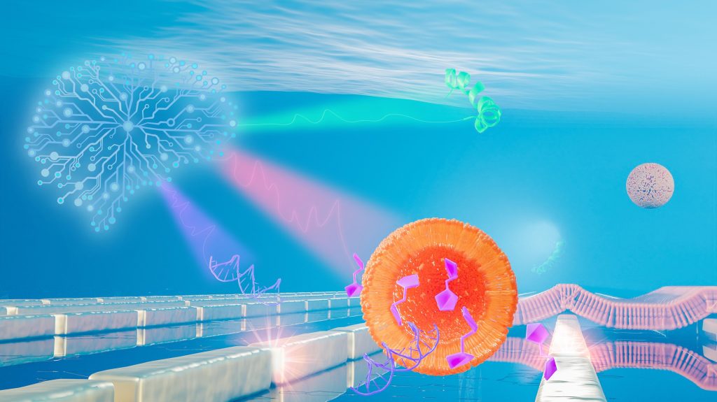
If you look at the big orange dot (representing the nanosensors?), you’ll see those purplish/fuschia objects resemble musical notes (biological molecules?). I think that brainlike object to the left and in light blue is the artificial intelligence (AI) component. (If anyone wants to correct my guesses or identify the bits I can’t, please feel free to add to the Comments for this blog.)

Getting back to my topic, keep the ‘musical notes’ in mind as you read about some of the latest research from l’École polytechnique fédérale de Lausanne (EPFL) in an April 7, 2021 news item on Nanowerk,

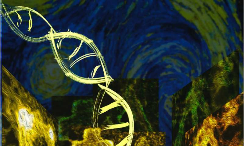
The tiny world of biomolecules is rich in fascinating interactions between a plethora of different agents such as intricate nanomachines (proteins), shape-shifting vessels (lipid complexes), chains of vital information (DNA) and energy fuel (carbohydrates). Yet the ways in which biomolecules meet and interact to define the symphony of life is exceedingly complex.

Scientists at the Bionanophotonic Systems Laboratory in EPFL’s School of Engineering have now developed a new biosensor that can be used to observe all major biomolecule classes of the nanoworld without disturbing them. Their innovative technique uses nanotechnology, metasurfaces, infrared light and artificial intelligence.

To each molecule its own melody

In this nano-sized symphony, perfect orchestration makes physiological wonders such as vision and taste possible, while slight dissonances can amplify into horrendous cacophonies leading to pathologies such as cancer and neurodegeneration.

An April 7, 2021 EPFL press release, which originated the news item, provides more detail,

“Tuning into this tiny world and being able to differentiate between proteins, lipids, nucleic acids and carbohydrates without disturbing their interactions is of fundamental importance for understanding life processes and disease mechanisms,” says Hatice Altug, the head of the Bionanophotonic Systems Laboratory. 

Light, and more specifically infrared light, is at the core of the biosensor developed by Altug’s team. Humans cannot see infrared light, which is beyond the visible light spectrum that ranges from blue to red. However, we can feel it in the form of heat in our bodies, as our molecules vibrate under the infrared light excitation.

Molecules consist of atoms bonded to each other and – depending on the mass of the atoms and the arrangement and stiffness of their bonds – vibrate at specific frequencies. This is similar to the strings on a musical instrument that vibrate at specific frequencies depending on their length. These resonant frequencies are molecule-specific, and they mostly occur in the infrared frequency range of the electromagnetic spectrum. 

“If you imagine audio frequencies instead of infrared frequencies, it’s as if each molecule has its own characteristic melody,” says Aurélian John-Herpin, a doctoral assistant at Altug’s lab and the first author of the publication. “However, tuning into these melodies is very challenging because without amplification, they are mere whispers in a sea of sounds. To make matters worse, their melodies can present very similar motifs making it hard to tell them apart.” 

Metasurfaces and artificial intelligence

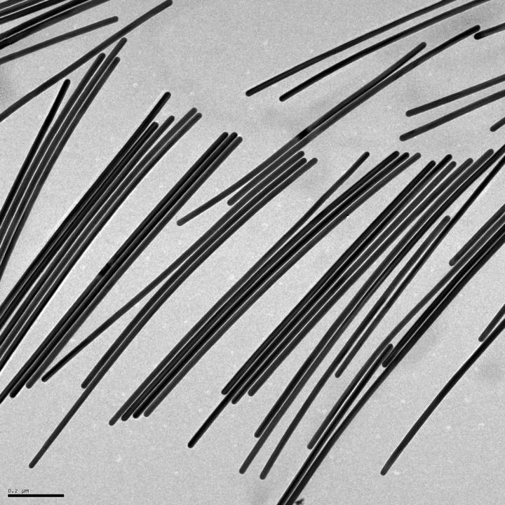
The scientists solved these two issues using metasurfaces and AI. Metasurfaces are man-made materials with outstanding light manipulation capabilities at the nano scale, thereby enabling functions beyond what is otherwise seen in nature. Here, their precisely engineered meta-atoms made out of gold nanorods act like amplifiers of light-matter interactions by tapping into the plasmonic excitations resulting from the collective oscillations of free electrons in metals. “In our analogy, these enhanced interactions make the whispered molecule melodies more audible,” says John-Herpin.

AI is a powerful tool that can be fed with more data than humans can handle in the same amount of time and that can quickly develop the ability to recognize complex patterns from the data. John-Herpin explains, “AI can be imagined as a complete beginner musician who listens to the different amplified melodies and develops a perfect ear after just a few minutes and can tell the melodies apart, even when they are played together – like in an orchestra featuring many instruments simultaneously.” 

The first biosensor of its kind

When the scientists’ infrared metasurfaces are augmented with AI, the new sensor can be used to analyze biological assays featuring multiple analytes simultaneously from the major biomolecule classes and resolving their dynamic interactions. 

“We looked in particular at lipid vesicle-based nanoparticles and monitored their breakage through the insertion of a toxin peptide and the subsequent release of vesicle cargos of nucleotides and carbohydrates, as well as the formation of supported lipid bilayer patches on the metasurface,” says Altug.

This pioneering AI-powered, metasurface-based biosensor will open up exciting perspectives for studying and unraveling inherently complex biological processes, such as intercellular communication via exosomesand the interaction of nucleic acids and carbohydrates with proteins in gene regulation and neurodegeneration. 

“We imagine that our technology will have applications in the fields of biology, bioanalytics and pharmacology – from fundamental research and disease diagnostics to drug development,” says Altug. 

Here’s a link to and a citation for the paper,

Infrared Metasurface Augmented by Deep Learning for Monitoring Dynamics between All Major Classes of Biomolecules by Aurelian John‐Herpin, Deepthy Kavungal. Lea von Mücke, Hatice Altug. Advanced Materials Volume 33, Issue 14 April 8, 2021 2006054 DOI: https://doi.org/10.1002/adma.202006054 First published: 22 February 2021

This paper is open access.

Chameleon skin (nanomaterial made of gold nanoparticles) for robots

A June 17, 2020 news item on Nanowerk trumpets research into how robots might be able to sport chameleon-like skin one day,

A new film made of gold nanoparticles changes color in response to any type of movement. Its unprecedented qualities could allow robots to mimic chameleons and octopi — among other futuristic applications.

Unlike other materials that try to emulate nature’s color changers, this one can respond to any type of movement, like bending or twisting. Robots coated in it could enter spaces that might be dangerous or impossible for humans, and offer information just based on the way they look.

For example, a camouflaged robot could enter tough-to-access underwater crevices. If the robot changes color, biologists could learn about the pressures facing animals that live in these environments.

Although some other color-changing materials can also respond to motion, this one can be printed and programmed to display different, complex patterns that are difficult to replicate.

This video from the University of California at Riverside researchers shows the material in action (Note: It gets more interesting after the first 20 secs.),

A June 15, 2020 University of California at Riverside (UCR) news release (also on EurekAlert but published on June 17, 2020) by Jules Bernstein, which originated the news item, delves further,

Nanomaterials are simply materials that have been reduced to an extremely small scale — tens of nanometers in width and length, or, about the size of a virus. When materials like silver or gold become smaller, their colors will change depending on their size, shape, and the direction they face.

“In our case, we reduced gold to nano-sized rods. We knew that if we could make the rods point in a particular direction, we could control their color,” said chemistry professor Yadong Yin. “Facing one way, they might appear red. Move them 45 degrees, and they change to green.”

The problem facing the research team was how to take millions of gold nanorods floating in a liquid solution and get them all to point in the same direction to display a uniform color.

Their solution was to fuse smaller magnetic nanorods onto the larger gold ones. The two different-sized rods were encapsulated in a polymer shield, so that they would remain side by side. That way, the orientation of both rods could be controlled by magnets.

“Just like if you hold a magnet over a pile of needles, they all point in the same direction. That’s how we control the color,” Yin said.

Once the nanorods are dried into a thin film, their orientation is fixed in place and they no longer respond to magnets. “But, if the film is flexible, you can bend and rotate it, and will still see different colors as the orientation changes,” Yin said.

Other materials, like butterfly wings, are shiny and colorful at certain angles, and can also change color when viewed at other angles. However, those materials rely on precisely ordered microstructures, which are difficult and expensive to make for large areas. But this new film can be made to coat the surface of any sized object just as easily as applying spray paint on a house.

Though futuristic robots are an ultimate application of this film, it can be used in many other ways. UC Riverside chemist Zhiwei Li, the first author on this paper, explained that the film can be incorporated into checks or cash as an authentication feature. Under normal lighting, the film is gray, but when you put on sunglasses and look at it through polarized lenses, elaborate patterns can be seen. In addition, the color contrast of the film may change dramatically if you twist the film.

The applications, in fact, are only limited by the imagination. “Artists could use this technology to create fascinating paintings that are wildly different depending on the angle from which they are viewed,” Li said. “It would be wonderful to see how the science in our work could be combined with the beauty of art.”

Here’s a link to and a citation for the paper,

Coupling magnetic and plasmonic anisotropy in hybrid nanorods for mechanochromic responses by Zhiwei Li, Jianbo Jin, Fan Yang, Ningning Song & Yadong Yin. Nature Communications volume 11, Article number: 2883 (2020) DOI: https://doi.org/10.1038/s41467-020-16678-8 Published: 08 June 2020

This paper is open access.

Vitamin C helps gold nanowires grow

This research gives new meaning to ‘Take your vitamin C’ as can be seen in a February 19, 2019 news item on Nanowerk,

A boost of vitamin C helped Rice University scientists turn small gold nanorods into fine gold nanowires.

Common, mild ascorbic acid is the not-so-secret sauce that helped the Rice lab of chemist Eugene Zubarev grow pure batches of nanowires from stumpy nanorods without the drawbacks of previous techniques.

“There’s no novelty per se in using vitamin C to make gold nanostructures because there are many previous examples,” Zubarev said. “But the slow and controlled reduction achieved by vitamin C is surprisingly suitable for this type of chemistry in producing extra-long nanowires.”

A February 19, 2019 Rice University news release (also on EurekAlert), which originated the news item, provides more technical detail about the research

The Rice lab’s nanorods are about 25 nanometers thick at the start of the process – and remain that way while their length grows to become long nanowires. Above 1,000 nanometers in length, the objects are considered nanowires, and that matters. The wires’ aspect ratio – length over width – dictates how they absorb and emit light and how they conduct electrons. Combined with gold’s inherent metallic properties, that could enhance their value for sensing, diagnostic, imaging and therapeutic applications.

Zubarev and lead author Bishnu Khanal, a Rice chemistry alumnus, succeeded in making their particles go far beyond the transition from nanorod to nanowire, theoretically to unlimited length.

The researchers also showed the process is fully controllable and reversible. That makes it possible to produce nanowires of any desired length, and thus the desired configuration for electronic or light-manipulating applications, especially those that involve plasmons, the light-triggered oscillation of electrons on a metal’s surface.

The nanowires’ plasmonic response can be tuned to emit light from visible to infrared and theoretically far beyond, depending on their aspect ratios.

The process is slow, so it takes hours to grow a micron-long nanowire. “In this paper, we only reported structures up to 4 to 5 microns in length,” Zubarev said. “But we’re working to make much longer nanowires.”

The growth process only appeared to work with pentahedrally twinned gold nanorods, which contain five linked crystals. These five-sided rods — “Think of a pencil, but with five sides instead of six,” Zubarev said — are stable along the flat surfaces, but not at the tips.

“The tips also have five faces, but they have a different arrangement of atoms,” he said. “The energy of those atoms is slightly lower, and when new atoms are deposited there, they don’t migrate anywhere else.”

That keeps the growing wires from gaining girth. Every added atom increases the wire’s length, and thus the aspect ratio.

The nanorods’ reactive tips get help from a surfactant, CTAB, that covers the flat surfaces of nanorods. “The surfactant forms a very dense, tight bilayer on the sides, but it cannot cover the tips effectively,” Zubarev said.

That leaves the tips open to an oxidation or reduction reaction. The ascorbic acid provides electrons that combine with gold ions and settle at the tips in the form of gold atoms. And unlike carbon nanotubes in a solution that easily aggregate, the nanowires keep their distance from one another.

“The most valuable feature is that it is truly one-dimensional elongation of nanorods to nanowires,” Zubarev said. “It does not change the diameter, so in principal we can take small rods with an aspect ratio of maybe two or three and elongate them to 100 times the length.”
He said the process should apply to other metal nanorods, including silver.

Here’s a link to and a citation for the paper,

Chemical Transformation of Nanorods to Nanowires: Reversible Growth and Dissolution of Anisotropic Gold Nanostructures by Bishnu P. Khanal and Eugene R. Zubarev. ACS Nano, 2019, 13 (2), pp 2370–2378 DOI: 10.1021/acsnano.8b09203 Publication Date (Web): February 12, 2019

Copyright © 2019 American Chemical Society

This paper is behind a paywall. Below you’ll find an image fo what I believe to be the vitamin C-enhanced gold nanowires.

Caption: Gold nanowires grown in the Rice University lab of chemist Eugene Zubarev promise to provide tunable plasmonic properties for optical and electronic applications. The wires can be controllably grown from nanorods, or reduced. Credit: Zubarev Research Group/Rice University

Eliminate cold storage for diagnostic tests?

There’s a nanoparticle coating that could eliminate the need for cold storage and/or refrigeration for diagnostic testing according to a Jan. 4, 2017 news item on Nanowerk,

Many diagnostic tests use antibodies to help confirm a myriad of medical conditions, from Zika infections to heart ailments and even some forms of cancer. Antibodies capture and help detect proteins, enzymes, bacteria and viruses present in injuries and illnesses, and must be kept at a constant low temperature to ensure their viability — often requiring refrigeration powered by electricity. This can make diagnostic testing in underdeveloped countries, disaster or remote areas and even war zones extremely expensive and difficult.

A team of engineers from Washington University in St. Louis and Air Force Research Laboratory have discovered an inexpensive work-around: a protective coating that could completely eliminate the need for cold storage and change the scope of medical diagnostic testing in places where it’s often needed the most.

“In many developing countries, electricity is not guaranteed,” said Srikanth Singamaneni, associate professor of mechanical engineering and materials science in Engineering & Applied Science at Washington University in St. Louis.

“So how do we best get them medical diagnostics? We did not know how to solve this problem previously.”

A Jan. 4, 2016 Washington University in St. Louis news release by Erika Ebsworth-Goold, which originated the news item, describes how previous research helped lead to a solution,

Singamaneni’s team previously used tiny gold nanorods in bio-diagnostic research, measuring changes in their optical properties to quantify protein concentrations in bio-fluids: the higher a concentration, the higher the likelihood of injury or disease.

In this new research, published in Advanced Materials, Singamaneni worked with faculty from Washington University’s School of Medicine and researchers from the Air Force Research Lab to grow metal-organic frameworks (MOFs) around antibodies attached to gold nanorods. The crystalline MOFs formed a protective layer around the antibodies and prevented them from losing activity at elevated temperatures. The protective effect lasted for a week even when the samples were stored at 60°C.

“This technology would allow point-of-care screening for biomarkers of diseases in urban and rural clinic settings where immediate patient follow-up is critical to treatment and wellbeing,” said Dr. Jeremiah J. Morrissey, professor of anesthesiology, Division of Clinical and Translational Research, Washington University School of Medicine and a co-author on the paper.

“On the spot testing eliminates the time lag in sending blood/urine samples to a central lab for testing and in tracking down patients to discuss test results. In addition, it may reduce costs associated with refrigerated shipping and storage.”

The protective MOF layer can be quickly and easily removed from the antibodies with a simple rinse of slightly acidic water, making a diagnostic strip or paper immediately ready to use. Singamaneni says this proof of concept research is now ready to be tested for clinical samples.

“As long as you are using antibodies, you can use this technology,” said Congzhou Wang, a postdoctoral researcher in Singamaneni’s lab and the paper’s lead author. “In bio-diagnostics from here on out, we will no longer need refrigeration.”

“The MOF-based protection of antibodies on sensor surfaces is ideal for preserving biorecognition abilities of sensors that are designed for deployment in the battlefield,” said Dr. Rajesh R. Naik, 711th Human Performance Wing of the Air Force Research Laboratory, Wright-Patterson Air Force Base, and a co-corresponding author of the paper.  “It provides remarkable stability and extremely easy to remove right before use.”

Here’s a link to and a citation for the paper,

Metal-Organic Framework as a Protective Coating for Biodiagnostic Chips by Congzhou Wang, Sirimuvva Tadepalli, Jingyi Luan, Keng-Ku Liu, Jeremiah J. Morrissey, Evan D. Kharasch, Rajesh R. Naik, and Srikanth Singamaneni. Advanced Materials DOI: 10.1002/adma.201604433 Version of Record online: 7 DEC 2016

© 2016 WILEY-VCH Verlag GmbH & Co. KGaA, Weinheim

This paper is behind a paywall.

A final observation, there’s at least one other project aimed at eliminating the need for refrigeration in the field of medical applications and that’s the nanopatch, a replacement for syringes used for liquid medications and vaccines (see my Dec. 16, 2016 posting for a description).

Nova Scotia’s (Canada) Sona Nanotech and its gold nanoparticles move

I hope one day to have at least one piece on nanotechnology for each province, the Yukon, and the territories. Unfortunately, today (Nov. 2, 2016) will not be the day I add one previously unsung province, etc. to the list as Nova Scotia has previously graced this blog with a nanotechnology story (my June 5, 2016 posting).

The latest nano news from Nova Scotia is found in a Nov. 1, 2016 article by James Risdon for the Chronicle Herald,

A Nova Scotia biotech startup with big plans for its super-small, non-toxic gold particles is looking to move its lab facilities to Halifax and expand.

Andrew McLeod, Sona Nanotech Ltd.’s president and chief operating officer, said Tuesday the company is already looking for lab space in Halifax and wants to hire three additional employees to handle production, research and business development.

Sona Nanotech has two products, its Gemini and Omni gold particles, intended to be used in the health-care industry for such things as the treatment of cancer and diagnostic testing.

These particles are measured in nanometres.

“You’re talking about something that’s on the order of millionths of the width of a human hair,” said McLeod. [The comparison measurements I’ve seen most frequestion for a single nanometre is 1/50,000 or 1/60,000 or 1/100,000 of a hair.]

While other players make gold particles, Sona Nanotech has developed a way to make its products so that they are free of a toxic chemical ,and that’s opening doors for the Nova Scotia startup whose products can be used inside the human body.

There’s already talk of Sona Nanotech teaming up with an as-yet-unnamed Canadian organization for a cancer research project, but McLeod was tight-lipped about the details.

Congratulations to Sona Nano!

For anyone curious about the business aspects of the story, I recommend reading Risdon’s article in its entirety.

Sona Nanotech’s website can be found here,

Sona Nanotech Ltd. has leveraged its team’s unique knowledge and experience with novel surface chemistry methods and surfactants to create a disruptive leap forward in metallic nanoparticle technology.

Co-founders Dr. Gerrard Marangoni, Dr. Kulbir Singh, and Dr. Michael McAlduff recognized the role that gold nanoparticles can play in a variety of life sciences applications, e.g.,  in-vivo 3-D imaging, GNR-enabled diagnostic test products and other cutting edge medical applications.  Gold nanorods can be enabling technologies for non-invasive targeted cell, tumor, tissue and organ treatments such as photothermal cancer cell destruction, and location specific drug and pain treatment.

The Problem
Gold nanorods have been made to date with toxic CTAB [cationic surfactant cethyltrimetylammonium bromide] which makes them much less attractive for in-vivo medical applications.

The Solution
100% CTAB-FREE – Gemini™ and Omni™ Patent-Pending Gold Nanorods – from Sona Nanotech Ltd.

The Problem
For a given colour contrast, large gold nanospheres are not as stable or mobile as gold nanorods (dip tests).

The Solution
Stable, high loading capacity GNRs [gold nanorods] from Sona Nanotech offer a broad range of rich, high contrast test color options.

So, there you have it.

Heat, evolution, and the shape of gold nanorods

A Feb. 23, 2015 news item on Azonano features gold nanorods and their shapeshifting ways when releasing heat,

Researchers at the U.S. Department of Energy’s Argonne National Laboratory have revealed previously unobserved behaviors that show how details of the transfer of heat at the nanoscale cause nanoparticles to change shape in ensembles.

The new findings depict three distinct stages of evolution in groups of gold nanorods, from the initial rod shape to the intermediate shape to a sphere-shaped nanoparticle. The research suggests new rules for the behavior of nanorod ensembles, providing insights into how to increase heat transfer efficiency in a nanoscale system.

A Feb. 18, 2015 Argonne National Laboratory news release by Justin H. S. Breaux, which originated the news item, provides more details about the work,

At the nanoscale, individual gold nanorods have unique electronic, thermal and optical properties. Understanding these properties and managing how collections of these elongated nanoparticles absorb and release this energy as heat will drive new research towards next-generation technologies such as water purification systems, battery materials and cancer research.

A good deal is known about how single nanorods behave—but little is known about how nanorods behave in ensembles of millions. Understanding how the individual behavior of each nanorod, including how its orientation and rate of transition differ from those around it, impacts the collective kinetics of the ensemble and is critical to using nanorods in future technologies.

“We started with a lot of questions,” said Argonne physicist Yuelin Li, “like ‘How much power can the particles sustain before losing functionality? How do individual changes at the nanoscale affect the overall functionality? How much heat is released to the surrounding area?’ Each nanorod is continuously undergoing a change in shape when heated beyond melting temperature, which means a change in the surface area and thus a change in its thermal and hydrodynamic properties.”

The researchers used a laser to heat the nanoparticles and X-rays to analyze their changing shapes. Generally, nanorods transition into nanospheres more quickly when supplied with a higher intensity of laser power. In this case, completely different ensemble behaviors were observed when this intensity increased incrementally. The intensity of the heat applied changes not only the nanoparticles’ shape at various rates but also affects their ability to efficiently absorb and release heat.

“For us, the key was to understand just how efficient the nanorods were at transferring light into heat in many different scenarios,” said nanoscientist Subramanian Sankaranarayanan of Argonne’s Center for Nanoscale Materials. “Then we had to determine the physics behind how heat was transferred and all the different ways these nanorods could transition into nanospheres.”

To observe how the rod makes this transition, researchers first shine a laser pulse at the nanorod suspended in a water solution at Argonne’s Advanced Photon Source. The laser lasts for less than a hundred femtoseconds, nearly one trillion times faster than a blink of the eye. What follows is a series of focused and rapid X-ray bursts using a technique called small angle X-ray scattering. The resulting data is used to determine the average shape of the particle as it changes over time.

In this way, scientists can reconstruct the minute changes occurring in the shape of the nanorod. However, to understand the physics underlying this phenomenon, the researchers needed to look deeper at how individual atoms vibrate and move during the transition. For this, they turned to the field of molecular dynamics using the supercomputing power of the 10-petaflop Mira supercomputer at the Argonne Leadership Computing Facility.

Mira used mathematical equations to pinpoint the individual movements of nearly two million of the nanorods’ atoms in the water. Using factors such as the shape, temperature and rate of change, the researchers built simulations of the nanorod in many different scenarios to see how the structure changes over time.

“In the end,” said Sankaranarayanan, “we discovered the heat transfer rates for shorter but wider nanospheres are lower than for their rod-shaped predecessors. This decrease in heat transfer efficiency at the nanoscale plays a key role in accelerating the transition from rod to sphere when heated beyond the melting temperature.”

Here’s a link to and a citation for the paper,

Femtosecond Laser Pulse Driven Melting in Gold Nanorod Aqueous Colloidal Suspension: Identification of a Transition from Stretched to Exponential Kinetics by Yuelin Li, Zhang Jiang, Xiao-Min Lin, Haidan Wen, Donald A. Walko, Sanket A. Deshmukh, Ram Subbaraman, Subramanian K. R. S. Sankaranarayanan, Stephen K. Gray, & Phay Ho. Scientific Reports 5, Article number: 8146 doi:10.1038/srep08146 Published 30 January 2015

This article is open access.

Gold nanorod instabilities

A Dec. 8, 2014 news item on Nanowerk focuses on research from Australia,

Researchers at Swinburne University of Technology [Melbourne, Australia]  have discovered an instability in gold nanoparticles that is critical for their application in future technology.

Gold nanorods are important building blocks for future applications in solar cells, cancer therapy and optical circuitry.

However their stability is under question due to their peculiar reshaping behaviour below melting points.

A Dec. 8, 2014 Swinburne University of Technology press release, which originated the news item, discusses melting points and shape instabilities in the context of this research,

A solid normally does not change its shape unless it reaches its melting point, or surface melting points. It is also known that the melting point for nanoparticles is suppressed due to their size.

PhD student Adam Taylor (now a postdoctoral researcher at Swinburne) said it came as a surprise that reshaping is observed well below these melting points. Until now, no one could explain this peculiar behaviour.

“In our work, we have discovered both theoretically and experimentally that the reshaping mechanism for nanoparticles below melting point is surface atom diffusion, rather than melting,” Mr Taylor said.

Surface atom diffusion is a process involving the motion of molecules at solid material surfaces that can generally be thought of in terms of particles jumping between adjacent adsorption sites on a surface.

“Surface atom diffusion always existed in bulk solids, but this is the first evidence that its effect is enhanced at the nano-size, dominating over the traditional theory of melting,” Associate Professor James Chon, who is supervising Mr Taylor’s research, said.

Mr Taylor said the more finely nanoparticles are shaped, the less stable they become.

“This is important, for example, for solar panel manufacturers as the more needle-like these nanoparticles are shaped the less stable they become. If you put these particles into a solar panel to concentrate light they may not last long in the sun before they degrade,” Mr Taylor said.

“This discovery will be crucial for future applications of gold nanorods, as people will need to reconsider their stability when applying them to solar cells, cancer therapeutic agents and optical circuitry.”

The researchers have provided an illustration of their work,

Courtesy Swinburne University of Technology

Courtesy Swinburne University of Technology

Here’s a link to and citation for the research,

Below Melting Point Photothermal Reshaping of Single Gold Nanorods Driven by Surface Diffusion by Adam B. Taylor, Arif M. Siddiquee, and James W. M. Chon. ACS Nano, Article ASAP DOI: 10.1021/nn5055283 Publication Date (Web): November 18, 2014

Copyright © 2014 American Chemical Society

This paper is behind a paywall but should you be in Australia and eligible to attend, there’s another opportunity to learn more; Taylor will be presenting his work at the Australian Institute of Physics conference on December 10, 2014 in Canberra.

Gold nanorods and mucus

Mucus can kill. Most of us are lucky enough to produce mucus appropriate for our bodies’ needs but people who have cystic fibrosis and other kinds of lung disease suffer greatly from mucus that is too thick to pass easily through the body. An Oct. 9, 2014 Optical Society of America (OSA) news release (also on EurekAlert) ‘shines’ a light on the topic of mucus and viscosity,

Some people might consider mucus an icky bodily secretion best left wrapped in a tissue, but to a group of researchers from the University of North Carolina at Chapel Hill, snot is an endlessly fascinating subject. The team has developed a way to use gold nanoparticles and light to measure the stickiness of the slimy substance that lines our airways.  The new method could help doctors better monitor and treat lung diseases such as cystic fibrosis and chronic obstructive pulmonary disease.

“People who are suffering from certain lung diseases have thickened mucus,” explained Amy Oldenburg, a physicist at the University of North Carolina at Chapel Hill whose research focuses on biomedical imaging systems. “In healthy adults, hair-like cell appendages called cilia line the airways and pull mucus out of the lungs and into the throat. But if the mucus is too viscous it can become trapped in the lungs, making breathing more difficult and also failing to remove pathogens that can cause chronic infections.”

Doctors can prescribe mucus-thinning drugs, but have no good way to monitor how the drugs affect the viscosity of mucus at various spots inside the body. This is where Oldenburg and her colleagues’ work may help.

The researchers placed coated gold nanorods on the surface of mucus samples and then tracked the rods’ diffusion into the mucus by illuminating the samples with laser light and analyzing the way the light bounced off the nanoparticles. The slower the nanorods diffused, the thicker the mucus. The team found this imaging method worked even when the mucus was sliding over a layer of cells—an important finding since mucus inside the human body is usually in motion.

“The ability to monitor how well mucus-thinning treatments are working in real-time may allow us to determine better treatments and tailor them for the individual,” said Oldenburg.

It will likely take five to 10 more years before the team’s mucus measuring method is tested on human patients, Oldenburg said. Gold is non-toxic, but for safety reasons the researchers would want to ensure that the gold nanorods would eventually be cleared from a patient’s system.

“This is a great example of interdisciplinary work in which optical scientists can meet a specific need in the clinic,” said Nozomi Nishimura, of Cornell University … . “As these types of optical technologies continue to make their way into medical practice, it will both expand the market for the technology as well as improve patient care.”

The team is also working on several lines of ongoing study that will some day help bring their monitoring device to the clinic. They are developing delivery methods for the gold nanorods, studying how their imaging system might be adapted to enter a patient’s airways, and further investigating how mucus flow properties differ throughout the body.

This work is being presented at:

The research team will present their work at The Optical Society’s (OSA) 98th Annual Meeting, Frontiers in Optics, being held Oct. 19-23 [2014] in Tucson, Arizona, USA.

Presentation FTu5F.2, “Imaging Gold Nanorod Diffusion in Mucus Using Polarization Sensitive OCT,” takes place Tuesday, Oct. 21 at 4:15 p.m. MST [Mountain Standard Time] in the Tucson Ballroom, Salon A at the JW Marriott Tucson Starr Pass Resort.

People with cystic fibrosis tend to have short lives (from the US National Library of Medicine MedLine Plus webpage on cystic fibrosis),

Most children with cystic fibrosis stay in good health until they reach adulthood. They are able to take part in most activities and attend school. Many young adults with cystic fibrosis finish college or find jobs.

Lung disease eventually worsens to the point where the person is disabled. Today, the average life span for people with CF who live to adulthood is about 37 years.

Death is most often caused by lung complications.

I hope this work proves helpful.

Vaccines that are pure gold give patients breathing space

I exaggerated a little bit, the vaccine isn’t pure gold but it does have gold nanoparticles which mimic a virus. From the June 25, 2013 news item on ScienceDaily,

Scientists in the US have developed a novel vaccination method that uses tiny gold particles to mimic a virus and carry specific proteins to the body’s specialist immune cells.

The technique differs from the traditional approach of using dead or inactive viruses as a vaccine and was demonstrated in the lab using a specific protein that sits on the surface of the respiratory syncytial virus (RSV).

The results have been published today, 26 June [2013], in IOP Publishing’s journal Nanotechnology by a team of researchers from Vanderbilt University.

The June 26, 2013 IOP [Institute of Physics] Publishing news release (perhaps the journal publishers posted their news release after it was published elsewhere?), which originated the news item, provides more details about RSV and the technique,

RSV is the leading viral cause of lower respiration tract infections, causing several hundred thousand deaths and an estimated 65 million infections a year, mainly in children and the elderly.

The detrimental effects of RSV come, in part, from a specific protein, called the F protein, which coats the surface of the virus. The protein enables the virus to enter into the cytoplasm of cells and also causes cells to stick together, making the virus harder to eliminate.

The body’s natural defence to RSV is therefore directed at the F protein; however, up until now, researchers have had difficulty creating a vaccine that delivers the F protein to the specialised immune cells in the body. If successful, the F protein could trigger an immune response which the body could ‘remember’ if a subject became infected with the real virus.

In this study the researchers created exceptionally small gold nanorods, just 21 nanometres wide and 57 nanometres long, which were almost exactly the same shape and size as the virus itself. The gold nanorods were successfully coated with the RSV F proteins and were bonded strongly thanks to the unique physical and chemical properties of the nanorods themselves.

The researchers then tested the ability of the gold nanorods to deliver the F protein to specific immune cells, known as dendritic cells, which were taken from adult blood samples.

Dendritic cells function as processing cells in the immune system, taking the important information from a virus, such as the F protein, and presenting it to cells that can perform an action against them―the T cells are just one example of a cell that can take action.

Once the F protein-coated nanorods were added to a sample of dendritic cells, the researchers analysed the proliferation of T cells as a proxy for an immune response. They found that the protein-coated nanorods caused the T cells to proliferate significantly more compared to non-coated nanorods and just the F protein alone.

Not only did this prove that the coated-nanorods were capable of mimicking the virus and stimulating an immune response, it also showed that they were not toxic to human cells, offering significant safety advantages and increasing their potential as a real-life human vaccine.

Lead author of the study, Professor James Crowe, said: “A vaccine for RSV, which is the major cause of viral pneumonia in children, is sorely needed. This study shows that we have developed methods for putting RSV F protein into exceptionally small particles and presenting it to immune cells in a format that physically mimics the virus. Furthermore, the particles themselves are not infectious.”

Due to the versatility of the gold nanorods, Professor Crowe believes that their potential use is not limited to RSV.

“This platform could be used to develop experimental vaccines for virtually any virus, and in fact other larger microbes such as bacteria and fungi.

“The studies we performed showed that the candidate vaccines stimulated human immune cells when they were interacted in the lab. The next steps to testing would be to test whether or not the vaccines work in vivo” Professor Crowe continued.

I look forward to hearing more about this new vaccine as they continue with the testing. Meanwhile, here’s a link to and a citation for the latest published work,

Gold nanorod vaccine for respiratory syncytial virus by John W Stone, Natalie J Thornburg, David L Blum, Sam J Kuhn, David W Wright, and James E Crowe Jr. Nanotechnology Volume 24 Number 29 or Nanotechnology 24 295102 doi:10.1088/0957-4484/24/29/295102

The article is open access.