Tag Archives: gold nanowires

Nanoscale tattoos for individual cells

It’s fascinating to read about a technique for applying ‘tattoos’ to living cells and I have two news items and news releases with different perspectives about this same research.

First out the door was the August 7, 2023 news item on ScienceDaily,

Engineers have developed nanoscale tattoos — dots and wires that adhere to live cells — in a breakthrough that puts researchers one step closer to tracking the health of individual cells.

The new technology allows for the first time the placement of optical elements or electronics on live cells with tattoo-like arrays that stick on cells while flexing and conforming to the cells’ wet and fluid outer structure.

“If you imagine where this is all going in the future, we would like to have sensors to remotely monitor and control the state of individual cells and the environment surrounding those cells in real time,” said David Gracias, a professor of chemical and biomolecular engineering at Johns Hopkins University who led the development of the technology. “If we had technologies to track the health of isolated cells, we could maybe diagnose and treat diseases much earlier and not wait until the entire organ is damaged.”

An August 7, 2023 Johns Hopkins University news release by (also on EurekAlert), which originated the news item, describes the research in an accessible fashion before delving into technical details,

Gracias, who works on developing  biosensor technologies that are nontoxic and noninvasive for the body, said the tattoos bridge the gap between living cells or tissue and conventional sensors and electronic materials. They’re essentially like barcodes or QR codes, he said.

“We’re talking about putting something like an electronic tattoo on a living object tens of times smaller than the head of a pin,” Gracias said. “It’s the first step towards attaching sensors and electronics on live cells.”

The structures were able to stick to soft cells for 16 hours even as the cells moved.

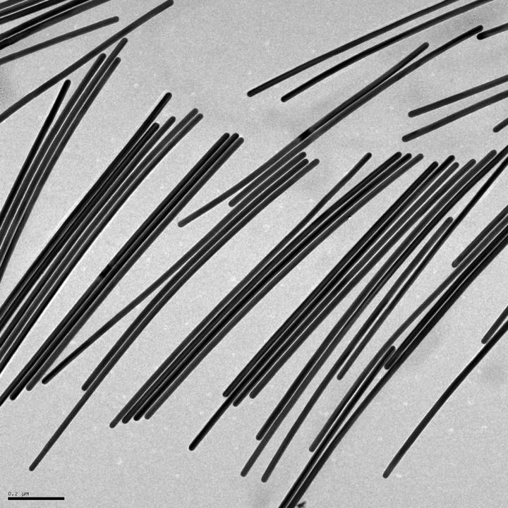
The researchers built the tattoos in the form of arrays with gold, a material known for its ability to prevent signal loss or distortion in electronic wiring. They attached the arrays to cells that make and sustain tissue in the human body, called fibroblasts. The arrays were then treated with  molecular glues and transferred onto the cells using an alginate hydrogel film, a gel-like laminate that can be dissolved after the gold adheres to the cell. The molecular glue on the array bonds to a film secreted by the cells called the extracellular matrix.

Previous research has demonstrated how to use hydrogels to stick nanotechnology onto human skin and internal animal organs. By showing how to adhere nanowires and nanodots onto single cells, Gracias’ team is addressing the long-standing challenge of making optical sensors and electronics compatible with biological matter at the single cell level. 

“We’ve shown we can attach complex nanopatterns to living cells, while ensuring that the cell doesn’t die,” Gracias said. “It’s a very important result that the cells can live and move with the tattoos because there’s often a significant incompatibility between living cells and the methods engineers use to fabricate electronics.”

The team’s ability to attach the dots and wires in an array form is also crucial. To use this technology to track bioinformation, researchers must be able to arrange sensors and wiring into specific patterns not unlike how they are arranged in electronic chips. 

“This is an array with specific spacing,” Gracias explained, “not a haphazard bunch of dots.”

The team plans to try to attach more complex nanocircuits that can stay in place for longer periods. They also want to experiment with different types of cells.

Other Johns Hopkins authors are Kam Sang Kwok, Yi Zuo, Soo Jin Choi, Gayatri J. Pahapale, and Luo Gu.

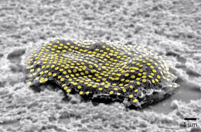
This looks more like a sea creature to me but it’s not,

Caption: False-colored gold nanodot array on a fibroblast cell. Credit: Kam Sang Kwok and Soo Jin Choi, Gracias Lab/Johns Hopkins University.[The measurement, i.e., what looks like a ‘u’ with a preceding tail, in the lower right corner of the image is one micron/one millionth add that to the ‘m’ and you have what’s commonly described as one micrometre.]

An August 10, 2023 news item on ScienceDaily offers a different perspective from the American Chemical Society (ACS) on this research,

For now, cyborgs exist only in fiction, but the concept is becoming more plausible as science progresses. And now, researchers are reporting in ACS’ Nano Letters that they have developed a proof-of-concept technique to “tattoo” living cells and tissues with flexible arrays of gold nanodots and nanowires. With further refinement, this method could eventually be used to integrate smart devices with living tissue for biomedical applications, such as bionics and biosensing.

An August 10, 2023 ACS news release (also on EurekAlert), which originated the news item, explains some of the issues with attaching electronics to living tissue,

Advances in electronics have enabled manufacturers to make integrated circuits and sensors with nanoscale resolution. More recently, laser printing and other techniques have made it possible to assemble flexible devices that can mold to curved surfaces. But these processes often use harsh chemicals, high temperatures or pressure extremes that are incompatible with living cells. Other methods are too slow or have poor spatial resolution. To avoid these drawbacks, David Gracias, Luo Gu and colleagues wanted to develop a nontoxic, high-resolution, lithographic method to attach nanomaterials to living tissue and cells.

The team used nanoimprint lithography to print a pattern of nanoscale gold lines or dots on a polymer-coated silicon wafer. The polymer was then dissolved to free the gold nanoarray so it could be transferred to a thin piece of glass. Next, the gold was functionalized with cysteamine and covered with a hydrogel layer, which, when peeled away, removed the array from the glass. The patterned side of this flexible array/hydrogel layer was coated with gelatin and attached to individual live fibroblast cells. In the final step, the hydrogel was degraded to expose the gold pattern on the surface of the cells. The researchers used similar techniques to apply gold nanoarrays to sheets of fibroblasts or to rat brains. Experiments showed that the arrays were biocompatible and could guide cell orientation and migration.

The researchers say their cost-effective approach could be used to attach other nanoscale components, such as electrodes, antennas and circuits, to hydrogels or living organisms, thereby opening up opportunities for the development of biohybrid materials, bionic devices and biosensors.

The authors acknowledge funding from the Air Force Office of Scientific Research, the National Institute on Aging, the National Science Foundation and the Johns Hopkins University Surpass Program.

Here’s a link to and a citation for the paper,

Toward Single Cell Tattoos: Biotransfer Printing of Lithographic Gold Nanopatterns on Live Cells by Kam Sang Kwok, Yi Zuo, Soo Jin Choi, Gayatri J. Pahapale, Luo Gu, and David H. Gracias. Nano Lett. 2023, 23, 16, 7477–7484 DOI: https://doi.org/10.1021/acs.nanolett.3c01960 Publication Date:August 1, 2023 Copyright © 2023 American Chemical Society

This paper is behind a paywall.

New ingredient for computers: water!

A July 25, 2019 news item on Nanowerk provides a description of Moore`s Law and some ‘watery’ research that may upend it,

Moore’s law – which says the number of components that could be etched onto the surface of a silicon wafer would double every two years – has been the subject of recent debate. The quicker pace of computing advancements in the past decade have led some experts to say Moore’s law, the brainchild of Intel co-founder Gordon Moore in the 1960s, no longer applies. Particularly of concern, next-generation computing devices require features smaller than 10 nanometers – driving unsustainable increases in fabrication costs.

Biology creates features at sub-10nm scales routinely, but they are often structured in ways that are not useful for applications like computing. A Purdue University group has found ways of transforming structures that occur naturally in cell membranes to create other architectures, like parallel 1nm-wide line segments, more applicable to computing.

Inspired by biological cell membranes, Purdue researchers in the Claridge Research Group have developed surfaces that act as molecular-scale blueprints for unpacking and aligning nanoscale components for next-generation computers. The secret ingredient? Water, in tiny amounts.

A July 25, 2019 Purdue University news release (also on EurekAlert), expands on the theme,

“Biology has an amazing tool kit for embedding chemical information in a surface,” said Shelley Claridge, a recently tenured faculty member in chemistry and biomedical engineering at Purdue, who leads a group of nanomaterials researchers. “What we’re finding is that these instructions can become even more powerful in nonbiological settings, where water is scarce.”

In work just published in Chem, sister journal to Cell, the group has found that stripes of lipids can unpack and order flexible gold nanowires with diameters of just 2 nm, over areas corresponding to many millions of molecules in the template surface.

“The real surprise was the importance of water,” Claridge said. “Your body is mostly water, so the molecules in your cell membranes depend on it to function. Even after we transform the membrane structure in a way that’s very nonbiological and dry it out, these molecules can pull enough water out of dry winter air to do their job.”

Their work aligns with Purdue’s Giant Leaps celebration, celebrating the global advancements in sustainability as part of Purdue’s 150th anniversary. Sustainability is one of the four themes of the yearlong celebration’s Ideas Festival, designed to showcase Purdue as an intellectual center solving real-world issues.

The research team is working with the Purdue Research Foundation Office of Technology Commercialization to patent their work. They are looking for partners for continued research and to take the technology to market. [emphasis mine]

I wonder how close they are to taking this work to market. Usually they say it will be five to 10 years but perhaps we’ll see water-based computers in the near future. In the meantime, here’s a link to and a citation for the paper,

1-nm-Wide Hydrated Dipole Arrays Regulate AuNW Assembly on Striped Monolayers in Nonpolar Solvent by Ashlin G. Porter, Tianhong Ouyang, Tyler R. Hayes, John Biechele-Speziale, Shane R. Russell, Shelley A. Claridge. Chem DOI: DOI:https://doi.org/10.1016/j.chempr.2019.07.002 Published online:July 25, 2019

This paper is behind a paywall.

Vitamin C helps gold nanowires grow

This research gives new meaning to ‘Take your vitamin C’ as can be seen in a February 19, 2019 news item on Nanowerk,

A boost of vitamin C helped Rice University scientists turn small gold nanorods into fine gold nanowires.

Common, mild ascorbic acid is the not-so-secret sauce that helped the Rice lab of chemist Eugene Zubarev grow pure batches of nanowires from stumpy nanorods without the drawbacks of previous techniques.

“There’s no novelty per se in using vitamin C to make gold nanostructures because there are many previous examples,” Zubarev said. “But the slow and controlled reduction achieved by vitamin C is surprisingly suitable for this type of chemistry in producing extra-long nanowires.”

A February 19, 2019 Rice University news release (also on EurekAlert), which originated the news item, provides more technical detail about the research

The Rice lab’s nanorods are about 25 nanometers thick at the start of the process – and remain that way while their length grows to become long nanowires. Above 1,000 nanometers in length, the objects are considered nanowires, and that matters. The wires’ aspect ratio – length over width – dictates how they absorb and emit light and how they conduct electrons. Combined with gold’s inherent metallic properties, that could enhance their value for sensing, diagnostic, imaging and therapeutic applications.

Zubarev and lead author Bishnu Khanal, a Rice chemistry alumnus, succeeded in making their particles go far beyond the transition from nanorod to nanowire, theoretically to unlimited length.

The researchers also showed the process is fully controllable and reversible. That makes it possible to produce nanowires of any desired length, and thus the desired configuration for electronic or light-manipulating applications, especially those that involve plasmons, the light-triggered oscillation of electrons on a metal’s surface.

The nanowires’ plasmonic response can be tuned to emit light from visible to infrared and theoretically far beyond, depending on their aspect ratios.

The process is slow, so it takes hours to grow a micron-long nanowire. “In this paper, we only reported structures up to 4 to 5 microns in length,” Zubarev said. “But we’re working to make much longer nanowires.”

The growth process only appeared to work with pentahedrally twinned gold nanorods, which contain five linked crystals. These five-sided rods — “Think of a pencil, but with five sides instead of six,” Zubarev said — are stable along the flat surfaces, but not at the tips.

“The tips also have five faces, but they have a different arrangement of atoms,” he said. “The energy of those atoms is slightly lower, and when new atoms are deposited there, they don’t migrate anywhere else.”

That keeps the growing wires from gaining girth. Every added atom increases the wire’s length, and thus the aspect ratio.

The nanorods’ reactive tips get help from a surfactant, CTAB, that covers the flat surfaces of nanorods. “The surfactant forms a very dense, tight bilayer on the sides, but it cannot cover the tips effectively,” Zubarev said.

That leaves the tips open to an oxidation or reduction reaction. The ascorbic acid provides electrons that combine with gold ions and settle at the tips in the form of gold atoms. And unlike carbon nanotubes in a solution that easily aggregate, the nanowires keep their distance from one another.

“The most valuable feature is that it is truly one-dimensional elongation of nanorods to nanowires,” Zubarev said. “It does not change the diameter, so in principal we can take small rods with an aspect ratio of maybe two or three and elongate them to 100 times the length.”
He said the process should apply to other metal nanorods, including silver.

Here’s a link to and a citation for the paper,

Chemical Transformation of Nanorods to Nanowires: Reversible Growth and Dissolution of Anisotropic Gold Nanostructures by Bishnu P. Khanal and Eugene R. Zubarev. ACS Nano, 2019, 13 (2), pp 2370–2378 DOI: 10.1021/acsnano.8b09203 Publication Date (Web): February 12, 2019

Copyright © 2019 American Chemical Society

This paper is behind a paywall. Below you’ll find an image fo what I believe to be the vitamin C-enhanced gold nanowires.

Caption: Gold nanowires grown in the Rice University lab of chemist Eugene Zubarev promise to provide tunable plasmonic properties for optical and electronic applications. The wires can be controllably grown from nanorods, or reduced. Credit: Zubarev Research Group/Rice University

Altered virus spins gold into beads

They’re not calling this synthetic biology but I’ m pretty sure that altering a virus gene so the virus can spin gold (Rumpelstiltskin anyone?) qualifies. From an August 24, 2018 news item on ScienceDaily,

The race is on to find manufacturing techniques capable of arranging molecular and nanoscale objects with precision.

Engineers at the University of California, Riverside, have altered a virus to arrange gold atoms into spheroids measuring a few nanometers in diameter. The finding could make production of some electronic components cheaper, easier, and faster.

An August 23, 2018 University of California at Riverside (UCR) news release (also on EurekAlett) by Holly Ober, which originated the news item, adds detail,

“Nature has been assembling complex, highly organized nanostructures for millennia with precision and specificity far superior to the most advanced technological approaches,” said Elaine Haberer, a professor of electrical and computer engineering in UCR’s Marlan and Rosemary Bourns College of Engineering and senior author of the paper describing the breakthrough. “By understanding and harnessing these capabilities, this extraordinary nanoscale precision can be used to tailor and build highly advanced materials with previously unattainable performance.”

Viruses exist in a multitude of shapes and contain a wide range of receptors that bind to molecules. Genetically modifying the receptors to bind to ions of metals used in electronics causes these ions to “stick” to the virus, creating an object of the same size and shape. This procedure has been used to produce nanostructures used in battery electrodes, supercapacitors, sensors, biomedical tools, photocatalytic materials, and photovoltaics.

The virus’ natural shape has limited the range of possible metal shapes. Most viruses can change volume under different scenarios, but resist the dramatic alterations to their basic architecture that would permit other forms.

The M13 bacteriophage, however, is more flexible. Bacteriophages are a type of virus that infects bacteria, in this case, gram-negative bacteria, such as Escherichia coli, which is ubiquitous in the digestive tracts of humans and animals. M13 bacteriophages genetically modified to bind with gold are usually used to form long, golden nanowires.

Studies of the infection process of the M13 bacteriophage have shown the virus can be converted to a spheroid upon interaction with water and chloroform. Yet, until now, the M13 spheroid has been completely unexplored as a nanomaterial template.

Haberer’s group added a gold ion solution to M13 spheroids, creating gold nanobeads that are spiky and hollow.

“The novelty of our work lies in the optimization and demonstration of a viral template, which overcomes the geometric constraints associated with most other viruses,” Haberer said. “We used a simple conversion process to make the M13 virus synthesize inorganic spherical nanoshells tens of nanometers in diameter, as well as nanowires nearly 1 micron in length.”

The researchers are using the gold nanobeads to remove pollutants from wastewater through enhanced photocatalytic behavior.

The work enhances the utility of the M13 bacteriophage as a scaffold for nanomaterial synthesis. The researchers believe the M13 bacteriophage template transformation scheme described in the paper can be extended to related bacteriophages.

Here’s a link to and a citation for the paper,

M13 bacteriophage spheroids as scaffolds for directed synthesis of spiky gold nanostructures by Tam-Triet Ngo-Duc, Joshua M. Plank, Gongde Chen, Reed E. S. Harrison, Dimitrios Morikis, Haizhou Liu, and Elaine D. Haberer. Nanoscale, 2018,10, 13055-13063 DOI: 10.1039/C8NR03229G First published on 25 Jun 2018

This paper is behind a paywall.

For another example of genetic engineering and synthetic biology, see my July 18, 2018 posting: Genetic engineering: an eggplant in Bangladesh and a synthetic biology grant at Concordia University (Canada).

For anyone unfamiliar with the Rumpelstiltskin fairytale about spinning straw into gold, see its Wikipedida entry.

Create gold nanoparticles and nanowires with water droplets.

For some reason it took a lot longer than usual to find this research paper despite having the journal (Nature Communications), the title (Spontaneous formation …), and the authors’ names. Thankfully, success was wrested from the jaws of defeat (I don’t care if that is trite; it’s how I felt) and links, etc. follow at the end as usual.

An April 19, 2018 Stanford University news release (also on EurekAlert) spins fascinating tale,

An experiment that, by design, was not supposed to turn up anything of note instead produced a “bewildering” surprise, according to the Stanford scientists who made the discovery: a new way of creating gold nanoparticles and nanowires using water droplets.

The technique, detailed April 19 [2018] in the journal Nature Communications, is the latest discovery in the new field of on-droplet chemistry and could lead to more environmentally friendly ways to produce nanoparticles of gold and other metals, said study leader Richard Zare, a chemist in the School of Humanities and Sciences and a co-founder of Stanford Bio-X.

“Being able to do reactions in water means you don’t have to worry about contamination. It’s green chemistry,” said Zare, who is the Marguerite Blake Wilbur Professor in Natural Science at Stanford.

Noble metal

Gold is known as a noble metal because it is relatively unreactive. Unlike base metals such as nickel and copper, gold is resistant to corrosion and oxidation, which is one reason it is such a popular metal for jewelry.

Around the mid-1980s, however, scientists discovered that gold’s chemical aloofness only manifests at large, or macroscopic, scales. At the nanometer scale, gold particles are very chemically reactive and make excellent catalysts. Today, gold nanostructures have found a role in a wide variety of applications, including bio-imaging, drug delivery, toxic gas detection and biosensors.

Until now, however, the only reliable way to make gold nanoparticles was to combine the gold precursor chloroauric acid with a reducing agent such as sodium borohydride.

The reaction transfers electrons from the reducing agent to the chloroauric acid, liberating gold atoms in the process. Depending on how the gold atoms then clump together, they can form nano-size beads, wires, rods, prisms and more.

A spritz of gold

Recently, Zare and his colleagues wondered whether this gold-producing reaction would proceed any differently with tiny, micron-size droplets of chloroauric acid and sodium borohydide. How large is a microdroplet? “It is like squeezing a perfume bottle and out spritzes a mist of microdroplets,” Zare said.

From previous experiments, the scientists knew that some chemical reactions proceed much faster in microdroplets than in larger solution volumes.

Indeed, the team observed that gold nanoparticle grew over 100,000 times faster in microdroplets. However, the most striking observation came while running a control experiment in which they replaced the reducing agent – which ordinarily releases the gold particles – with microdroplets of water.

“Much to our bewilderment, we found that gold nanostructures could be made without any added reducing agents,” said study first author Jae Kyoo Lee, a research associate.

Viewed under an electron microscope, the gold nanoparticles and nanowires appear fused together like berry clusters on a branch.

The surprise finding means that pure water microdroplets can serve as microreactors for the production of gold nanostructures. “This is yet more evidence that reactions in water droplets can be fundamentally different from those in bulk water,” said study coauthor Devleena Samanta, a former graduate student in Zare’s lab and co-author on the paper.

If the process can be scaled up, it could eliminate the need for potentially toxic reducing agents that have harmful health side effects or that can pollute waterways, Zare said.

It’s still unclear why water microdroplets are able to replace a reducing agent in this reaction. One possibility is that transforming the water into microdroplets greatly increases its surface area, creating the opportunity for a strong electric field to form at the air-water interface, which may promote the formation of gold nanoparticles and nanowires.

“The surface area atop a one-liter beaker of water is less than one square meter. But if you turn the water in that beaker into microdroplets, you will get about 3,000 square meters of surface area – about the size of half a football field,” Zare said.

The team is exploring ways to utilize the nanostructures for various catalytic and biomedical applications and to refine their technique to create gold films.

“We observed a network of nanowires that may allow the formation of a thin layer of nanowires,” Samanta said.

Here’s a link and a citation for the paper,

Spontaneous formation of gold nanostructures in aqueous microdroplets by Jae Kyoo Lee, Devleena Samanta, Hong Gil Nam, & Richard N. Zare. Nature Communicationsvolume 9, Article number: 1562 (2018) doi:10.1038/s41467-018-04023-z Published online: 19 April 2018

Not unsurprisingly given Zare’s history as recounted in the news release, this paper is open access.

Acoustic nanomotors deliver Cas9-sgRNA complex to the cell

The gene editing tool .CRISPR (clustered regularly interspaced short palindromic repeats) does feature in this story but only as a minor character; the real focus is on the delivery system. From a February 9, 2018 news item on Nanowerk ()Note: A link has been removed),

In cancer research, the “Cas-9–sgRNA” complex is an effective genomic editing tool, but its delivery across the cell membrane to the target (tumor) genome has not yet been satisfactorily solved.

American and Danish scientists have now developed an active nanomotor for the efficient transport, delivery, and release of this gene scissoring system. As detailed in their paper in the journal Angewandte Chemie (“Active Intracellular Delivery of a Cas9/sgRNA Complex Using Ultrasound-Propelled Nanomotors”), their nanovehicle is propelled towards its target by ultrasound.

The publisher (Wiley) has made this image illustrating the work available,

Courtesy: Wiley

A February 9, 2018 Wiley Publications news release (also on EurekAlert), which originated the news item, provides more information,

Genomic engineering as a promising cancer therapeutic approach has experienced a tremendous surge since the discovery of the adaptive bacterial immune defense system “CRISPR” and its potential as a gene editing tool over a decade ago. Engineered CRISPR systems for gene editing now contain two main components, a single guide RNA or sgRNA and Cas-9 nuclease. While the sgRNA guides the nuclease to the specified gene sequence, Cas-9 nuclease performs its editing with surgical efficiency. However, the delivery of the large machinery to the target genome is still problematic. The authors of the Angewandte Chemie study, Liangfang Zhang and Joseph Wang from the University of California San Diego, and their colleagues now propose ultrasound-propelled gold nanowires as an active transport/release vehicle for the Cas9-sgRNA complex over the membrane.

Gold nanowires may cross a membrane passively, but thanks to their rod- or wirelike asymmetric shape, active motion can be triggered by ultrasound. “The asymmetric shape of the gold nanowire motor, given by the fabrication process, is essential for the acoustic propulsion,” the authors remarked. They assembled the vehicle by attaching the Cas-9 protein/RNA complex to the gold nanowire through sulfide bridges. These reduceable linkages have the advantage that inside the tumor cell, the bonds would be broken by glutathione, a natural reducing compound enriched in tumor cells. The Cas9-sgRNA would be released and sent to the nucleus to do its editing work, for, example, the knockout of a gene.

As a test system, the scientists monitored the suppression of fluorescence emitted by green fluorescence protein expressing melanoma B16F10 cells. Ultrasound was applied for five minutes, which accelerated the nanomotor carrying the Cas9-sgRNA complex across the membrane, accelerating it even inside the cell, as the authors noted. Moreover, they observed their Cas9-sgRNA complex effectively suppressing fluorescence with only tiny concentrations of the complex needed.

Thus, both the effective use of an acoustic nanomotor as an active transporter and the small payload needed for efficient gene knockout are intriguing results of the study. The simplicity of the system, which uses only few and readily available components, is another remarkable achievement.

Here’s a link to and a citation for the paper,

Active Intracellular Delivery of a Cas9/sgRNA Complex Using Ultrasound-Propelled Nanomotors by Malthe Hansen-Bruhn, Dr. Berta Esteban-Fernández de Ávila, Dr. Mara Beltrán-Gastélum, Prof. Jing Zhao, Dr. Doris E. Ramírez-Herrera, Pavimol Angsantikul, Prof. Kurt Vesterager Gothelf, Prof. Liangfang Zhang, and Prof. Joseph Wang. Angewandte Chemie International Edition Vol. 57 Issue 7 DOI: 10.1002/anie.201713082 Version of Record online: 6 FEB 2018

© 2018 Wiley-VCH Verlag GmbH & Co. KGaA, Weinheim

This paper is behind a paywall.

Growing and sharpening gold

An Oct. 19, 2016 news item on phys.org compares nanogold to a snowflake,

Grown like a snowflake and sharpened with a sewing machine, a novel device by Kansas State University researchers may benefit biomedical professionals and the patients they serve during electrode and organ transplant procedures.

The device uses gold nanowires and was developed by Bret Flanders, associate professor of physics, and Govind Paneru, former graduate research assistant in physics, to manipulate and sense characteristics of individual cells in medical procedures. The gold nanowires are 1,000 times smaller than a human hair.

An Oct. 19, 2016 Kansas State University news release (also on EurekAlert) by Tiffany Roney, which originated the news item, expands on the theme,

“Conventional surgical tools, including electrodes that are implanted in people’s tissue, are unfavorably large on the cellular level,” Flanders said. “Working at the individual cellular level is of increasing importance in areas such as neurosurgery. Potentially, this sleek device, made from gold nanowires, could get in close and do the job.”

Flanders said the size of the nanowires is what makes their device so unique.

Each wire is less than 100 nanometers in diameter. Cells in skin and hair are about 10-20 micrometers in diameter, while red blood cells measure about 7 micrometers. Because the wire is so small, it can pierce a biological cell to stimulate the cell membrane and investigate its interior.

The nanowires are electrochemically grown, meaning they do not grow by a lengthening or enlarging an existing wire, but rather by accumulating particles from solution into a new wire.

In heavily zoomed video footage the nanowire appears to grow out of the micrometer-thick electrode. Actually, the nanowire forms similarly to how a snowflake is assembled in the sky when water vapor molecules in the air condense onto the surface of pollen or dust and grow non-uniformly until they become a recognizable snowflake.

“We start with a sharp microelectrode on a microscope stage,” Flanders said. “Similar to snowflake formation, the gold atoms condense onto its sharp tip. Like the water condensing onto the snowflake seed, the golden solution condenses onto the gold ‘seed,’ or the microelectrode.”

The researchers developed sharp electrodes with an unconventional tool not found in many laboratories: a sewing machine.

“It’s like putting the wire in a pencil sharpener, where you turn the crank to sharpen it, except we don’t do it mechanically with a pencil sharpener — we do it with a common salt solution and a sewing machine,” Flanders said. “This turned out to be the approach that worked the best, and the sewing machine cost only $10 at the Salvation Army.”

The sewing machine oscillates the microelectrode up and down in a beaker of potassium chloride solution. Application of a voltage dissolves the tip of the microelectrode.

“The process sharpens the electrode because the tip is in the solution longer than any other point,” Flanders said. “If we did not oscillate the wire, the whole wire would dissolve. Instead, dipping the tip in and out causes the tip to dissolve the most, thereby sharpening it.”

The sharpened electrode allows the nanowire to grow. The researchers then dismount the nanowire from the electrode and ship it to collaborators across the country, including a nanofabrication company that may incorporate the invention into a pre-existing device to provide it with greater power.

There are two published pieces associated with the research but they are older. Here’s a link to and a citation for each,

Single-step growth and low resistance interconnecting of gold nanowires by Birol Ozturk, Bret N Flanders, Daniel R Grischkowsky, and Tetsuya D Mishima. Nanotechnology, Volume 18, Number 17 doi:10.1088/0957-4484/18/17/175707 Published 2 April 2007
Directed growth of single-crystal indium wires by Ishan Talukdar, Birol Ozturk, Bret N. Flanders, and Tetsuya D. Mishima. Appl. Phys. Lett. 88, 221907 (2006); http://dx.doi.org/10.1063/1.2208431 Published online 31 May 2006

Both papers are behind paywalls.

Glucose-sensing contact lens invented by US and Korean researchers

Blood tests for glucose levels may one day be a feature of the past according to an Oct. 3, 2016 news item on ScienceDaily,

Blood testing is the standard option for checking glucose levels, but a new technology could allow non-invasive testing via a contact lens that samples glucose levels in tears.

“There’s no noninvasive method to do this,” said Wei-Chuan Shih, a researcher with the University of Houston [UH] who worked with colleagues at UH and in Korea to develop the project, described in the high-impact journal Advanced Materials. “It always requires a blood draw. This is unfortunately the state of the art.”

A Sept. 27, 2016 UH news release (also on EurekAlert) by Jeannie Kever, which originated the news item, describes the proposed technology,

… glucose is a good target for optical sensing, and especially for what is known as surface-enhanced Raman scattering spectroscopy [also known as surface-enhanced Raman scattering or surface-enhanced Raman spectroscopy, and SERS], said Shih, an associate professor of electrical and computer engineering whose lab, the NanoBioPhotonics Group, works on optical biosensing enabled by nanoplasmonics.

This is an alternative approach, in contrast to a Raman spectroscopy-based noninvasive glucose sensor Shih developed as a Ph.D. student at the Massachusetts Institute of Technology. He holds two patents for technologies related to directly probing skin tissue using laser light to extract information about glucose concentrations.

The paper describes the development of a tiny device, built from multiple layers of gold nanowires stacked on top of a gold film and produced using solvent-assisted nanotransfer printing, which optimized the use of surface-enhanced Raman scattering to take advantage of the technique’s ability to detect small molecular samples.

Surface-enhanced Raman scattering – named for Indian physicist C.V. Raman [Raman scattering; SERS history begins in 1973 according to its Wikipedia entry], who discovered the effect in 1928 – uses information about how light interacts with a material to determine properties of the molecules that make up the material.

The device enhances the sensing properties of the technique by creating “hot spots,” or narrow gaps within the nanostructure which intensified the Raman signal, the researchers said.

Researchers created the glucose sensing contact lens to demonstrate the versatility of the technology. The contact lens concept isn’t unheard of – Google has submitted a patent for a multi-sensor contact lens, which the company says can also detect glucose levels in tears – but the researchers say this technology would also have a number of other applications.

“It should be noted that glucose is present not only in the blood but also in tears, and thus accurate monitoring of the glucose level in human tears by employing a contact-lens-type sensor can be an alternative approach for noninvasive glucose monitoring,” the researchers wrote.

“Everyone knows tears have a lot to mine,” Shih said. “The question is, whether you have a detector that is capable of mining it, and how significant is it for real diagnostics.”

In addition to Shih, authors on the paper include Yeon Sik Jung, Jae Won Jeong and Kwang-Min Baek, all with the Korea Advanced Institute of Science and Technology; Seung Yong Lee of the Korea Institute of Science and Technology, and Md Masud Parvez Arnob of UH.

Although non-invasive glucose sensing is just one potential application of the technology, Shih said it provided a good way to prove the technology. “It’s one of the grand challenges to be solved,” he said. “It’s a needle in a haystack challenge.”

Scientists know that glucose is present in tears, but Shih said how tear glucose levels correlate with blood glucose levels hasn’t been established. The more important finding, he said, is that the structure is an effective mechanism for using surface-enhanced Raman scattering spectroscopy.

Although traditional nanofabrication techniques rely on a hard substrate – usually glass or a silicon wafer – Shih said researchers wanted a flexible nanostructure, which would be more suited to wearable electronics. The layered nanoarray was produced on a hard substrate but lifted off and printed onto a soft contact, he said.

Here’s a link to and a citation for the paper,

Wafer Scale Phase-Engineered 1T- and 2H-MoSe2/Mo Core–Shell 3D-Hierarchical Nanostructures toward Efficient Electrocatalytic Hydrogen Evolution Reaction by Yindong Qu, Henry Medina, Sheng-Wen Wang, Yi-Chung Wang, Chia-Wei Chen, Teng-Yu Su, Arumugam Manikandan, Kuangye Wang, Yu-Chuan Shih, Je-Wei Chang, Hao-Chung Kuo, Chi-Yung Lee, Shih-Yuan Lu, Guozhen Shen, Zhiming M. Wang, and Yu-Lun Chueh. Advanced Materials DOI: 10.1002/adma.201602697 Version of Record online: 26 SEP 2016

© 2016 WILEY-VCH Verlag GmbH & Co. KGaA, Weinheim

This paper is behind a paywall.

Why asbestos and carbon nanotubes are so dangerous to cells

Sphere or spear? Apparently cells can’t tell that an asbestos fibre or long carbon nanotube are spears due to their rounded tips according to researchers at Brown University. From the Sept. 18, 2011 news item on Nanowerk,

Through molecular simulations and experiments, the team reports in Nature Nanotechnology that certain nanomaterials, such as carbon nanotubes, enter cells tip-first and almost always at a 90-degree angle. The orientation ends up fooling the cell; by taking in the rounded tip first, the cell mistakes the particle for a sphere, rather than a long cylinder. By the time the cell realizes the material is too long to be fully ingested, it’s too late.

Here’s a representation of what the scientists mean,


Something perpendicular this way comes Cells ingest things by engulfing them. When a long perpendicular fiber comes near, the cell senses only its tip, mistakes it for a sphere, and begins engulfing something too long to handle. Credit: Gao Lab/Brown University

Here’s what happens when a cell encounters a carbon nanotube, asbestos fibre, gold nanowires, and other materials that are long and perpendicular with rounded tips,

Like asbestos fibers, commercially available carbon nanotubes and gold nanowires have rounded tips that often range from 10 to 100 nanometers in diameter. Size is important here; the diameter fits well within the cell’s parameters for what it can handle. Brushing up against the nanotube, special proteins called receptors on the cell spring into action, clustering and bending the membrane wall to wrap the cell around the nanotube tip in a sequence that the authors call “tip recognition.” As this occurs, the nanotube is tipped to a 90-degree angle, which reduces the amount of energy needed for the cell to engulf the particle.

Once the engulfing — endocytosis — begins, there is no turning back. Within minutes, the cell senses it can’t fully engulf the nanostructure and essentially dials 911. “At this stage, it’s too late,” Gao [Huajian Gao] said. “It’s in trouble and calls for help, triggering an immune response that can cause repeated inflammation.”

I gather this is the starting point for mesothelioma. Here’s a description of the process (from the Brown University Sept. 18, 2011 news release,

“We thought the tube was going to lie on the cell membrane to obtain more binding sites. However, our simulations revealed the tube steadily rotating to a high-entry degree, with its tip being fully wrapped,” said Xinghua Shi, first author on the paper who earned his doctorate at Brown and is at the Chinese Academy of Sciences in Beijing. “It is counter-intuitive and is mainly due to the bending energy release as the membrane is wrapping the tube.”

Here’s a video from Brown illustrating the process,

Cells bite off more than they can chew from Brown PAUR on Vimeo.

The whole thing has me wondering about long vs. short carbon nanotubes. Does this mean that short carbon nanotubes can be ingested successfully? If so, at what point does short become too long to ingest? It doesn’t seem like my questions are going to be answered too soon since the team would like to go in this direction (from the Brown news release),

The team would like to study whether nanotubes without rounded tips — or less rigid nanomaterials such as nanoribbons — pose the same dilemma for cells.

“Interestingly, if the rounded tip of a carbon nanotube is cut off (meaning the tube is open and hollow), the tube lies on the cell membrane, instead of entering the cell at a high-degree-angle,” Shi said.