Tag Archives: heart attack

Repairing a ‘broken’ heart with a 3D printed patch

The idea of using stem cells to help heal your heart so you don’t have scar tissue seems to be a step closer to reality. From an April 14, 2017 news item on ScienceDaily which announces the research and explains why scar tissue in your heart is a problem,

A team of biomedical engineering researchers, led by the University of Minnesota, has created a revolutionary 3D-bioprinted patch that can help heal scarred heart tissue after a heart attack. The discovery is a major step forward in treating patients with tissue damage after a heart attack.

According to the American Heart Association, heart disease is the No. 1 cause of death in the U.S. killing more than 360,000 people a year. During a heart attack, a person loses blood flow to the heart muscle and that causes cells to die. Our bodies can’t replace those heart muscle cells so the body forms scar tissue in that area of the heart, which puts the person at risk for compromised heart function and future heart failure.

An April 13, 2017 University of Minnesota news release (also on EurekAlert but dated April 14, 2017), which originated the news item, describes the work in more detail,

In this study, researchers from the University of Minnesota-Twin Cities, University of Wisconsin-Madison, and University of Alabama-Birmingham used laser-based 3D-bioprinting techniques to incorporate stem cells derived from adult human heart cells on a matrix that began to grow and beat synchronously in a dish in the lab.

When the cell patch was placed on a mouse following a simulated heart attack, the researchers saw significant increase in functional capacity after just four weeks. Since the patch was made from cells and structural proteins native to the heart, it became part of the heart and absorbed into the body, requiring no further surgeries.

“This is a significant step forward in treating the No. 1 cause of death in the U.S.,” said Brenda Ogle, an associate professor of biomedical engineering at the University of Minnesota. “We feel that we could scale this up to repair hearts of larger animals and possibly even humans within the next several years.”

Ogle said that this research is different from previous research in that the patch is modeled after a digital, three-dimensional scan of the structural proteins of native heart tissue.  The digital model is made into a physical structure by 3D printing with proteins native to the heart and further integrating cardiac cell types derived from stem cells.  Only with 3D printing of this type can we achieve one micron resolution needed to mimic structures of native heart tissue.

“We were quite surprised by how well it worked given the complexity of the heart,” Ogle said.  “We were encouraged to see that the cells had aligned in the scaffold and showed a continuous wave of electrical signal that moved across the patch.”

Ogle said they are already beginning the next step to develop a larger patch that they would test on a pig heart, which is similar in size to a human heart.

The researchers has made this video of beating heart cells in a petri dish available,

Date: Published on Apr 14, 2017

Caption: Researchers used laser-based 3D-bioprinting techniques to incorporate stem cells derived from adult human heart cells on a matrix that began to grow and beat synchronously in a dish in the lab. Credit: Brenda Ogle, University of Minnesota

Here’s a link to and a citation for the paper,

Myocardial Tissue Engineering With Cells Derived From Human-Induced Pluripotent Stem Cells and a Native-Like, High-Resolution, 3-Dimensionally Printed Scaffold by Ling Gao, Molly E. Kupfer, Jangwook P. Jung, Libang Yang, Patrick Zhang, Yong Da Sie, Quyen Tran, Visar Ajeti, Brian T. Freeman, Vladimir G. Fast, Paul J. Campagnola, Brenda M. Ogle, Jianyi Zhang. Circulation Research April 14, 2017, Volume 120, Issue 8 https://doi.org/10.1161/CIRCRESAHA.116.310277 Circulation Research. 2017;120:1318-1325 Originally published online] January 9, 2017

This paper appears to be open access.

York University (Toronto, Ontario, Canada) research team creates 3D beating heart and matters of the heart at the Ontario Institute for Regenerative Medicine

I have two items about cardiac research in Ontario. Not strictly speaking about nanotechnology, the two items do touch on topics covered here before, 3D organs and stem cells.

York University and its 3D beating heart

A Feb. 9, 2017 York University news release (also on EurekAlert), describe an innovative approach to creating 3D heart tissue,

Matters of the heart can be complicated, but York University scientists have found a way to create 3D heart tissue that beats in synchronized harmony, like a heart in love, that will lead to better understanding of cardiac health, and improved treatments.

York U chemistry Professor Muhammad Yousaf and his team of grad students have devised a way to stick three different types of cardiac cells together, like Velcro, to make heart tissue that beats as one.

Until now, most 2D and 3D in vitro tissue did not beat in harmony and required scaffolding for the cells to hold onto and grow, causing limitations. In this research, Yousaf and his team made a scaffold free beating tissue out of three cell types found in the heart – contractile cardiac muscle cells, connective tissue cells and vascular cells.

The researchers believe this is the first 3D in vitro cardiac tissue with three cell types that can beat together as one entity rather than at different intervals.

“This breakthrough will allow better and earlier drug testing, and potentially eliminate harmful or toxic medications sooner,” said Yousaf of York U’s Faculty of Science.

In addition, the substance used to stick cells together (ViaGlue), will provide researchers with tools to create and test 3D in vitro cardiac tissue in their own labs to study heart disease and issues with transplantation. Cardiovascular associated diseases are the leading cause of death globally and are responsible for 40 per cent of deaths in North America.

“Making in vitro 3D cardiac tissue has long presented a challenge to scientists because of the high density of cells and muscularity of the heart,” said Dmitry Rogozhnikov, a chemistry PhD student at York. “For 2D or 3D cardiac tissue to be functional it needs the same high cellular density and the cells must be in contact to facilitate synchronized beating.”

Although the 3D cardiac tissue was created at a millimeter scale, larger versions could be made, said Yousaf, who has created a start-up company OrganoLinX to commercialize the ViaGlue reagent and to provide custom 3D tissues on demand.

Here’s a link to and a citation for the paper,

Scaffold Free Bio-orthogonal Assembly of 3-Dimensional Cardiac Tissue via Cell Surface Engineering by Dmitry Rogozhnikov, Paul J. O’Brien, Sina Elahipanah, & Muhammad N. Yousaf. Scientific Reports 6, Article number: 39806 (2016) doi:10.1038/srep39806 Published online: 23 December 2016

This paper is open access.

Ontario Institute for Regenerative Medicine and its heart stem cell research

Steven Erwood has written about how Toronto has become a centre for certain kinds of cardiac research by focusing on specific researchers in a Feb. 13, 2017 posting on the Ontario Institute for Regenerative Medicine’s expression blog (Note: Links have been removed),

You may have heard that Paris is the city of love, but you might not know that Toronto specializes in matters of the heart, particularly broken hearts.

Dr. Ren Ke Li, an investigator with the Ontario Institute for Regenerative Medicine, established his lab at the Toronto General Hospital Research Institute in 1993 hoping to find a way to replace the muscle cells, or cardiomyocytes, that are lost after a heart attack. Specifically, Li hoped to transplant a collection of cells, called stem cells, into a heart damaged by a heart attack. Stem cells have the power to differentiate into virtually any cell type, so if Li could coax them to become cardiomyocytes, they could theoretically reverse the damage caused by the heart attack.

Over the years, Li’s experiments using stem cells to regenerate and repair damaged heart tissue, which progressed all the way through to human clinical trials, pushed Li to rethink his approach to heart repair. Most of the transplanted cells failed to engraft to the host tissue and many of those that did successfully integrate into the patient’s heart remained non-contractile, sitting still beside the rest of the beating heart muscle. Despite this, the treatments were still proving beneficial — albeit less beneficial than Li had hoped. These cells weren’t replacing the lost cardiomyocytes, but they were still helping the patient recover. Li was then just beginning to reveal something that is now well described: transplanting exogenous stem cells (originating outside the patient) onto damaged tissue stimulated the endogenous stem cells to repair that damage. These transplanted stem cells were changing the behaviour of the patient’s own stem cells, enhancing their response to injury.

Li calls this process “rejuvenation” — arguing that the reason older populations can’t recover from cardiac injury is because they have fewer stem cells, and those stem cells have lost their ability to repair and regenerate damaged tissue over time. Li argues that the positive effects he was seeing in his experiments and clinical trials was a restoration or reversal of age-related deterioration in repair capability — a rejuvenation of the aged heart.

Li, alongside fellow OIRM [Ontario Institute for Regenerative Medicine] researcher and cardiac surgeon at Toronto General Hospital, Dr. Richard Weisel, dedicated a large part of their research effort to understanding this process. Weisel explains, “We put young cells into old animals, and we can get them to respond to a heart attack like a young person — which is remarkable!”

A team of researchers led by the duo published an article in Basic Research in Cardiology last month describing a new method to rejuvenate the aged heart, and characterizing this rejuvenation at the molecular and cellular level.

Successfully advancing this research to the clinic is where Weisel thinks Toronto provides a unique advantage. “We have the ability to do the clinical trials — the same people who are working on these projects [in the lab], can also take them into the clinic, and a lot of other places in the world [the clinicians and the researchers] are separate. We’ve been doing that for all the areas of stem cell research.” This unique set of circumstances, Weisel argues, more readily allows for a successful transition from research to clinical practice.

But an integrated research and clinical environment isn’t all the city has to offer to those looking to make substantial progress in stem cell therapies. Dr. Michael Laflamme, OIRM researcher and a leading authority on stem cell therapies for cardiac repair, called his decision to relocate to Toronto from the University of Washington in Seattle “a no-brainer”.

Laflamme focuses on improving the existing approaches to exogenous stem cell transplantation in cardiac repair and believes that solving the problems Li faced in his early experiments is just a matter of finding the right cell type. Laflamme, in an ongoing preclinical trial funded by OIRM, is differentiating stem cells in a bioreactor into ventricular cardiomyocytes, the specific type of cell lost after a heart attack, and delivering those cells directly to the scar tissue in hopes of turning it back into muscle. Laflamme is optimistic these ventricular cardiomyocytes might be just the cell type he’s looking for. Using these cells in animal models, although in a mixture of other cardiac cell types, Laflamme explains, “We’ve shown that those cells will stably engraft and they actually become electrically integrated with the rest of the tissue — they will [beat] in synchrony with the rest of the heart.”

Laflamme states that “Toronto is the place where we can get this stuff done better and we can get it done faster,” citing the existing Toronto-based expertise in both the differentiation of stem cells and the biotechnological means to scale these processes as being unparalleled elsewhere in the world.

It’s not only academic researchers and clinicians that recognize Toronto’s potential to advance regenerative medicine and stem cell therapy. Pharmaceutical giant Bayer, partnered with San Francisco-based venture capital firm Versant Ventures, announced last December a USD 225 million investment in a stem cell biotechnology company called BlueRock Therapeutics — the second largest investment of it’s kind in the history of the biotechnology industry. …

There’s substantially to more Erwood’s piece in the original posting.

One final thought, I wonder if there is a possibility that York University’s ViaGlue might be useful in the work talking place at Ontario Institute for Regenerative Medicine. I realize the two institutions are in the same city but do the researchers even know about each other’s work?
Nanotechnology delivery system for skin disease therapies

A Feb. 29, 2016 news item on ScienceDaily announces a new development concerning free radicals that may be helpful with skin diseases and pathologies,

Researchers at The Hebrew University of Jerusalem have developed a nanotechnology-based delivery system containing a protective cellular pathway inducer that activates the body’s natural defense against free radicals efficiently, a development that could control a variety of skin pathologies and disorders.

A Feb. 29, 2016 Hebrew University of Jerusalem press release on EurekAlert, which originated the news item, expands on the theme,

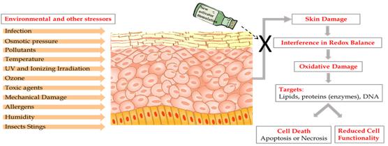
The human skin is constantly exposed to various pollutants, UV rays, radiation and other stressors that exist in our day-to-day environment. When they filter into the body they can create Reactive Oxygen Species (ROS) – oxygen molecules known as Free Radicals, which are able to damage and destroy cells, including lipids, proteins and DNA.

In the skin – the largest organ of the body – an excess of ROS can lead to various skin conditions, including inflammatory diseases, pigmenting disorders, wrinkles and some types of skin cancer, and can also affect internal organs. This damage is known as Oxidative Stress.

The body is naturally equipped with defense mechanisms to counter oxidative stress. It has anti-oxidants and, more importantly, anti-oxidant enzymes that attack the ROS before they cause damage.

In a review article published in the journal Cosmetics, a PhD student from The Hebrew University of Jerusalem, working in collaboration with researchers at the Technion – Israel Institute of Technology, suggested an innovative way to invigorate the body to produce antioxidant enzymes, while maintaining skin cell redox balance – a gentle equilibrium between Reactive Oxygen Species and their detoxification.

“The approach of using the body’s own defense system is very effective. We showed that activation of the body’s defense system with the aid of a unique delivery system is feasible, and may leverage dermal cure,” said Hebrew University researcher Maya Ben-Yehuda Greenwald.

Ben-Yehuda Greenwald showed that applying nano-size droplets of microemulsion liquids containing a cellular protective pathway inducer into the skin activates the natural skin defense systems.

“Currently, there are many scientific studies supporting the activation of the body’s defense mechanisms. However, none of these studies has demonstrated the use of a nanotechnology-based delivery system to do so,” Ben-Yehuda Greenwald said.

Production of antioxidant enzymes in the body is signaled in the DNA by activation of Nrf2 – a powerful protein that exists in every cell in our body. This Nrf2 cellular-protective signaling pathway is a major intersection of many other signaling pathways affecting each other and determining cell functionality and fate. Nrf2 is capable of coordinating the cellular response to internal as well as external stressors by tight regulation of phase-II protective enzymes, such as the antioxidant enzymes.

Ben-Yehuda Greenwald has also discovered a new family of compounds capable of activating the Nrf2 pathway. Moreover, by incorporating them into the unique delivery system she has developed, she managed to efficiently stimulate the activation of the Nrf2 pathway and mimic the activity of the body’s’ natural way of coping with a variety of stress conditions.

“The formula we have created could be used in topical medication for treating skin conditions. Our formula could be used both as preventive means and for treatment of various skin conditions, such as infections, over-exposure to UV irradiation, inflammatory conditions, and also internal disease,” she said.

While the researchers focused on the skin, the formulation could prove to be effective in enhancing the body’s natural protection against the damaging effects of ROS in other parts of the body, such as inflammation in cardiovascular diseases, heart attack, cancer, multiple sclerosis and Alzheimer’s.

Here’s an image provided by Ben-Yehuda Greenwald illustrating the team’s work,

Caption: These are the consequences of skin exposure to stressors. Credit: Maya Ben-Yehuda Greenwald

Caption: These are the consequences of skin exposure to stressors. Credit: Maya Ben-Yehuda Greenwald

Here’s a link to and a citation for the paper,

Skin Redox Balance Maintenance: The Need for an Nrf2-Activator Delivery System by Maya Ben-Yehuda Greenwald, Shmuel Ben-Sasson, Havazelet Bianco-Peled, and Ron Kohen. Cosmetics 2016, 3(1), 1; doi:10.3390/cosmetics3010001 Published: 15 January 2016

This paper appears to be open access.