Tag Archives: HIV

CRISPR/Cas9 used successfully to edit SIV (simian immunodeficiency virus, which is similar to HIV) out of monkey genome

Before reading further please note, the research discussed in this posting is based on animal testing, which many people find highly disturbing.

CRISPR (clustered regularly interspaced short palindromic repeats)/Cas9 (CRISPR-associated protein 9), or more familiarly CRISPR/Cas9, has been been used to edit simian immunodeficiency virus from infected monkeys’ cells according to a December 2, 2020 article by Matthew Rozsa for Salon.com (Note: Links have been removed),

With multiple coronavirus vaccines being produced as we speak, the COVID-19 pandemic appears to have an end in sight, though the HIV pandemic continues after more than 40 years. That might seem like a head-scratcher: why is HIV, a virus we’ve known about for decades, so much harder to cure than a virus discovered just last year? Part of the reason is that HIV, as a retrovirus, is a more complex virus to vaccinate against than SARS-CoV-2 — hence why a vaccine or other cure has eluded scientists for decades. 

Now, a surprising new study on a related retrovirus shows incredible promise for the potential to develop a cure for HIV, or human immunodeficiency virus. In an article published in the scientific journal Nature Communications, scientists revealed that they had used CRISPR – a genetic technology that can alter DNA and whose developers won the 2020 Nobel Prize in Chemistry [specifically, Jennifer Doudna and Emanuelle Charpentier received the Nobel for developing CRISPR-cas9 or CRISPR/Cas9 not CRISPR alone) — to successfully edit SIV (simian immunodeficiency virus), a virus similar to HIV, out of the genomes of non-human primates.  Specifically, the scientists were able to edit out the SIV genome from rhesus macaque monkeys’ infected cells.

For anyone who’s interested in how CRISPR was developed and the many contributions which have led to the current state-of-the-art for CRISPR gene editing, see the History subsection of Wikipedia’s CRISPR entry.

Getting back to Rozsa’s December 2, 2020 article,

“This study used the CRISPR CaS9 system, which has been described as molecular scissors,” Andrew G. MacLean, PhD, wrote to Salon. MacLean is an associate professor at the Tulane National Primate Research Center and the Department of Microbiology and Immunology at Tulane University School of Medicine and was a senior co-investigator of the study. “It uses a highly specific targeting system to cut out a specific portion of DNA that is necessary for HIV to be able to produce more virus.”

He added, “Our collaborators at in the Khalili Lab at Temple University have developed a method of ‘packaging’ this within a single so-called vector. A vector is a non-disease causing virus that is used as a carrier for the CRISPR CaS9 scissors to get it into the tissues of interest.”

The experiments with SIV are considered to be a gateway to understanding HIV, as HIV is believed to have evolved from SIV, and is genetically similar.

“The rhesus macaque model of HIV/AIDS is the most valuable model to test efficacy of new interventions or approaches for preventing or treating HIV infection, prior to human clinical trials,” Binhua Ling, PhD, associate professor at the Southwest National Primate Research Center, Texas Biomedical Research Institute, wrote to Salon. “This first proof-of-principal [emphasis mine] study on the rhesus macaque model indicates that this virus-vehicle-delivered-CRISPR system can reach many tissue sites of the body, and is able to effectively delete virus DNA in infected cells. This paves the way for applying the same technology to the human body, which could lead to a cure for HIV infection.”

Tricia H. Burdo, PhD, another senior co-investigator on the new study who works at the Lewis Katz School of Medicine at Temple University, explained to Salon by email that “HIV is in a class of viruses (retroviruses) that inserts itself into the DNA of the host, so you can really think of this now as a genetic disease” — in other words, the kind of thing that would be ripe for CRISPR’s scissors-like ability to remove errant or unwanted genetic material. Burdo notes that the CRISPR technology discussed in the article “cuts out this foreign viral gene.”

The study was conducted on eight Rhesus macaque monkeys. That is a very small number to start with and not all of the monkeys received the CRISPR/Cas9 treatment. From the ‘Animals used in the study and ethical statement‘ subsection of the study, “Animals were sacrificed for tissue collection 3 weeks after … .” Leaving aside how anyone may feel about ‘sacrificing …’, three weeks is not a long time for observation.

Should you be interested, there is a November 30, 2020 Tulane University news release announcing the research.

If you want to read the whole study, here’s a link and a citation,

CRISPR based editing of SIV proviral DNA in ART treated non-human primates by Pietro Mancuso, Chen Chen, Rafal Kaminski, Jennifer Gordon, Shuren Liao, Jake A. Robinson, Mandy D. Smith, Hong Liu, Ilker K. Sariyer, Rahsan Sariyer, Tiffany A. Peterson, Martina Donadoni, Jaclyn B. Williams, Summer Siddiqui, Bruce A. Bunnell, Binhua Ling, Andrew G. MacLean, Tricia H. Burdo & Kamel Khalili. Nature Communications volume 11, Article number: 6065 (2020) DOI: https://doi.org/10.1038/s41467-020-19821-7 Published: 27 November 2020

This paper is open access.

As Rozsa notes in his December 2, 2020 article, the Joint United Nations Programme on HIV/AIDS estimates that 32.7 million [24.8 million–42.2 million] people have died from AIDS-related illnesses since the start (1981?) of the epidemic to the end of 2019.

Gold nanoparticle loaded with CRISPR used to edit genes

CRISPR (clustered regularly interspaced short palindromic repeats) gene editing is usually paired with a virus (9, 12a, etc.) but this time scientists are using a gold nanoparticle. From a May 27, 2019 news item on Nanowerk (Note: Links have been removed),

Scientists at Fred Hutchinson Cancer Research Center took a step toward making gene therapy more practical by simplifying the way gene-editing instructions are delivered to cells. Using a gold nanoparticle instead of an inactivated virus, they safely delivered gene-editing tools in lab models of HIV and inherited blood disorders, as reported in Nature Materials (“Targeted homology-directed repair in blood stem and progenitor cells with CRISPR nanoformulations”).

A May 27, 2019 Fred Hutchinson Cancer Research Center news release (also on EurekAlert) by Jake Siegel, which originated the news item, expands on the theme, provides more detail,

It’s the first time that a gold nanoparticle loaded with CRISPR has been used to edit genes in a rare but powerful subset of blood stem cells, the source of all blood cells. The CRISPR-carrying gold nanoparticle led to successful gene editing in blood stem cells with no toxic effects.

“As gene therapies make their way through clinical trials and become available to patients, we need a more practical approach,” said senior author Dr. Jennifer Adair, an assistant member of the Clinical Research Division at Fred Hutch, adding that current methods of performing gene therapy are inaccessible to millions of people around the world. “I wanted to find something simpler, something that would passively deliver gene editing to blood stem cells.”

While CRISPR has made it faster and easier to precisely deliver genetic modifications to the genome, it still has challenges. Getting cells to accept CRISPR gene-editing tools involves a small electric shock that can damage and even kill the cells. And if precise gene edits are required, then additional molecules must be engineered to deliver them –adding cost and time.

Gold nanoparticles are a promising alternative because the surface of these tiny spheres (around 1 billionth the size of a grain of table salt) allows other molecules to easily stick to them and stay adhered.

“We engineered the gold nanoparticles to quickly cross the cell membrane, dodge cell organelles that seek to destroy them and go right to the cell nucleus to edit genes,” said Dr. Reza Shahbazi, a Fred Hutch postdoctoral researcher who has worked with gold nanoparticles for drug and gene delivery for seven years.

Shahbazi made the gold particles from laboratory-grade gold that is purified and comes as a liquid in a small lab bottle. He mixed the purified gold into a solution that causes the individual gold ions to form tiny particles, which the researchers then measured for size.

They found that a particular size – 19 nanometers wide – was the best for being big and sticky enough to add gene-editing materials to the surface of the particles, while still being small enough for cells to absorb them.

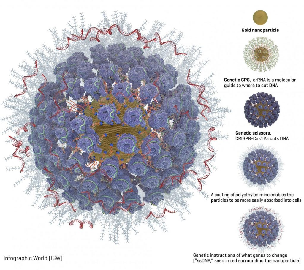
Packed onto the gold particles, the Fred Hutch team added these gene-editing components (diagram available [see below]):

A type of molecular guide called crRNA acts as a genetic GPS to show the CRISPR complex where in the genome to make the cut.

CRISPR nuclease protein, often called “genetic scissors,” makes the cut in the DNA. The CRISPR nuclease protein most often used is Cas9. But the Fred Hutch researchers also studied Cas12a (formerly called Cpf1) because Cas12a makes a staggered cut in DNA. The researchers hoped this would allow the cells to more efficiently repair the cut and while so doing embed the new genetic instructions into the cell. Another advantage of Cas12a over Cas9 is that it only requires one molecular guide, which is important because of space constraints on the nanoparticles. Cas9 requires two molecular guides.

Instructions for what genetic changes to make (“ssDNA”). The Fred Hutch team chose two inherited genetic changes that bestow protection from disease: CCR5, which protects against HIV, and gamma hemoglobin, which protects against blood disorders such as sickle cell disease and thalassemia.

A coating of a polyethylenimine swarms the surface of the particles to give them a more positive charge, which enables them to more readily be absorbed into cells. This is an improvement over another method of getting cells to take up gene editing tools, called electroporation, which involves lightly shocking the cells to get them to open and allow the genetic instructions to enter.

Then the researchers isolated blood stem cells with a protein marker on their surface called CD34. These CD34-positive cells contain the blood-making progenitor cells that give rise to the entire blood and immune system.

“These cells replenish blood in the body every day, making them a good candidate for one-time gene therapy because it will last a lifetime as the cells replace themselves,” Adair said.

Observing human blood stem cells in a lab dish, the researchers found that their fully loaded gold nanoparticles were taken up naturally by cells within six hours of being added and within 24 to 48 hours they could see gene editing happening. They observed that the Cas12a CRISPR protein partner was better at delivering very precise genetic edits to the cells than the more commonly used cas9 protein partner.

The gene-editing effect reached a peak eight weeks after the researchers injected the cells into mouse models; 22 weeks after injection the edited cells were still there. The Fred Hutch researchers also found edited cells in the bone marrow, spleen and thymus of the mouse models, a sign that the dividing blood cells in those organs could carry on the treatment without the mice having to be treated again.

“We believe we have a good candidate for two diseases — HIV and hemoglobinopathies — though we are also evaluating other disease targets where small genetic changes can have a big impact, as well as ways to make bigger genetic changes,” Adair said. “The next step is to increase how much gene editing happens in each cell, which is definitely doable. That will make it closer to being an effective therapy.”

In the study, the researchers report 10 to 20 percent of cells took on the gene edits, which is a promising start, but the researchers would like to aim for 50% or more of the cells being edited, which they believe will have a good chance of combatting these diseases.

###

Adair and Shahbazi are looking for commercial partners to develop the technology into therapies for people. They hope to begin clinical trials within a few years.

Here’s the diagram of a gold nanoparticle loaded with CRISPR,

Caption: Graphic of a fully loaded gold nanoparticle with CRISPR and other gene editing tools. Credit: Image courtesy of the Adair lab at Fred Hutch.

Here’s a link to and a citation for the paper,

Targeted homology-directed repair in blood stem and progenitor cells with CRISPR nanoformulations by Reza Shahbazi, Gabriella Sghia-Hughes, Jack L. Reid, Sara Kubek, Kevin G. Haworth, Olivier Humbert, Hans-Peter Kiem & Jennifer E. Adair. Nature Materials (2019) DOI https://doi.org/10.1038/s41563-019-0385-5Published 27 May 2019

This paper is behind a paywall.

Greater mortality for the CRISPR twins Lulu and Nana?

Every time I think this CRISPR (clustered regularly interspaced short palindromic repeats) story is winding down, something new happens. The latest (I think) is in a June 3, 2019 news item on ScienceDaily,

A genetic mutation that a Chinese scientist attempted to create in twin babies born last year, ostensibly to help them fend off HIV infection, is also associated with a 21% increase in mortality in later life, according to an analysis by University of California, Berkeley, scientists.

The researchers scanned more than 400,000 genomes and associated health records contained in a British database, UK Biobank, and found that people who had two mutated copies of the gene had a significantly higher death rate between ages 41 and 78 than those with one or no copies.

Sarah Zhang’s June 3, 2019 article for The Atlantic provides an overview of the situation before exploring the current controversy,

In the 1990s, virologists in New York learned of a genetic mutation that would become one of the most famous ever discovered. They found it in a man who could not be infected with HIV. He turned out to be missing just 32 letters in a gene called CCR5, and remarkably, it was enough to make him resistant to the virus killing so many others. About 1 percent of people of European descent carry two copies of this mutation, now known as CCR5-Δ32.

In 2018, a Chinese scientist named He Jiankui made the mutation infamous when he attempted to use CRISPR to edit CCR5-Δ32 (pronounced “CCR5-delta-32”) into human embryos. He chose this mutation, he said, because the babies’ father was HIV-positive, and he wanted to make the resulting twin girls resistant to the virus. CCR5-Δ32 is also, after all, one of the most studied mutations.

He’s work immediately provoked outrage among scientists, who knew enough to know how much they did not know about the risks of altering CCR5. And now a new study suggests that CCR5-Δ32 is indeed harmful overall.

The girls’ CCR5 genes were altered, according to data He presented, but they do not exactly match the 32-letter deletion; it’s unclear whether either of them is actually resistant to HIV. Even if they were unable to get HIV, a body of research already suggested that CCR5-Δ32 made people more vulnerable to the flu and West Nile virus. A “good” mutation in the context of HIV can be “bad” in another context. No one knew, exactly, the net effect of a CCR5-Δ32 mutation.

For some reason, Zhang makes no mention of the possibly enhanced cognitive abilities that the twins may have as a consequence of the gene editing assuming that He Jiankui successfully edited the genes. (To my knowledge, the results and data have not been released for review by colleagues.)

Regardless, Zhang’s article provides a handy overview and update.

For anyone who’s interested in more detail about this latest research into mortality and CCR5, there’s a June 3, 2019 University of California at Berkeley news release (also on EurekAlert) by Robert Sanders, which also originated the ScienceDaily news item, details the latest research,

Previous studies have associated two mutated copies of the gene, CCR5, with a fourfold increase in the death rate after influenza infection, and the higher overall mortality rate may reflect this greater susceptibility to death from the flu. But the researchers say there could be any number of explanations, since the protein that CCR5 codes for, and which no longer works in those having the mutation in both copies of the gene, is involved in many body functions.

“Beyond the many ethical issues involved with the CRISPR babies, the fact is that, right now, with current knowledge, it is still very dangerous to try to introduce mutations without knowing the full effect of what those mutations do,” said Rasmus Nielsen, a UC Berkeley professor of integrative biology. “In this case, it is probably not a mutation that most people would want to have. You are actually, on average, worse off having it.”

“Because one gene could affect multiple traits, and because, depending on the environment, the effects of a mutation could be quite different, I think there can be many uncertainties and unknown effects in any germline editing,” said postdoctoral fellow Xinzhu “April” Wei.
Wei is first author and Nielsen is senior author of a paper describing the research that will appear online on Monday, June 3, in the journal Nature Medicine.

Mutation prevents HIV infection

The gene CCR5 codes for a protein that, among other things, sits on the surface of immune cells and helps some strains of HIV, including the most common ones, to enter and infect them. Jiankui He, the Chinese scientist who last November shocked the world by announcing he had experimented with CCR5 on at least two babies, said he wanted to introduce a mutation in the gene that would prevent this. Naturally-occurring mutations that disable the protein are rare in Asians, but a mutation found in about 11% of Northern Europeans protects them against HIV infection.

The genetic mutation, ∆32 (Delta 32), refers to a missing 32-base-pair segment in the CCR5 gene. This mutation interferes with the localization on the cell surface of the protein for which CCR5 codes, thwarting HIV binding and infection. He was unable to duplicate the natural mutation, but appears to have generated a similar deletion that would also inactivate the protein. One of the twin babies reportedly had one copy of CCR5 modified by CRISPR-Cas9 gene editing, while the other baby had both copies edited.

But inactivating a protein found in all humans and most animals is likely to have negative effects, Nielsen said, especially when done to both copies of the gene — a so-called homozygous mutation

“Here is a functional protein that we know has an effect in the organism, and it is well-conserved among many different species, so it is likely that a mutation that destroys the protein is, on average, not good for you,” he said. “Otherwise, evolutionary mechanisms would have destroyed that protein a long time ago.”

After He’s experiment became public, Nielsen and Wei, who study current genetic variation to understand the origin of human, animal and plant traits, decided to investigate the effect of the CCR5-∆32 mutation using data from UK Biobank. The database houses genomic information on a half million U.K. citizens that is linked to their medical records. The genomic information is much like that acquired by Ancestry.com and 23andMe: details on nearly a million individual variations in the genetic sequence, so-called single nucleotide polymorphisms (SNPs).

Two independent measures indicated a higher mortality rate for those with two mutated genes. Fewer people than expected with two mutations enrolled in the database, indicating that they had died at a higher rate than the general population. And fewer than expected survived from ages 40 to 78.

“Both the proportions before enrollment and the survivorship after enrollment tell the same story, which is that you have lower survivability or higher mortality if you have two copies of the mutation,” Nielsen said. “There is simply a deficiency of individuals with two copies.”

Because the ∆32 mutation is relatively common in Northern Europeans, it must have been favored by natural selection at some point, Nielsen said, though probably not to protect against HIV, since the virus has circulated among humans only since the 1980s.

Wei said that some evidence links the mutation to increased survival after stroke and protection against smallpox and flaviviruses, a group that includes the dengue, Zika and West Nile viruses.

Despite these possible benefits, the potential unintended effects of creating genetic mutations, in both adult somatic cells and in embryonic, germline cells, argue for caution, the researchers said.

“I think there are a lot of things that are unknown at the current stage about genes’ functions,” Wei said. “The CRISPR technology is far too dangerous to use right now for germline editing.”

Here’s a link to and a citation for the latest paper,

CCR5-∆32 is deleterious in the homozygous state in humans by Xinzhu Wei & Rasmus Nielsen. Nature Medicine (2019) DOI: https://doi.org/10.1038/s41591-019-0459-6 Published 03 June 2019

This paper is behind a paywall.

For those who have an insatiable appetite for detail, there’s my November 28, 2018 posting which covers what happened when the CRISPR twins, Lulu and Nana, was first announced, along with a few updates to January 23, 2019. The May 17, 2019 posting covers the news of possible cognitive advantages for the CCR5-Δ32 gene-edited twins and explores some of the social implications.

The CRISPR ((clustered regularly interspaced short palindromic repeats)-CAS9 gene-editing technique may cause new genetic damage kerfuffle

Setting the stage

Not unexpectedly, CRISPR-Cas9  or clustered regularly interspaced short palindromic repeats-CRISPR-associated protein 9 can be dangerous as these scientists note in a July 16, 2018 news item on phys.org,

Scientists at the Wellcome Sanger Institute have discovered that CRISPR/Cas9 gene editing can cause greater genetic damage in cells than was previously thought. These results create safety implications for gene therapies using CRISPR/Cas9 in the future as the unexpected damage could lead to dangerous changes in some cells.

Reported today (16 July 2018) in the journal Nature Biotechnology, the study also revealed that standard tests for detecting DNA changes miss finding this genetic damage, and that caution and specific testing will be required for any potential gene therapies.

This CRISPR-Cas9 image reminds me of popcorn,

CRISPR-associated protein Cas9 (white) from Staphylococcus aureus based on Protein Database ID 5AXW. Credit: Thomas Splettstoesser (Wikipedia, CC BY-SA 4.0)[ downloaded from https://phys.org/news/2018-07-genome-crisprcas9-gene-higher-thought.html#jCp]

A July 16, 2018 Wellcome Sanger Institute press release (also on EurekAlert), which originated the news item, offers a little more explanation,

CRISPR/Cas9 is one of the newest genome editing tools. It can alter sections of DNA in cells by cutting at specific points and introducing changes at that location. Already extensively used in scientific research, CRISPR/Cas9 has also been seen as a promising way to create potential genome editing treatments for diseases such as HIV, cancer or sickle cell disease. Such therapeutics could inactivate a disease-causing gene, or correct a genetic mutation. However, any potential treatments would have to prove that they were safe.

Previous research had not shown many unforeseen mutations from CRISPR/Cas9 in the DNA at the genome editing target site. To investigate this further the researchers carried out a full systematic study in both mouse and human cells and discovered that CRISPR/Cas9 frequently caused extensive mutations, but at a greater distance from the target site.

The researchers found many of the cells had large genetic rearrangements such as DNA deletions and insertions. These could lead to important genes being switched on or off, which could have major implications for CRISPR/Cas9 use in therapies. In addition, some of these changes were too far away from the target site to be seen with standard genotyping methods.

Prof Allan Bradley, corresponding author on the study from the Wellcome Sanger Institute, said: “This is the first systematic assessment of unexpected events resulting from CRISPR/Cas9 editing in therapeutically relevant cells, and we found that changes in the DNA have been seriously underestimated before now. It is important that anyone thinking of using this technology for gene therapy proceeds with caution, and looks very carefully to check for possible harmful effects.”

Michael Kosicki, the first author from the Wellcome Sanger Institute, said: “My initial experiment used CRISPR/Cas9 as a tool to study gene activity, however it became clear that something unexpected was happening. Once we realised the extent of the genetic rearrangements we studied it systematically, looking at different genes and different therapeutically relevant cell lines, and showed that the CRISPR/Cas9 effects held true.”

The work has implications for how CRISPR/Cas9 is used therapeutically and is likely to re-spark researchers’ interest in finding alternatives to the standard CRISPR/Cas9 method for gene editing.

Prof Maria Jasin, an independent researcher from Memorial Slone Kettering Cancer Centre, New York, who was not involved in the study said: “This study is the first to assess the repertoire of genomic damage arising at a CRISPR/Cas9 cleavage site. While it is not known if genomic sites in other cell lines will be affected in the same way, this study shows that further research and specific testing is needed before CRISPR/Cas9 is used clinically.”

For anyone who’d like to better understand the terms gene editing and CRISPR-Cas9, the Wellcome Sanger Institute provides these explanatory webpages, What is genome editing? and What is CRISPR-Cas9?

For the more advanced, here’s a link and a citation for the paper,

Repair of double-strand breaks induced by CRISPR–Cas9 leads to large deletions and complex rearrangements by Michael Kosicki, Kärt Tomberg, & Allan Bradley. Nature Biotechnology DOI: https://doi.org/10.1038/nbt.4192 Published 16 July 2018

This paper appears to be open access.

The kerfuffle

It seems this news has affected the CRISPR market. From a July 16, 2018 article by Cale Guthrie Weissman for Fast Company,

… CRISPR could unknowingly delete or alter non-targeted genes, which could lead to myriad unintended consequences. This is especially frightening, since the technology is going to be used in human clinical trials.

Meanwhile, other scientists working with CRISPR are trying to downplay the findings, telling STAT [a life sciences and business journalism website] that there have been no reported adverse effects similar to what the study describes. The news, however, has brought about a market reaction–at least three publicly traded companies that focus on CRISPR-based therapies are in stock nosedive. Crispr Therapeutics is down by over 6%; Editas fell by over 3%; and Intellia Therapeutics dropped by over 5%. [emphasis mine]

Damage control

Gaetan Burgio (geneticist, Australian National University)  in a July 16, 2018 essay on phys.org (originating from The Conversation) suggests some calm (Note: Links have been removed),

But a new study has called into question the precision of the technique [CRISPR gene editing technology].

The hope for gene editing is that it will be able to cure and correct diseases. To date, many successes have been reported, including curing deafness in mice, and in altering cells to cure cancer.

Some 17 clinical trials in human patients are registered [emphasis mine] testing gene editing on leukaemias, brain cancers and sickle cell anaemia (where red blood cells are misshaped, causing them to die). Before implementing CRISPR technology in clinics to treat cancer or congenital disorders, we must address whether the technique is safe and accurate.

There are a few options for getting around this problem. One option is to isolate the cells we wish to edit from the body and reinject only the ones we know have been correctly edited.

For example, lymphocytes (white blood cells) that are crucial to killing cancer cells could be taken out of the body, then modified using CRISPR to heighten their cancer-killing properties. The DNA of these cells could be sequenced in detail, and only the cells accurately and specifically gene-modified would be selected and delivered back into the body to kill the cancer cells.

While this strategy is valid for cells we can isolate from the body, some cells, such as neurons and muscles, cannot be removed from the body. These types of cells might not be suitable for gene editing using Cas9 scissors.

Fortunately, researchers have discovered other forms of CRISPR systems that don’t require the DNA to be cut. Some CRISPR systems only cut the RNA, not the DNA (DNA contains genetic instructions, RNA convey the instructions on how to synthesise proteins).

As RNA [ribonucleic acid] remains in our cells only for a specific period of time before being degraded, this would allow us to control the timing and duration of the CRISPR system delivery and reverse it (so the scissors are only functional for a short period of time).

This was found to be successful for dementia in mice. Similarly, some CRISPR systems simply change the letters of the DNA, rather than cutting them. This was successful for specific mutations causing diseases such as hereditary deafness in mice.

I agree with Burgio’s conclusion (not included here) that we have a lot more to learn and I can’t help wondering why there are 17 registered human clinical trials at this point.

Yes! Art, genetic modifications, gene editing, and xenotransplantation at the Vancouver Biennale (Canada)

Patricia Piccinini’s Curious Imaginings Courtesy: Vancouver Biennale [downloaded from http://dailyhive.com/vancouver/vancouver-biennale-unsual-public-art-2018/]

Up to this point, I’ve been a little jealous of the Art/Sci Salon’s (Toronto, Canada) January 2018 workshops for artists and discussions about CRISPR ((clustered regularly interspaced short palindromic repeats))/Cas9 and its social implications. (See my January 10, 2018 posting for more about the events.) Now, it seems Vancouver may be in line for its ‘own’ discussion about CRISPR and the implications of gene editing. The image you saw (above) represents one of the installations being hosted by the 2018 – 2020 edition of the Vancouver Biennale.

While this posting is mostly about the Biennale and Piccinini’s work, there is a ‘science’ subsection featuring the science of CRISPR and xenotransplantation. Getting back to the Biennale and Piccinini: A major public art event since 1988, the Vancouver Biennale has hosted over 91 outdoor sculptures and new media works by more than 78 participating artists from over 25 countries and from 4 continents.

Quickie description of the 2018 – 2020 Vancouver Biennale

The latest edition of the Vancouver Biennale was featured in a June 6, 2018 news item on the Daily Hive (Vancouver),

The Vancouver Biennale will be bringing new —and unusual— works of public art to the city beginning this June.

The theme for this season’s Vancouver Biennale exhibition is “re-IMAGE-n” and it kicks off on June 20 [2018] in Vanier Park with Saudi artist Ajlan Gharem’s Paradise Has Many Gates.

Gharem’s architectural chain-link sculpture resembles a traditional mosque, the piece is meant to challenge the notions of religious orthodoxy and encourages individuals to image a space free of Islamophobia.

Melbourne artist Patricia Piccinini’s Curious Imaginings is expected to be one of the most talked about installations of the exhibit. Her style of “oddly captivating, somewhat grotesque, human-animal hybrid creature” is meant to be shocking and thought-provoking.

Piccinini’s interactive [emphasis mine] experience will “challenge us to explore the social impacts of emerging biotechnology and our ethical limits in an age where genetic engineering and digital technologies are already pushing the boundaries of humanity.”

Piccinini’s work will be displayed in the 105-year-old Patricia Hotel in Vancouver’s Strathcona neighbourhood. The 90-day ticketed exhibition [emphasis mine] is scheduled to open this September [2018].

Given that this blog is focused on nanotechnology and other emerging technologies such as CRISPR, I’m focusing on Piccinini’s work and its art/science or sci-art status. This image from the GOMA Gallery where Piccinini’s ‘Curious Affection‘ installation is being shown from March 24 – Aug. 5, 2018 in Brisbane, Queensland, Australia may give you some sense of what one of her installations is like,

Courtesy: Queensland Art Gallery | Gallery of Modern Art (QAGOMA)

I spoke with Serena at the Vancouver Biennale office and asked about the ‘interactive’ aspect of Piccinini’s installation. She suggested the term ‘immersive’ as an alternative. In other words, you won’t be playing with the sculptures or pressing buttons and interacting with computer screens or robots. She also noted that the ticket prices have not been set yet and they are currently developing events focused on the issues raised by the installation. She knew that 2018 is the 200th anniversary of the publication of Mary Shelley’s Frankenstein but I’m not sure how the Biennale folks plan (or don’t plan)  to integrate any recognition of the novle’s impact on the discussions about ‘new’ technologies .They expect Piccinini will visit Vancouver. (Note 1: Piccinini’s work can  also be seen in a group exhibition titled: Frankenstein’s Birthday Party at the Hosfselt Gallery in San Francisco (California, US) from June 23 – August 11, 2018.  Note 2: I featured a number of international events commemorating the 200th anniversary of the publication of Mary Shelley’s novel, Frankenstein, in my Feb. 26, 2018 posting. Note 3: The term ‘Frankenfoods’ helped to shape the discussion of genetically modified organisms and food supply on this planet. It was a wildly successful campaign for activists affecting legislation in some areas of research. Scientists have not been as enthusiastic about the effects. My January 15, 2009 posting briefly traces a history of the term.)

The 2018 – 2020 Vancouver Biennale and science

A June 7, 2018 Vancouver Biennale news release provides more detail about the current series of exhibitions,

The Biennale is also committed to presenting artwork at the cutting edge of discussion and in keeping with the STEAM (science, technology, engineering, arts, math[ematics]) approach to integrating the arts and sciences. In August [2018], Colombian/American visual artist Jessica Angel will present her monumental installation Dogethereum Bridge at Hinge Park in Olympic Village. Inspired by blockchain technology, the artwork’s design was created through the integration of scientific algorithms, new developments in technology, and the arts. This installation, which will serve as an immersive space and collaborative hub for artists and technologists, will host a series of activations with blockchain as the inspirational jumping-off point.

In what is expected to become one of North America’s most talked-about exhibitions of the year, Melbourne artist Patricia Piccinini’s Curious Imaginings will see the intersection of art, science, and ethics. For the first time in the Biennale’s fifteen years of creating transformative experiences, and in keeping with the 2018-2020 theme of “re-IMAGE-n,” the Biennale will explore art in unexpected places by exhibiting in unconventional interior spaces.  The hyperrealist “world of oddly captivating, somewhat grotesque, human-animal hybrid creatures” will be the artist’s first exhibit in a non-museum setting, transforming a wing of the 105-year-old Patricia Hotel. Situated in Vancouver’s oldest neighbourbood of Strathcona, Piccinini’s interactive experience will “challenge us to explore the social impacts of emerging bio-technology and our ethical limits in an age where genetic engineering and digital technologies are already pushing the boundaries of humanity.” In this intimate hotel setting located in a neighborhood continually undergoing its own change, Curious Imaginings will empower visitors to personally consider questions posed by the exhibition, including the promises and consequences of genetic research and human interference. …

There are other pieces being presented at the Biennale but my special interest is in the art/sci pieces and, at this point, CRISPR.

Piccinini in more depth

You can find out more about Patricia Piccinini in her biography on the Vancouver Biennale website but I found this Char Larsson April 7, 2018 article for the Independent (UK) more informative (Note: A link has been removed),

Patricia Piccinini’s sculptures are deeply disquieting. Walking through Curious Affection, her new solo exhibition at Brisbane’s Gallery of Modern Art, is akin to entering a science laboratory full of DNA experiments. Made from silicone, fibreglass and even human hair, her sculptures are breathtakingly lifelike, however, we can’t be sure what life they are like. The artist creates an exuberant parallel universe where transgenic experiments flourish and human evolution has given way to genetic engineering and DNA splicing.

Curious Affection is a timely and welcome recognition of Piccinini’s enormous contribution to reaching back to the mid-1990s. Working across a variety of mediums including photography, video and drawing, she is perhaps best known for her hyperreal creations.

As a genre, hyperrealism depends on the skill of the artist to create the illusion of reality. To be truly successful, it must convince the spectator of its realness. Piccinini acknowledges this demand, but with a delightful twist. The excruciating attention to detail deliberately solicits our desire to look, only to generate unease, as her sculptures are imbued with a fascinating otherness. Part human, part animal, the works are uncannily familiar, but also alarmingly “other”.

Inspired by advances in genetically modified pigs to generate replacement organs for humans [also known as xenotransplantation], we are reminded that Piccinini has always been at the forefront of debates concerning the possibilities of science, technology and DNA cloning. She does so, however, with a warm affection and sense of humour, eschewing the hysterical anxiety frequently accompanying these scientific developments.

Beyond the astonishing level of detail achieved by working with silicon and fibreglass, there is an ethics at work here. Piccinini is asking us not to avert our gaze from the other, and in doing so, to develop empathy and understanding through the encounter.

I encourage anyone who’s interested to read Larsson’s entire piece (April 7, 2018 article).

According to her Wikipedia entry, Piccinini works in a variety of media including video, sound, sculpture, and more. She also has her own website.

Gene editing and xenotransplantation

Sarah Zhang’s June 8, 2018 article for The Atlantic provides a peek at the extraordinary degree of interest and competition in the field of gene editing and CRISPR ((clustered regularly interspaced short palindromic repeats))/Cas9 research (Note: A link has been removed),

China Is Genetically Engineering Monkeys With Brain Disorders

Guoping Feng applied to college the first year that Chinese universities reopened after the Cultural Revolution. It was 1977, and more than a decade’s worth of students—5.7 million—sat for the entrance exams. Feng was the only one in his high school to get in. He was assigned—by chance, essentially—to medical school. Like most of his contemporaries with scientific ambitions, he soon set his sights on graduate studies in the United States. “China was really like 30 to 50 years behind,” he says. “There was no way to do cutting-edge research.” So in 1989, he left for Buffalo, New York, where for the first time he saw snow piled several feet high. He completed his Ph.D. in genetics at the State University of New York at Buffalo.

Feng is short and slim, with a monk-like placidity and a quick smile, and he now holds an endowed chair in neuroscience at MIT, where he focuses on the genetics of brain disorders. His 45-person lab is part of the McGovern Institute for Brain Research, which was established in 2000 with the promise of a $350 million donation, the largest ever received by the university. In short, his lab does not lack for much.

Yet Feng now travels to China several times a year, because there, he can pursue research he has not yet been able to carry out in the United States. [emphasis mine] …

Feng had organized a symposium at SIAT [Shenzhen Institutes of Advanced Technology], and he was not the only scientist who traveled all the way from the United States to attend: He invited several colleagues as symposium speakers, including a fellow MIT neuroscientist interested in tree shrews, a tiny mammal related to primates and native to southern China, and Chinese-born neuroscientists who study addiction at the University of Pittsburgh and SUNY Upstate Medical University. Like Feng, they had left China in the ’80s and ’90s, part of a wave of young scientists in search of better opportunities abroad. Also like Feng, they were back in China to pursue a type of cutting-edge research too expensive and too impractical—and maybe too ethically sensitive—in the United States.

Here’s what precipitated Feng’s work in China, (from Zhang’s article; Note: Links have been removed)

At MIT, Feng’s lab worked on genetically engineering a monkey species called marmosets, which are very small and genuinely bizarre-looking. They are cheaper to keep due to their size, but they are a relatively new lab animal, and they can be difficult to train on lab tasks. For this reason, Feng also wanted to study Shank3 on macaques in China. Scientists have been cataloging the social behavior of macaques for decades, making it an obvious model for studies of disorders like autism that have a strong social component. Macaques are also more closely related to humans than marmosets, making their brains a better stand-in for those of humans.

The process of genetically engineering a macaque is not trivial, even with the advanced tools of CRISPR. Researchers begin by dosing female monkeys with the same hormones used in human in vitro fertilization. They then collect and fertilize the eggs, and inject the resulting embryos with CRISPR proteins using a long, thin glass needle. Monkey embryos are far more sensitive than mice embryos, and can be affected by small changes in the pH of the injection or the concentration of CRISPR proteins. Only some of the embryos will have the desired mutation, and only some will survive once implanted in surrogate mothers. It takes dozens of eggs to get to just one live monkey, so making even a few knockout monkeys required the support of a large breeding colony.

The first Shank3 macaque was born in 2015. Four more soon followed, bringing the total to five.

To visit his research animals, Feng now has to fly 8,000 miles across 12 time zones. It would be a lot more convenient to carry out his macaque research in the United States, of course, but so far, he has not been able to.

He originally inquired about making Shank3 macaques at the New England Primate Research Center, one of eight national primate research centers then funded by the National Institutes of Health in partnership with a local institution (Harvard Medical School, in this case). The center was conveniently located in Southborough, Massachusetts, just 20 miles west of the MIT campus. But in 2013, Harvard decided to shutter the center.

The decision came as a shock to the research community, and it was widely interpreted as a sign of waning interest in primate research in the United States. While the national primate centers have been important hubs of research on HIV, Zika, Ebola, and other diseases, they have also come under intense public scrutiny. Animal-rights groups like the Humane Society of the United States have sent investigators to work undercover in the labs, and the media has reported on monkey deaths in grisly detail. Harvard officially made its decision to close for “financial” reasons. But the announcement also came after the high-profile deaths of four monkeys from improper handling between 2010 and 2012. The deaths sparked a backlash; demonstrators showed up at the gates. The university gave itself two years to wind down their primate work, officially closing the center in 2015.

“They screwed themselves,” Michael Halassa, the MIT neuroscientist who spoke at Feng’s symposium, told me in Shenzhen. Wei-Dong Yao, another one of the speakers, chimed in, noting that just two years later CRISPR has created a new wave of interest in primate research. Yao was one of the researchers at Harvard’s primate center before it closed; he now runs a lab at SUNY Upstate Medical University that uses genetically engineered mouse and human stem cells, and he had come to Shenzhen to talk about restarting his addiction research on primates.

Here’s comes the competition (from Zhang’s article; Note: Links have been removed),

While the U.S. government’s biomedical research budget has been largely flat, both national and local governments in China are eager to raise their international scientific profiles, and they are shoveling money into research. A long-rumored, government-sponsored China Brain Project is supposed to give neuroscience research, and primate models in particular, a big funding boost. Chinese scientists may command larger salaries, too: Thanks to funding from the Shenzhen local government, a new principal investigator returning from overseas can get 3 million yuan—almost half a million U.S. dollars—over his or her first five years. China is even finding success in attracting foreign researchers from top U.S. institutions like Yale.

In the past few years, China has seen a miniature explosion of genetic engineering in monkeys. In Kunming, Shanghai, and Guangzhou, scientists have created monkeys engineered to show signs of Parkinson’s, Duchenne muscular dystrophy, autism, and more. And Feng’s group is not even the only one in China to have created Shank3 monkeys. Another group—a collaboration primarily between researchers at Emory University and scientists in China—has done the same.

Chinese scientists’ enthusiasm for CRISPR also extends to studies of humans, which are moving much more quickly, and in some cases under less oversight, than in the West. The first studies to edit human embryos and first clinical trials for cancer therapies using CRISPR have all happened in China. [emphases mine]

Some ethical issues are also covered (from Zhang’s article),

Parents with severely epileptic children had asked him if it would be possible to study the condition in a monkey. Feng told them what he thought would be technically possible. “But I also said, ‘I’m not sure I want to generate a model like this,’” he recalled. Maybe if there were a drug to control the monkeys’ seizures, he said: “I cannot see them seizure all the time.”

But is it ethical, he continued, to let these babies die without doing anything? Is it ethical to generate thousands or millions of mutant mice for studies of brain disorders, even when you know they will not elucidate much about human conditions?

Primates should only be used if other models do not work, says Feng, and only if a clear path forward is identified. The first step in his work, he says, is to use the Shank3 monkeys to identify the changes the mutations cause in the brain. Then, researchers might use that information to find targets for drugs, which could be tested in the same monkeys. He’s talking with the Oregon National Primate Research Center about carrying out similar work in the United States. ….[Note: I have a three-part series about CRISPR and germline editing* in the US, precipitated by research coming out of Oregon, Part 1, which links to the other parts, is here.]

Zhang’s June 8, 2018 article is excellent and I highly recommend reading it.

I touched on the topic of xenotransplanttaion in a commentary on a book about the science  of the television series, Orphan Black in a January 31,2018 posting (Note: A chimera is what you use to incubate a ‘human’ organ for transplantation or, more accurately, xenotransplantation),

On the subject of chimeras, the Canadian Broadcasting Corporation (CBC) featured a January 26, 2017 article about the pig-human chimeras on its website along with a video,

The end

I am very excited to see Piccinini’s work come to Vancouver. There have been a number of wonderful art and art/science installations and discussions here but this is the first one (I believe) to tackle the emerging gene editing technologies and the issues they raise. (It also fits in rather nicely with the 200th anniversary of the publication of Mary Shelley’s Frankenstein which continues to raise issues and stimulate discussion.)

In addition to the ethical issues raised in Zhang’s article, there are some other philosophical questions:

  • what does it mean to be human
  • if we are going to edit genes to create hybrid human/animals, what are they and how do they fit into our current animal/human schema
  • are you still human if you’ve had an organ transplant where the organ was incubated in a pig

There are also going to be legal issues. In addition to any questions about legal status, there are also fights about intellectual property such as the one involving Harvard & MIT’s [Massachusetts Institute of Technology] Broad Institute vs the University of California at Berkeley (March 15, 2017 posting)..

While I’m thrilled about the Piccinini installation, it should be noted the issues raised by other artworks hosted in this version of the Biennale are important. Happily, they have been broached here in Vancouver before and I suspect this will result in more nuanced  ‘conversations’ than are possible when a ‘new’ issue is introduced.

Bravo 2018 – 2020 Vancouver Biennale!

* Germline editing is when your gene editing will affect subsequent generations as opposed to editing out a mutated gene for the lifetime of a single individual.

Art/sci and CRISPR links

This art/science posting may prove of some interest:

The connectedness of living things: an art/sci project in Saskatchewan: evolutionary biology (February 16, 2018)

A selection of my CRISPR posts:

CRISPR and editing the germline in the US (part 1 of 3): In the beginning (August 15, 2017)

NOTE: An introductory CRISPR video describing how CRISPR/Cas9 works was embedded in part1.

Why don’t you CRISPR yourself? (January 25, 2018)

Editing the genome with CRISPR ((clustered regularly interspaced short palindromic repeats)-carrying nanoparticles (January 26, 2018)

Immune to CRISPR? (April 10, 2018)
Hit and run gene therapy?

The approach looks promising but there’s a still long way to go before this ‘simpler, gentler’ approach to gene therapy will make its way into any treatments. From an August 30, 2017 news item on Nanowerk,

A new biomedical tool using nanoparticles that deliver transient gene changes to targeted cells could make therapies for a variety of diseases — including cancer, diabetes and HIV — faster and cheaper to develop, and more customizable.

The tool, developed by researchers at Fred Hutchinson Cancer Research Center and tested in preclinical models, is described in a paper published August 30 [2017] in Nature Communications.

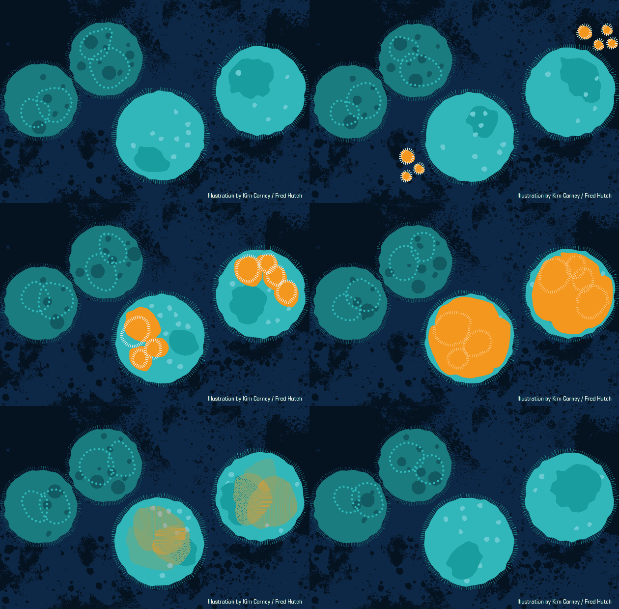
This animation demonstrates the approach,

Biodegradable nanoparticles (orange) carry short-lived gene therapy to specific cells (light teal). Animation by Kimberly Carney / Fred Hutch News Service

An August 30, 2017 Fred Hutchinson Cancer Research Center (Fred Hutch) news release (from news release received via email; also on EurekAlert) by Sabrina Richards, which originated the news item, elucidates further (Note: Some links and notes have been removed),

“Our goal is to streamline the manufacture of cell-based therapies,” said lead author DR. MATTHIAS STEPHAN [6], a faculty member in the Fred Hutch Clinical Research Division and an expert in developing biomaterials. “In this study, we created a product where you just add it to cultured cells and that’s it — no additional manufacturing steps.”

Stephan and his colleagues developed a nanoparticle delivery system to extend the therapeutic potential of messenger RNA, which delivers molecular instructions from DNA to cells in the body, directing them to make proteins to prevent or fight disease.

The researchers’ approach was designed to zero in on specific cell types — T cells of the immune system and blood stem cells — and deliver mRNA directly to the cells, triggering short-term gene expression. It’s called “hit-and-run” genetic programming because the transient effect of mRNA does not change the DNA, but it is enough to make a permanent impact on the cells’ therapeutic potential.

Stephan and colleagues used three examples in the Nature Communications paper to demonstrate their technology:

* Nanoparticles carried a gene-editing tool to T cells of the immune system that snipped out their natural T-cell receptors, and then was paired with genes encoding a “chimeric antigen receptor” or CAR, a synthetic molecule designed to attack cancer.
* Targeted to blood stem cells, nanoparticles were equipped with mRNA that enabled the stem cells to multiply and replace blood cancer cells with healthy cells when used in bone marrow transplants.
* Nanoparticles targeted to CAR-T cells and containing foxo1 mRNA, which signals the anti-cancer T cells to develop into a type of “memory” cell that is more aggressive and destroys tumor cells more effectively and maintains anti-tumor activity longer.

Other attempts to engineer mRNA into disease-fighting cells have been tricky. The large messenger molecule degrades quickly before it can have an effect, and the body’s immune system recognizes it as foreign — not coming from DNA in the nucleus of the cell — and destroys it.

Stephan and his Fred Hutch collaborators devised a workaround to those hurdles.

“We developed a nanocarrier that binds and condenses synthetic mRNA and protects it from degradation,” Stephan said. The researchers surrounded the nanoparticle with a negatively charged envelope with a targeting ligand attached to the surface so that the particle selectively homes in and binds to a particular cell type.

The cells swallow up the tiny carrier, which can be loaded with different types of manmade mRNA. “If you know from the scientific literature that a signaling pathway works in synergy, you could co-deliver mRNA in a single nanoparticle,” Stephan said. “Every cell that takes up the nanoparticle can express both.”

The approach involves mixing the freeze-dried nanoparticles with water and a sample of cells. Within four hours, cells start showing signs that the editing has taken effect. Boosters can be given if needed. Made from a dissolving biomaterial, the nanoparticles are removed from the body like other cell waste.

“Just add water to our freeze-dried product,” Stephan said. Since it’s built on existing technologies and doesn’t require knowledge of nanotechnology, he intends for it to be an off-the-shelf way for cell-therapy engineers to develop new approaches to treating a variety of diseases.

The approach could replace labor-intensive electroporation, a multistep cell-manufacturing technique that requires specialized equipment and clean rooms. All the handling ends up destroying many of the cells, which limits the amount that can be used in treatments for patients.

Gentler to cells, the nanoparticle system developed by the Fred Hutch team showed that up to 60 times more cells survive the process compared with electroporation. This is a critical feature for ensuring enough cells are viable when transferred to patients.

“You can imagine taking the nanoparticles, injecting them into a patient and then you don’t have to culture cells at all anymore,” he said.

Stephan has tested the technology is cultured cells in the lab, and it’s not yet available as a treatment. Stephan is looking for commercial partners to move the technology toward additional applications and into clinical trials where it could be developed into a therapy.

Here’s a link to and a citation for the paper,

Hit-and-run programming of therapeutic cytoreagents using mRNA nanocarriers by H. F. Moffett, M. E. Coon, S. Radtke, S. B. Stephan, L. McKnight, A. Lambert, B. L. Stoddard, H. P. Kiem, & M. T. Stephan. Nature Communications 8, Article number: 389 (2017) doi:10.1038/s41467-017-00505-8 Published online: 30 August 2017

This paper is open access.

Faster diagnostics with nanoparticles and magnetic phenomenon discovered 170 years ago

A Jan. 19, 2017 news item on ScienceDaily announces some new research from the University of Central Florida (UCF),

A UCF researcher has combined cutting-edge nanoscience with a magnetic phenomenon discovered more than 170 years ago to create a method for speedy medical tests.

The discovery, if commercialized, could lead to faster test results for HIV, Lyme disease, syphilis, rotavirus and other infectious conditions.

“I see no reason why a variation of this technique couldn’t be in every hospital throughout the world,” said Shawn Putnam, an assistant professor in the University of Central Florida’s College of Engineering & Computer Science.

A Jan. 19, 2017 UCF news release by Mark Schlueb, which originated the news item,  provides more technical detail,

At the core of the research recently published in the academic journal Small are nanoparticles – tiny particles that are one-billionth of a meter. Putnam’s team coated nanoparticles with the antibody to BSA, or bovine serum albumin, which is commonly used as the basis of a variety of diagnostic tests.

By mixing the nanoparticles in a test solution – such as one used for a blood test – the BSA proteins preferentially bind with the antibodies that coat the nanoparticles, like a lock and key.

That reaction was already well known. But Putnam’s team came up with a novel way of measuring the quantity of proteins present. He used nanoparticles with an iron core and applied a magnetic field to the solution, causing the particles to align in a particular formation. As proteins bind to the antibody-coated particles, the rotation of the particles becomes sluggish, which is easy to detect with laser optics.

The interaction of a magnetic field and light is known as Faraday rotation, a principle discovered by scientist Michael Faraday in 1845. Putnam adapted it for biological use.

“It’s an old theory, but no one has actually applied this aspect of it,” he said.

Other antigens and their unique antibodies could be substituted for the BSA protein used in the research, allowing medical tests for a wide array of infectious diseases.

The proof of concept shows the method could be used to produce biochemical immunology test results in as little as 15 minutes, compared to several hours for ELISA, or enzyme-linked immunosorbent assay, which is currently a standard approach for biomolecule detection.

Here’s a link to and a citation for the paper,

High-Throughput, Protein-Targeted Biomolecular Detection Using Frequency-Domain Faraday Rotation Spectroscopy by Richard J. Murdock, Shawn A. Putnam, Soumen Das, Ankur Gupta, Elyse D. Z. Chase, and Sudipta Seal. Small DOI: 10.1002/smll.201602862 Version of Record online: 16 JAN 2017

© 2017 WILEY-VCH Verlag GmbH & Co. KGaA, Weinheim

This paper is behind a paywall.

Nano-decoy for human influenza A virus

While the implications for this research are exciting, keep in mind that so far they’ve been testing immune-compromised mice. An Oct. 24, 2016 news item on Nanowerk announces the research,

To infect its victims, influenza A heads for the lungs, where it latches onto sialic acid on the surface of cells. So researchers created the perfect decoy: A carefully constructed spherical nanoparticle coated in sialic acid lures the influenza A virus to its doom. When misted into the lungs, the nanoparticle traps influenza A, holding it until the virus self-destructs.

An Oct. 24, 2015 Rensselaer Polytechnic Institute press release by Mary L. Martialay, which originated the news item, describes the research (Note: Links have been removed),

In a study on immune-compromised mice, the treatment reduced influenza A mortality from 100 percent to 25 percent over 14 days. The novel approach, which is radically different from existing influenza A vaccines, and treatments based on neuraminidase inhibitors, could be extended to a host of viruses that use a similar approach to infecting humans, such as Zika, HIV, and malaria. …

“Instead of blocking the virus, we mimicked its target – it’s a completely novel approach,” said Robert Linhardt, a glycoprotein expert and Rensselaer Polytechnic Institute professor who led the research. “It is effective with influenza and we have reason to believe it will function with many other viruses. This could be a therapeutic in cases where vaccine is not an option, such as exposure to an unanticipated strain, or with immune-compromised patients.”

The project is a collaboration between researchers within the Center for Biotechnology and Interdisciplinary Studies (CBIS) at Rensselaer and several institutions in South Korea including Kyungpook National University. Lead author Seok-Joon Kwon, a CBIS research scientist, coordinated the project across borders, enabling the South Korean institutions to test a drug designed and characterized at Rensselaer. …

To access the interior of a cell and replicate itself, influenza A must first bind to the cell surface, and then cut itself free. It binds with the protein hemagglutinin, and severs that tie with the enzyme neuraminidase. Influenza A produces numerous variations each of hemagglutinin and neuraminidase, all of which are antigens within the pathogen that provoke an immune system response. Strains of influenza A are characterized according to the variation of hemagglutinin and neuraminidase they carry, thus the origin of the familiar H1N1 or H3N2 designations.

Medications to counter the virus do exist, but all are vulnerable to the continual antigenic evolution of the virus. A yearly vaccine is effective only if it matches the strain of virus that infects the body. And the virus has shown an ability to develop resistance to a class of therapeutics based on neuraminidase inhibitors, which bind to and block neuraminidase.

The new solution targets an aspect of infection that does not change: all hemagglutinin varieties of influenza A must bind to human sialic acid. To trap the virus, the team designed a dendrimer, a spherical nanoparticle with treelike branches emanating from its core. On the outermost branches, they attached molecules, or “ligands,” of sialic acid.

The research found that the size of the dendrimer and the spacing between the ligands is integral to the function of the nanoparticle. Hemagglutinin occurs in clusters of three, or “trimers,” on the surface of the virus, and researchers found that a spacing of 3 nanometers between ligands resulted in the strongest binding to the trimers. Once bound to the densely packed dendrimer, viral neuraminidase is unable to sever the link. The coat of the virus contains millions of trimers, but the research revealed that only a few links provokes the virus to discharge its genetic cargo and ultimately self-destruct.

A different approach, using a less structured nanoparticle, had been previously tested in unrelated research, but the nanoparticle selected proved both toxic, and could be inactivated by neuraminidase. The new approach is far more promising.

“The major accomplishment was in designing an architecture that is optimized to bind so tightly to the hemagglutinin, the neuraminidase can’t squeeze in and free the virus,” said Linhardt. “It’s trapped.”

Here’s a link to and a citation for the paper,

Nanostructured glycan architecture is important in the inhibition of influenza A virus infection by Seok-Joon Kwon, Dong Hee Na, Jong Hwan Kwak, Marc Douaisi, Fuming Zhang, Eun Ji Park, Jong-Hwan Park, Hana Youn, Chang-Seon Song, Ravi S. Kane, Jonathan S. Dordick, Kyung Bok Lee, & Robert J. Linhardt. Nature Nanotechnology (2016)  doi:10.1038/nnano.2016.181 Published online 24 October 2016

This paper is behind a paywall.

Possible nanoparticle-based vaccine/microbiocide for herpes simplex virus-2

An April 27, 2016 news item on ScienceDaily describes a new therapeutic and preventative technology for herpes,

An effective vaccine against the virus that causes genital herpes has evaded researchers for decades. But now, researchers from the University of Illinois at Chicago [UIC] working with scientists from Germany have shown that zinc-oxide nanoparticles shaped like jacks can prevent the virus from entering cells, and help natural immunity to develop.

“We call the virus-trapping nanoparticle a microbivac, because it possesses both microbicidal and vaccine-like properties,” says corresponding author Deepak Shukla, professor of ophthalmology and microbiology & immunology in the UIC College of Medicine. “It is a totally novel approach to developing a vaccine against herpes, and it could potentially also work for HIV and other viruses,” he said.

The particles could serve as a powerful active ingredient in a topically-applied vaginal cream that provides immediate protection against herpes virus infection while simultaneously helping stimulate immunity to the virus for long-term protection, explained Shukla.

An April 27, 2016 UIC news release (also on EurekAlert), which originated the news item, provides more context for the work,

Herpes simplex virus-2, which causes serious eye infections in newborns and immunocompromised patients as well as genital herpes, is one of the most common human viruses. According to the Centers for Disease Control and Prevention, about 15 percent of people from ages 14-49 carry HSV-2, which can hide out for long periods of time in the nervous system. The genital lesions caused by the virus increase the risk for acquiring human immunodeficiency virus, or HIV.

“Your chances of getting HIV are three to four times higher if you already have genital herpes, which is a very strong motivation for developing new ways of preventing herpes infection,” Shukla said.

Treatments for HSV-2 include daily topical medications to suppress the virus and shorten the duration of outbreaks, when the virus is active and genital lesions are present. However, drug resistance is common, and little protection is provided against further infections. Efforts to develop a vaccine have been unsuccessful because the virus does not spend much time in the bloodstream, where most traditional vaccines do their work.

The news release goes on to provide technical details,

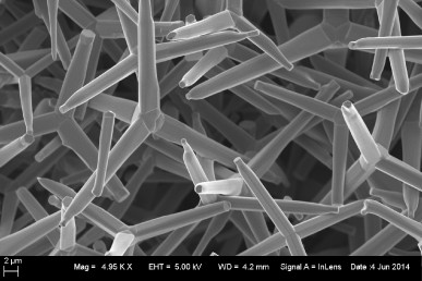
The tetrapod-shaped zinc-oxide nanoparticles, called ZOTEN, have negatively charged surfaces that attract the HSV-2 virus, which has positively charged proteins on its outer envelope. ZOTEN nanoparticles were synthesized using technology developed by material scientists at Germany’s Kiel University and protected under a joint patent with UIC.

When bound to the nanoparticles, HSV-2 cannot infect cells. But the bound virus remains susceptible to processing by immune cells called dendritic cells that patrol the vaginal lining. The dendritic cells “present” the virus to other immune cells that produce antibodies. The antibodies cripple the virus and trigger the production of customized killer cells that identify infected cells and destroy them before the virus can take over and spread.

The researchers showed that female mice swabbed with HSV-2 and an ointment containing ZOTEN had significantly fewer genital lesions than mice treated with a cream lacking ZOTEN. Mice treated with ZOTEN also had less inflammation in the central nervous system, where the virus can hide out.

The researchers were able to watch immune cells pry the virus off the nanoparticles for immune processing, using high-resolution fluorescence microscopy.

“It’s very clear that ZOTEN facilitates the development of immunity by holding the virus and letting the dendritic cells get to it,” Shukla said.

If found safe and effective in humans, a ZOTEN-containing cream ideally would be applied vaginally just prior to intercourse, Shukla said. But if a woman who had been using it regularly missed an application, he said, she may have already developed some immunity and still have some protection. Shukla hopes to further develop the nanoparticles to work against HIV, which like HSV-2 also has positively charged proteins embedded in its outer envelope.

ZOTEN particles are uniform in size and shape, making them attractive for use in other biomedical applications. The novel flame transport synthesis technology used to make them allows large-scale production, said Rainer Adelung, professor of nanomaterials at Kiel University. And, because no chemicals are used, the production process is green.

Adelung hopes to begin commercial production of ZOTEN through a startup company that will be run jointly with his colleagues at UIC.

Here’s an image of the particles, courtesy of UIC,

Zinc oxide tetrapod nanoparticles. Credit: Deepak Shukla

Zinc oxide tetrapod nanoparticles. Credit: Deepak Shukla

Here’s a link to and a citation for the paper,

Intravaginal Zinc Oxide Tetrapod Nanoparticles as Novel Immunoprotective Agents against Genital Herpes by Thessicar E. Antoine, Satvik R. Hadigal, Abraam M. Yakoub, Yogendra Kumar Mishra, Palash Bhattacharya, Christine Haddad, Tibor Valyi-Nagy, Rainer Adelung, Bellur S. Prabhakar, and Deepak Shukla. The Journal of Immunology April 27, 2016 1502373  doi: 10.4049/jimmunol.1502373 Published online before print April 27, 2016

This paper is behind a paywall.

One final comment, it’s a long from a mouse vagina in this study to a human one.

Lab tests show silver nanoparticles in cream blocks HIV entry for up to 72 hours

Since at least 2005 (the article reference will be given later in this posting), researchers have been aware that silver nanoparticles can block the HIV virus from entering a cell. The latest work in this area has resulted in a vaginal cream laced with silver nanoparticles according to a Jan. 28, 2014 news item on ScienceDaily,

Lara Villegas [Humberto Lara Villegas, specialist in nanoparticles and virology from the University of Monterrey, Mexico (UDEM)] explained that HIV makes its entry to immune cells (CD4) of the organism with the aid of a protein known as GP120, which allows the virus adherence to the cells. This same principle is used by silver nanoparticles to attach themselves to this protein and block it, turning the virus inactive.

The Mexican researcher informed that the cream has been tested in samples of human tissue and has proven the efficiency of silver nanoparticles to avoid the transmission of the virus through cervical mucous membrane.

The Jan. 28, 2014 Investigación y Desarrollo news release (on the Alpha Gallileo website), which originated the news item, provides additional details from Lara Villegas’ perspective,

The researcher from UDEM, who has worked in Israel and The United States, assured that after applied, the cream starts to work in less than a minute, and has an effective protection of up to 72 hours.

Given that the function of this product is the inactivation of the virus, although this is a vaginal cream, will also protect the sexual partner.

“Normally – he highlighted-, the medication used against the virus act within the cell to avoid its replication. This is a very different case, given that the nanoparticle goes directly against the HIV and no longer allows its entry to the cell”.

So far, no toxicity of the silver nanoparticles has been reported, although he added that research is yet to be performed to evaluate the possible side effects of silver properties.

“Right now, I am certain that this microbicide is going to avoid the virus entering the organism, but I cannot yet assure that is totally harmless, because the clinical trials are a long and expensive process”, the researched added.

He exposed that the use of gels are usually accompanied by irritation, which favors the entry of the virus, which is why the cream was enriched with an anti-inflammatory effect.

Currently, with the obtained results, researchers will proceed to perform experimentation in mice that accept human cells, to later begin with human clinical trials.

He added that this cream could prevent the transmition of other sexually acquired virus like the Human Papilloma Virus (HPV). Likewise, he considered that silver nanoparticles could be used to combat bacteria transmitted the same way.

As promised here’s a citation for and a link to the 2005 paper; I haven’t found any references in my admittedly brief search for a paper about this latest work,,

Interaction of silver nanoparticles with HIV-1 by Jose Luis Elechiguerra, Justin L Burt, Jose R Morones, Alejandra Camacho-Bragado, Xiaoxia Gao, Humberto H Lara, and Miguel Jose Yacaman. Journal of Nanobiotechnology 2005, 3:6  doi:10.1186/1477-3155-3-6

This paper is open access.

Here’s  the Investigación y Desarrollo website which seems to act as a hub for research in Mexico. Note: You will need Spanish language skills to fully utilize this site.