Tag Archives: human brains

Revival of dead pig brains raises moral questions about life and death

The line between life and death may not be what we thought it was according to some research that was reported in April 2019. Ed Wong’s April 17, 2019 article (behind a paywall) for The Atlantic was my first inkling about the life-death questions raised by some research performed at Yale University, (Note: Links have been removed)

The brain, supposedly, cannot long survive without blood. Within seconds, oxygen supplies deplete, electrical activity fades, and unconsciousness sets in. If blood flow is not restored, within minutes, neurons start to die in a rapid, irreversible, and ultimately fatal wave.

But maybe not? According to a team of scientists led by Nenad Sestan at Yale School of Medicine, this process might play out over a much longer time frame, and perhaps isn’t as inevitable or irreparable as commonly believed. Sestan and his colleagues showed this in dramatic fashion—by preserving and restoring signs of activity in the isolated brains of pigs that had been decapitated four hours earlier.

The team sourced 32 pig brains from a slaughterhouse, placed them in spherical chambers, and infused them with nutrients and protective chemicals, using pumps that mimicked the beats of a heart. This system, dubbed BrainEx, preserved the overall architecture of the brains, preventing them from degrading. It restored flow in their blood vessels, which once again became sensitive to dilating drugs. It stopped many neurons and other cells from dying, and reinstated their ability to consume sugar and oxygen. Some of these rescued neurons even started to fire. “Everything was surprising,” says Zvonimir Vrselja, who performed most of the experiments along with Stefano Daniele.

… “I don’t see anything in this report that should undermine confidence in brain death as a criterion of death,” says Winston Chiong, a neurologist at the University of California at San Francisco. The matter of when to declare someone dead has become more controversial since doctors began relying more heavily on neurological signs, starting around 1968, when the criteria for “brain death” were defined. But that diagnosis typically hinges on the loss of brainwide activity—a line that, at least for now, is still final and irreversible. After MIT Technology Review broke the news of Sestan’s work a year ago, he started receiving emails from people asking whether he could restore brain function to their loved ones. He very much cannot. BrainEx isn’t a resurrection chamber.

“It’s not going to result in human brain transplants,” adds Karen Rommelfanger, who directs Emory University’s neuroethics program. “And I don’t think this means that the singularity is coming, or that radical life extension is more possible than before.”

So why do the study? “There’s potential for using this method to develop innovative treatments for patients with strokes or other types of brain injuries, and there’s a real need for those kinds of treatments,” says L. Syd M Johnson, a neuroethicist at Michigan Technological University. The BrainEx method might not be able to fully revive hours-dead brains, but Yama Akbari, a critical-care neurologist at the University of California at Irvine, wonders whether it would be more successful if applied minutes after death. Alternatively, it could help to keep oxygen-starved brains alive and intact while patients wait to be treated. “It’s an important landmark study,” Akbari says.

Yong notes that the study still needs to be replicated in his article which also probes some of the ethical issues associated with the latest neuroscience research.

Nature published the Yale study,

Restoration of brain circulation and cellular functions hours post-mortem by Zvonimir Vrselja, Stefano G. Daniele, John Silbereis, Francesca Talpo, Yury M. Morozov, André M. M. Sousa, Brian S. Tanaka, Mario Skarica, Mihovil Pletikos, Navjot Kaur, Zhen W. Zhuang, Zhao Liu, Rafeed Alkawadri, Albert J. Sinusas, Stephen R. Latham, Stephen G. Waxman & Nenad Sestan. Nature 568, 336–343 (2019) DOI: https://doi.org/10.1038/s41586-019-1099-1 Published 17 April 2019 Issue Date 18 April 2019

This paper is behind a paywall.

Two neuroethicists had this to say (link to their commentary in Nature follows) as per an April 71, 2019 news release from Case Western Reserve University (also on EurekAlert), Note: Links have been removed,

The brain is more resilient than previously thought. In a groundbreaking experiment published in this week’s issue of Nature, neuroscientists created an artificial circulation system that successfully restored some functions and structures in donated pig brains–up to four hours after the pigs were butchered at a USDA food processing facility. Though there was no evidence of restored consciousness, brains from the pigs were without oxygen for hours, yet could still support key functions provided by the artificial system. The result challenges the notion that mammalian brains are fully and irreversibly damaged by a lack of oxygen.

“The assumptions have always been that after a couple minutes of anoxia, or no oxygen, the brain is ‘dead,'” says Stuart Youngner, MD, who co-authored a commentary accompanying the study with Insoo Hyun, PhD, both professors in the Department of Bioethics at Case Western Reserve University School of Medicine. “The system used by the researchers begs the question: How long should we try to save people?”

In the pig experiment, researchers used an artificial perfusate (a type of cell-free “artificial blood”), which helped brain cells maintain their structure and some functions. Resuscitative efforts in humans, like CPR, are also designed to get oxygen to the brain and stave off brain damage. After a period of time, if a person doesn’t respond to resuscitative efforts, emergency medical teams declare them dead.

The acceptable duration of resuscitative efforts is somewhat uncertain. “It varies by country, emergency medical team, and hospital,” Youngner said. Promising results from the pig experiment further muddy the waters about the when to stop life-saving efforts.

At some point, emergency teams must make a critical switch from trying to save a patient, to trying to save organs, said Youngner. “In Europe, when emergency teams stop resuscitation efforts, they declare a patient dead, and then restart the resuscitation effort to circulate blood to the organs so they can preserve them for transplantation.”

The switch can involve extreme means. In the commentary, Youngner and Hyun describe how some organ recovery teams use a balloon to physically cut off blood circulation to the brain after declaring a person dead, to prepare the organs for transplantation.

The pig experiment implies that sophisticated efforts to perfuse the brain might maintain brain cells. If technologies like those used in the pig experiment could be adapted for humans (a long way off, caution Youngner and Hyun), some people who, today, are typically declared legally dead after a catastrophic loss of oxygen could, tomorrow, become candidates for brain resuscitation, instead of organ donation.

Said Youngner, “As we get better at resuscitating the brain, we need to decide when are we going to save a patient, and when are we going to declare them dead–and save five or more who might benefit from an organ.”

Because brain resuscitation strategies are in their infancy and will surely trigger additional efforts, the scientific and ethics community needs to begin discussions now, says Hyun. “This study is likely to raise a lot of public concerns. We hoped to get ahead of the hype and offer an early, reasoned response to this scientific advance.”

Both Youngner and Hyun praise the experiment as a “major scientific advancement” that is overwhelmingly positive. It raises the tantalizing possibility that the grave risks of brain damage caused by a lack of oxygen could, in some cases, be reversible.
“Pig brains are similar in many ways to human brains, which makes this study so compelling,” Hyun said. “We urge policymakers to think proactively about what this line of research might mean for ongoing debates around organ donation and end of life care.”

Here’s a link to and a citation to the Nature commentary,

Pig experiment challenges assumptions around brain damage in people by Stuart Youngner and Insoo Hyun. Nature 568, 302-304 (2019) DOI: 10.1038/d41586-019-01169-8 April 17, 2019

This paper is open access.

I was hoping to find out more about BrainEx, but this April 17, 2019 US National Institute of Mental Health news release is all I’ve been able to find in my admittedly brief online search. The news release offers more celebration than technical detail.

Quick comment

Interestingly, there hasn’t been much of a furor over this work. Not yet.

Artificial synapse based on tantalum oxide from Korean researchers

This memristor story comes from South Korea as we progress on the way to neuromorphic computing (brainlike computing). A Sept. 7, 2018 news item on ScienceDaily makes the announcement,

A research team led by Director Myoung-Jae Lee from the Intelligent Devices and Systems Research Group at DGIST (Daegu Gyeongbuk Institute of Science and Technology) has succeeded in developing an artificial synaptic device that mimics the function of the nerve cells (neurons) and synapses that are response for memory in human brains. [sic]

Synapses are where axons and dendrites meet so that neurons in the human brain can send and receive nerve signals; there are known to be hundreds of trillions of synapses in the human brain.

This chemical synapse information transfer system, which transfers information from the brain, can handle high-level parallel arithmetic with very little energy, so research on artificial synaptic devices, which mimic the biological function of a synapse, is under way worldwide.

Dr. Lee’s research team, through joint research with teams led by Professor Gyeong-Su Park from Seoul National University; Professor Sung Kyu Park from Chung-ang University; and Professor Hyunsang Hwang from Pohang University of Science and Technology (POSTEC), developed a high-reliability artificial synaptic device with multiple values by structuring tantalum oxide — a trans-metallic material — into two layers of Ta2O5-x and TaO2-x and by controlling its surface.

A September 7, 2018 DGIST press release (also on EurekAlert), which originated the news item, delves further into the work,

The artificial synaptic device developed by the research team is an electrical synaptic device that simulates the function of synapses in the brain as the resistance of the tantalum oxide layer gradually increases or decreases depending on the strength of the electric signals. It has succeeded in overcoming durability limitations of current devices by allowing current control only on one layer of Ta2O5-x.

In addition, the research team successfully implemented an experiment that realized synapse plasticity [or synaptic plasticity], which is the process of creating, storing, and deleting memories, such as long-term strengthening of memory and long-term suppression of memory deleting by adjusting the strength of the synapse connection between neurons.

The non-volatile multiple-value data storage method applied by the research team has the technological advantage of having a small area of an artificial synaptic device system, reducing circuit connection complexity, and reducing power consumption by more than one-thousandth compared to data storage methods based on digital signals using 0 and 1 such as volatile CMOS (Complementary Metal Oxide Semiconductor).

The high-reliability artificial synaptic device developed by the research team can be used in ultra-low-power devices or circuits for processing massive amounts of big data due to its capability of low-power parallel arithmetic. It is expected to be applied to next-generation intelligent semiconductor device technologies such as development of artificial intelligence (AI) including machine learning and deep learning and brain-mimicking semiconductors.

Dr. Lee said, “This research secured the reliability of existing artificial synaptic devices and improved the areas pointed out as disadvantages. We expect to contribute to the development of AI based on the neuromorphic system that mimics the human brain by creating a circuit that imitates the function of neurons.”

Here’s a link to and a citation for the paper,

Reliable Multivalued Conductance States in TaOx Memristors through Oxygen Plasma-Assisted Electrode Deposition with in Situ-Biased Conductance State Transmission Electron Microscopy Analysis by Myoung-Jae Lee, Gyeong-Su Park, David H. Seo, Sung Min Kwon, Hyeon-Jun Lee, June-Seo Kim, MinKyung Jung, Chun-Yeol You, Hyangsook Lee, Hee-Goo Kim, Su-Been Pang, Sunae Seo, Hyunsang Hwang, and Sung Kyu Park. ACS Appl. Mater. Interfaces, 2018, 10 (35), pp 29757–29765 DOI: 10.1021/acsami.8b09046 Publication Date (Web): July 23, 2018

Copyright © 2018 American Chemical Society

This paper is open access.

You can find other memristor and neuromorphic computing stories here by using the search terms I’ve highlighted,  My latest (more or less) is an April 19, 2018 posting titled, New path to viable memristor/neuristor?

Finally, here’s an image from the Korean researchers that accompanied their work,

Caption: Representation of neurons and synapses in the human brain. The magnified synapse represents the portion mimicked using solid-state devices. Credit: Daegu Gyeongbuk Institute of Science and Technology(DGIST)

Brainy and brainy: a novel synaptic architecture and a neuromorphic computing platform called SpiNNaker

I have two items about brainlike computing. The first item hearkens back to memristors, a topic I have been following since 2008. (If you’re curious about the various twists and turns just enter  the term ‘memristor’ in this blog’s search engine.) The latest on memristors is from a team than includes IBM (US), École Politechnique Fédérale de Lausanne (EPFL; Swizterland), and the New Jersey Institute of Technology (NJIT; US). The second bit comes from a Jülich Research Centre team in Germany and concerns an approach to brain-like computing that does not include memristors.

Multi-memristive synapses

In the inexorable march to make computers function more like human brains (neuromorphic engineering/computing), an international team has announced its latest results in a July 10, 2018 news item on Nanowerk,

Two New Jersey Institute of Technology (NJIT) researchers, working with collaborators from the IBM Research Zurich Laboratory and the École Polytechnique Fédérale de Lausanne, have demonstrated a novel synaptic architecture that could lead to a new class of information processing systems inspired by the brain.

The findings are an important step toward building more energy-efficient computing systems that also are capable of learning and adaptation in the real world. …

A July 10, 2018 NJIT news release (also on EurekAlert) by Tracey Regan, which originated by the news item, adds more details,

The researchers, Bipin Rajendran, an associate professor of electrical and computer engineering, and S. R. Nandakumar, a graduate student in electrical engineering, have been developing brain-inspired computing systems that could be used for a wide range of big data applications.

Over the past few years, deep learning algorithms have proven to be highly successful in solving complex cognitive tasks such as controlling self-driving cars and language understanding. At the heart of these algorithms are artificial neural networks – mathematical models of the neurons and synapses of the brain – that are fed huge amounts of data so that the synaptic strengths are autonomously adjusted to learn the intrinsic features and hidden correlations in these data streams.

However, the implementation of these brain-inspired algorithms on conventional computers is highly inefficient, consuming huge amounts of power and time. This has prompted engineers to search for new materials and devices to build special-purpose computers that can incorporate the algorithms. Nanoscale memristive devices, electrical components whose conductivity depends approximately on prior signaling activity, can be used to represent the synaptic strength between the neurons in artificial neural networks.

While memristive devices could potentially lead to faster and more power-efficient computing systems, they are also plagued by several reliability issues that are common to nanoscale devices. Their efficiency stems from their ability to be programmed in an analog manner to store multiple bits of information; however, their electrical conductivities vary in a non-deterministic and non-linear fashion.

In the experiment, the team showed how multiple nanoscale memristive devices exhibiting these characteristics could nonetheless be configured to efficiently implement artificial intelligence algorithms such as deep learning. Prototype chips from IBM containing more than one million nanoscale phase-change memristive devices were used to implement a neural network for the detection of hidden patterns and correlations in time-varying signals.

“In this work, we proposed and experimentally demonstrated a scheme to obtain high learning efficiencies with nanoscale memristive devices for implementing learning algorithms,” Nandakumar says. “The central idea in our demonstration was to use several memristive devices in parallel to represent the strength of a synapse of a neural network, but only chose one of them to be updated at each step based on the neuronal activity.”

Here’s a link to and a citation for the paper,

Neuromorphic computing with multi-memristive synapses by Irem Boybat, Manuel Le Gallo, S. R. Nandakumar, Timoleon Moraitis, Thomas Parnell, Tomas Tuma, Bipin Rajendran, Yusuf Leblebici, Abu Sebastian, & Evangelos Eleftheriou. Nature Communications volume 9, Article number: 2514 (2018) DOI: https://doi.org/10.1038/s41467-018-04933-y Published 28 June 2018

This is an open access paper.

Also they’ve got a couple of very nice introductory paragraphs which I’m including here, (from the June 28, 2018 paper in Nature Communications; Note: Links have been removed),

The human brain with less than 20 W of power consumption offers a processing capability that exceeds the petaflops mark, and thus outperforms state-of-the-art supercomputers by several orders of magnitude in terms of energy efficiency and volume. Building ultra-low-power cognitive computing systems inspired by the operating principles of the brain is a promising avenue towards achieving such efficiency. Recently, deep learning has revolutionized the field of machine learning by providing human-like performance in areas, such as computer vision, speech recognition, and complex strategic games1. However, current hardware implementations of deep neural networks are still far from competing with biological neural systems in terms of real-time information-processing capabilities with comparable energy consumption.

One of the reasons for this inefficiency is that most neural networks are implemented on computing systems based on the conventional von Neumann architecture with separate memory and processing units. There are a few attempts to build custom neuromorphic hardware that is optimized to implement neural algorithms2,3,4,5. However, as these custom systems are typically based on conventional silicon complementary metal oxide semiconductor (CMOS) circuitry, the area efficiency of such hardware implementations will remain relatively low, especially if in situ learning and non-volatile synaptic behavior have to be incorporated. Recently, a new class of nanoscale devices has shown promise for realizing the synaptic dynamics in a compact and power-efficient manner. These memristive devices store information in their resistance/conductance states and exhibit conductivity modulation based on the programming history6,7,8,9. The central idea in building cognitive hardware based on memristive devices is to store the synaptic weights as their conductance states and to perform the associated computational tasks in place.

The two essential synaptic attributes that need to be emulated by memristive devices are the synaptic efficacy and plasticity. …

It gets more complicated from there.

Now onto the next bit.

SpiNNaker

At a guess, those capitalized N’s are meant to indicate ‘neural networks’. As best I can determine, SpiNNaker is not based on the memristor. Moving on, a July 11, 2018 news item on phys.org announces work from a team examining how neuromorphic hardware and neuromorphic software work together,

A computer built to mimic the brain’s neural networks produces similar results to that of the best brain-simulation supercomputer software currently used for neural-signaling research, finds a new study published in the open-access journal Frontiers in Neuroscience. Tested for accuracy, speed and energy efficiency, this custom-built computer named SpiNNaker, has the potential to overcome the speed and power consumption problems of conventional supercomputers. The aim is to advance our knowledge of neural processing in the brain, to include learning and disorders such as epilepsy and Alzheimer’s disease.

A July 11, 2018 Frontiers Publishing news release on EurekAlert, which originated the news item, expands on the latest work,

“SpiNNaker can support detailed biological models of the cortex–the outer layer of the brain that receives and processes information from the senses–delivering results very similar to those from an equivalent supercomputer software simulation,” says Dr. Sacha van Albada, lead author of this study and leader of the Theoretical Neuroanatomy group at the Jülich Research Centre, Germany. “The ability to run large-scale detailed neural networks quickly and at low power consumption will advance robotics research and facilitate studies on learning and brain disorders.”

The human brain is extremely complex, comprising 100 billion interconnected brain cells. We understand how individual neurons and their components behave and communicate with each other and on the larger scale, which areas of the brain are used for sensory perception, action and cognition. However, we know less about the translation of neural activity into behavior, such as turning thought into muscle movement.

Supercomputer software has helped by simulating the exchange of signals between neurons, but even the best software run on the fastest supercomputers to date can only simulate 1% of the human brain.

“It is presently unclear which computer architecture is best suited to study whole-brain networks efficiently. The European Human Brain Project and Jülich Research Centre have performed extensive research to identify the best strategy for this highly complex problem. Today’s supercomputers require several minutes to simulate one second of real time, so studies on processes like learning, which take hours and days in real time are currently out of reach.” explains Professor Markus Diesmann, co-author, head of the Computational and Systems Neuroscience department at the Jülich Research Centre.

He continues, “There is a huge gap between the energy consumption of the brain and today’s supercomputers. Neuromorphic (brain-inspired) computing allows us to investigate how close we can get to the energy efficiency of the brain using electronics.”

Developed over the past 15 years and based on the structure and function of the human brain, SpiNNaker — part of the Neuromorphic Computing Platform of the Human Brain Project — is a custom-built computer composed of half a million of simple computing elements controlled by its own software. The researchers compared the accuracy, speed and energy efficiency of SpiNNaker with that of NEST–a specialist supercomputer software currently in use for brain neuron-signaling research.

“The simulations run on NEST and SpiNNaker showed very similar results,” reports Steve Furber, co-author and Professor of Computer Engineering at the University of Manchester, UK. “This is the first time such a detailed simulation of the cortex has been run on SpiNNaker, or on any neuromorphic platform. SpiNNaker comprises 600 circuit boards incorporating over 500,000 small processors in total. The simulation described in this study used just six boards–1% of the total capability of the machine. The findings from our research will improve the software to reduce this to a single board.”

Van Albada shares her future aspirations for SpiNNaker, “We hope for increasingly large real-time simulations with these neuromorphic computing systems. In the Human Brain Project, we already work with neuroroboticists who hope to use them for robotic control.”

Before getting to the link and citation for the paper, here’s a description of SpiNNaker’s hardware from the ‘Spiking neural netowrk’ Wikipedia entry, Note: Links have been removed,

Neurogrid, built at Stanford University, is a board that can simulate spiking neural networks directly in hardware. SpiNNaker (Spiking Neural Network Architecture) [emphasis mine], designed at the University of Manchester, uses ARM processors as the building blocks of a massively parallel computing platform based on a six-layer thalamocortical model.[5]

Now for the link and citation,

Performance Comparison of the Digital Neuromorphic Hardware SpiNNaker and the Neural Network Simulation Software NEST for a Full-Scale Cortical Microcircuit Model by
Sacha J. van Albada, Andrew G. Rowley, Johanna Senk, Michael Hopkins, Maximilian Schmidt, Alan B. Stokes, David R. Lester, Markus Diesmann, and Steve B. Furber. Neurosci. 12:291. doi: 10.3389/fnins.2018.00291 Published: 23 May 2018

As noted earlier, this is an open access paper.

Transparent graphene electrode technology and complex brain imaging

Michael Berger has written a May 24, 2018 Nanowerk Spotlight article about some of the latest research on transparent graphene electrode technology and the brain (Note: A link has been removed),

In new work, scientists from the labs of Kuzum [Duygu Kuzum, an Assistant Professor of Electrical and Computer Engineering at the University of California, San Diego {UCSD}] and Anna Devor report a transparent graphene microelectrode neural implant that eliminates light-induced artifacts to enable crosstalk-free integration of 2-photon microscopy, optogenetic stimulation, and cortical recordings in the same in vivo experiment. The new class of transparent brain implant is based on monolayer graphene. It offers a practical pathway to investigate neuronal activity over multiple spatial scales extending from single neurons to large neuronal populations.

Conventional metal-based microelectrodes cannot be used for simultaneous measurements of multiple optical and electrical parameters, which are essential for comprehensive investigation of brain function across spatio-temporal scales. Since they are opaque, they block the field of view of the microscopes and generate optical shadows impeding imaging.

More importantly, they cause light induced artifacts in electrical recordings, which can significantly interfere with neural signals. Transparent graphene electrode technology presented in this paper addresses these problems and allow seamless and crosstalk-free integration of optical and electrical sensing and manipulation technologies.

In their work, the scientists demonstrate that by careful design of key steps in the fabrication process for transparent graphene electrodes, the light-induced artifact problem can be mitigated and virtually artifact-free local field potential (LFP) recordings can be achieved within operating light intensities.

“Optical transparency of graphene enables seamless integration of imaging, optogenetic stimulation and electrical recording of brain activity in the same experiment with animal models,” Kuzum explains. “Different from conventional implants based on metal electrodes, graphene-based electrodes do not generate any electrical artifacts upon interacting with light used for imaging or optogenetics. That enables crosstalk free integration of three modalities: imaging, stimulation and recording to investigate brain activity over multiple spatial scales extending from single neurons to large populations of neurons in the same experiment.”

The team’s new fabrication process avoids any crack formation in the transfer process, resulting in a 95-100% yield for the electrode arrays. This fabrication quality is important for expanding this technology to high-density large area transparent arrays to monitor brain-scale cortical activity in large animal models or humans.

“Our technology is also well-suited for neurovascular and neurometabolic studies, providing a ‘gold standard’ neuronal correlate for optical measurements of vascular, hemodynamic, and metabolic activity,” Kuzum points out. “It will find application in multiple areas, advancing our understanding of how microscopic neural activity at the cellular scale translates into macroscopic activity of large neuron populations.”

“Combining optical techniques with electrical recordings using graphene electrodes will allow to connect the large body of neuroscience knowledge obtained from animal models to human studies mainly relying on electrophysiological recordings of brain-scale activity,” she adds.

Next steps for the team involve employing this technology to investigate coupling and information transfer between different brain regions.

This work is part of the US BRAIN (Brain Research through Advancing Innovative Neurotechnologies) initiative and there’s more than one team working with transparent graphene electrodes. John Hewitt in an Oct. 21, 2014 posting on ExtremeTech describes two other teams’ work (Note: Links have been removed),

The solution [to the problems with metal electrodes], now emerging from multiple labs throughout the universe is to build flexible, transparent electrode arrays from graphene. Two studies in the latest issue of Nature Communications, one from the University of Wisconsin-Madison and the other from Penn [University of Pennsylvania], describe how to build these devices.

The University of Wisconsin researchers are either a little bit smarter or just a little bit richer, because they published their work open access. It’s a no-brainer then that we will focus on their methods first, and also in more detail. To make the arrays, these guys first deposited the parylene (polymer) substrate on a silicon wafer, metalized it with gold, and then patterned it with an electron beam to create small contact pads. The magic was to then apply four stacked single-atom-thick graphene layers using a wet transfer technique. These layers were then protected with a silicon dioxide layer, another parylene layer, and finally molded into brain signal recording goodness with reactive ion etching.

PennTransparentelectrodeThe researchers went with four graphene layers because that provided optimal mechanical integrity and conductivity while maintaining sufficient transparency. They tested the device in opto-enhanced mice whose neurons expressed proteins that react to blue light. When they hit the neurons with a laser fired in through the implant, the protein channels opened and fired the cell beneath. The masterstroke that remained was then to successfully record the electrical signals from this firing, sit back, and wait for the Nobel prize office to call.

The Penn State group [Note: Every reearcher mentioned in the paper Hewitt linked to is from the University of Pennsylvania] in the  used a similar 16-spot electrode array (pictured above right), and proceeded — we presume — in much the same fashion. Their angle was to perform high-resolution optical imaging, in particular calcium imaging, right out through the transparent electrode arrays which simultaneously recorded in high-temporal-resolution signals. They did this in slices of the hippocampus where they could bring to bear the complex and multifarious hardware needed to perform confocal and two-photon microscopy. These latter techniques provide a boost in spatial resolution by zeroing in over narrow planes inside the specimen, and limiting the background by the requirement of two photons to generate an optical signal. We should mention that there are voltage sensitive dyes available, in addition to standard calcium dyes, which can almost record the fastest single spikes, but electrical recording still reigns supreme for speed.

What a mouse looks like with an optogenetics system plugged in

What a mouse looks like with an optogenetics system plugged in

One concern of both groups in making these kinds of simultaneous electro-optic measurements was the generation of light-induced artifacts in the electrical recordings. This potential complication, called the Becqueral photovoltaic effect, has been known to exist since it was first demonstrated back in 1839. When light hits a conventional metal electrode, a photoelectrochemical (or more simply, a photovoltaic) effect occurs. If present in these recordings, the different signals could be highly disambiguatable. The Penn researchers reported that they saw no significant artifact, while the Wisconsin researchers saw some small effects with their device. In particular, when compared with platinum electrodes put into the opposite side cortical hemisphere, the Wisconsin researchers found that the artifact from graphene was similar to that obtained from platinum electrodes.

Here’s a link to and a citation for the latest research from UCSD,

Deep 2-photon imaging and artifact-free optogenetics through transparent graphene microelectrode arrays by Martin Thunemann, Yichen Lu, Xin Liu, Kıvılcım Kılıç, Michèle Desjardins, Matthieu Vandenberghe, Sanaz Sadegh, Payam A. Saisan, Qun Cheng, Kimberly L. Weldy, Hongming Lyu, Srdjan Djurovic, Ole A. Andreassen, Anders M. Dale, Anna Devor, & Duygu Kuzum. Nature Communicationsvolume 9, Article number: 2035 (2018) doi:10.1038/s41467-018-04457-5 Published: 23 May 2018

This paper is open access.

You can find out more about the US BRAIN initiative here and if you’re curious, you can find out more about the project at UCSD here. Duygu Kuzum (now at UCSD) was at  the University of Pennsylvania in 2014 and participated in the work mentioned in Hewitt’s 2014 posting.

Injectable bandages for internal bleeding and hydrogel for the brain

This injectable bandage could be a gamechanger (as they say) if it can be taken beyond the ‘in vitro’ (i.e., petri dish) testing stage. A May 22, 2018 news item on Nanowerk makes the announcement (Note: A link has been removed),

While several products are available to quickly seal surface wounds, rapidly stopping fatal internal bleeding has proven more difficult. Now researchers from the Department of Biomedical Engineering at Texas A&M University are developing an injectable hydrogel bandage that could save lives in emergencies such as penetrating shrapnel wounds on the battlefield (Acta Biomaterialia, “Nanoengineered injectable hydrogels for wound healing application”).

A May 22, 2018 US National Institute of Biomedical Engineering and Bioengiineering news release, which originated the news item, provides more detail (Note: Links have been removed),

The researchers combined a hydrogel base (a water-swollen polymer) and nanoparticles that interact with the body’s natural blood-clotting mechanism. “The hydrogel expands to rapidly fill puncture wounds and stop blood loss,” explained Akhilesh Gaharwar, Ph.D., assistant professor and senior investigator on the work. “The surface of the nanoparticles attracts blood platelets that become activated and start the natural clotting cascade of the body.”

Enhanced clotting when the nanoparticles were added to the hydrogel was confirmed by standard laboratory blood clotting tests. Clotting time was reduced from eight minutes to six minutes when the hydrogel was introduced into the mixture. When nanoparticles were added, clotting time was significantly reduced, to less than three minutes.

In addition to the rapid clotting mechanism of the hydrogel composite, the engineers took advantage of special properties of the nanoparticle component. They found they could use the electric charge of the nanoparticles to add growth factors that efficiently adhered to the particles. “Stopping fatal bleeding rapidly was the goal of our work,” said Gaharwar. “However, we found that we could attach growth factors to the nanoparticles. This was an added bonus because the growth factors act to begin the body’s natural wound healing process—the next step needed after bleeding has stopped.”

The researchers were able to attach vascular endothelial growth factor (VEGF) to the nanoparticles. They tested the hydrogel/nanoparticle/VEGF combination in a cell culture test that mimics the wound healing process. The test uses a petri dish with a layer of endothelial cells on the surface that create a solid skin-like sheet. The sheet is then scratched down the center creating a rip or hole in the sheet that resembles a wound.

When the hydrogel containing VEGF bound to the nanoparticles was added to the damaged endothelial cell wound, the cells were induced to grow back and fill-in the scratched region—essentially mimicking the healing of a wound.

“Our laboratory experiments have verified the effectiveness of the hydrogel for initiating both blood clotting and wound healing,” said Gaharwar. “We are anxious to begin tests in animals with the hope of testing and eventual use in humans where we believe our formulation has great potential to have a significant impact on saving lives in critical situations.”

The work was funded by grant EB023454 from the National Institute of Biomedical Imaging and Bioengineering (NIBIB), and the National Science Foundation. The results were reported in the February issue of the journal Acta Biomaterialia.

The paper was published back in April 2018 and there was an April 2, 2018 Texas A&M University news release on EurekAlert making the announcement (and providing a few unique details),

A penetrating injury from shrapnel is a serious obstacle in overcoming battlefield wounds that can ultimately lead to death.Given the high mortality rates due to hemorrhaging, there is an unmet need to quickly self-administer materials that prevent fatality due to excessive blood loss.

With a gelling agent commonly used in preparing pastries, researchers from the Inspired Nanomaterials and Tissue Engineering Laboratory have successfully fabricated an injectable bandage to stop bleeding and promote wound healing.

In a recent article “Nanoengineered Injectable Hydrogels for Wound Healing Application” published in Acta Biomaterialia, Dr. Akhilesh K. Gaharwar, assistant professor in the Department of Biomedical Engineering at Texas A&M University, uses kappa-carrageenan and nanosilicates to form injectable hydrogels to promote hemostasis (the process to stop bleeding) and facilitate wound healing via a controlled release of therapeutics.

“Injectable hydrogels are promising materials for achieving hemostasis in case of internal injuries and bleeding, as these biomaterials can be introduced into a wound site using minimally invasive approaches,” said Gaharwar. “An ideal injectable bandage should solidify after injection in the wound area and promote a natural clotting cascade. In addition, the injectable bandage should initiate wound healing response after achieving hemostasis.”

The study uses a commonly used thickening agent known as kappa-carrageenan, obtained from seaweed, to design injectable hydrogels. Hydrogels are a 3-D water swollen polymer network, similar to Jell-O, simulating the structure of human tissues.

When kappa-carrageenan is mixed with clay-based nanoparticles, injectable gelatin is obtained. The charged characteristics of clay-based nanoparticles provide hemostatic ability to the hydrogels. Specifically, plasma protein and platelets form blood adsorption on the gel surface and trigger a blood clotting cascade.

“Interestingly, we also found that these injectable bandages can show a prolonged release of therapeutics that can be used to heal the wound” said Giriraj Lokhande, a graduate student in Gaharwar’s lab and first author of the paper. “The negative surface charge of nanoparticles enabled electrostatic interactions with therapeutics thus resulting in the slow release of therapeutics.”

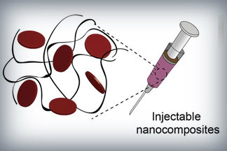
Nanoparticles that promote blood clotting and wound healing (red discs), attached to the wound-filling hydrogel component (black) form a nanocomposite hydrogel. The gel is designed to be self-administered to stop bleeding and begin wound-healing in emergency situations. Credit: Lokhande, et al. 1

Here’s a link to and a citation for the paper,

Nanoengineered injectable hydrogels for wound healing application by Giriraj Lokhande, James K. Carrow, Teena Thakur, Janet R. Xavier, Madasamy Parani, Kayla J. Bayless, Akhilesh K. Gaharwar. Acta Biomaterialia Volume 70, 1 April 2018, Pages 35-47
https://doi.org/10.1016/j.actbio.2018.01.045

This paper is behind a paywall.

Hydrogel and the brain

It’s been an interesting week for hydrogels. On May 21, 2018 there was a news item on ScienceDaily about a bioengineered hydrogel which stimulated brain tissue growth after a stroke (mouse model),

In a first-of-its-kind finding, a new stroke-healing gel helped regrow neurons and blood vessels in mice with stroke-damaged brains, UCLA researchers report in the May 21 issue of Nature Materials.

“We tested this in laboratory mice to determine if it would repair the brain in a model of stroke, and lead to recovery,” said Dr. S. Thomas Carmichael, Professor and Chair of neurology at UCLA. “This study indicated that new brain tissue can be regenerated in what was previously just an inactive brain scar after stroke.”

The brain has a limited capacity for recovery after stroke and other diseases. Unlike some other organs in the body, such as the liver or skin, the brain does not regenerate new connections, blood vessels or new tissue structures. Tissue that dies in the brain from stroke is absorbed, leaving a cavity, devoid of blood vessels, neurons or axons, the thin nerve fibers that project from neurons.

After 16 weeks, stroke cavities in mice contained regenerated brain tissue, including new neural networks — a result that had not been seen before. The mice with new neurons showed improved motor behavior, though the exact mechanism wasn’t clear.

Remarkable stuff.

Crowdsourcing brain research at Princeton University to discover 6 new neuron types

Spritely music!

There were already 1/4M registered players as of May 17, 2018 but I’m sure there’s room for more should you be inspired. A May 17, 2018 Princeton University news release (also on EurekAlert) reveals more about the game and about the neurons,

With the help of a quarter-million video game players, Princeton researchers have created and shared detailed maps of more than 1,000 neurons — and they’re just getting started.

“Working with Eyewirers around the world, we’ve made a digital museum that shows off the intricate beauty of the retina’s neural circuits,” said Sebastian Seung, the Evnin Professor in Neuroscience and a professor of computer science and the Princeton Neuroscience Institute (PNI). The related paper is publishing May 17 [2018] in the journal Cell.

Seung is unveiling the Eyewire Museum, an interactive archive of neurons available to the general public and neuroscientists around the world, including the hundreds of researchers involved in the federal Brain Research through Advancing Innovative Neurotechnologies (BRAIN) Initiative.

“This interactive viewer is a huge asset for these larger collaborations, especially among people who are not physically in the same lab,” said Amy Robinson Sterling, a crowdsourcing specialist with PNI and the executive director of Eyewire, the online gaming platform for the citizen scientists who have created this data set.

“This museum is something like a brain atlas,” said Alexander Bae, a graduate student in electrical engineering and one of four co-first authors on the paper. “Previous brain atlases didn’t have a function where you could visualize by individual cell, or a subset of cells, and interact with them. Another novelty: Not only do we have the morphology of each cell, but we also have the functional data, too.”

The neural maps were developed by Eyewirers, members of an online community of video game players who have devoted hundreds of thousands of hours to painstakingly piecing together these neural cells, using data from a mouse retina gathered in 2009.

Eyewire pairs machine learning with gamers who trace the twisting and branching paths of each neuron. Humans are better at visually identifying the patterns of neurons, so every player’s moves are recorded and checked against each other by advanced players and Eyewire staffers, as well as by software that is improving its own pattern recognition skills.

Since Eyewire’s launch in 2012, more than 265,000 people have signed onto the game, and they’ve collectively colored in more than 10 million 3-D “cubes,” resulting in the mapping of more than 3,000 neural cells, of which about a thousand are displayed in the museum.

Each cube is a tiny subset of a single cell, about 4.5 microns across, so a 10-by-10 block of cubes would be the width of a human hair. Every cell is reviewed by between 5 and 25 gamers before it is accepted into the system as complete.

“Back in the early years it took weeks to finish a single cell,” said Sterling. “Now players complete multiple neurons per day.” The Eyewire user experience stays focused on the larger mission — “For science!” is a common refrain — but it also replicates a typical gaming environment, with achievement badges, a chat feature to connect with other players and technical support, and the ability to unlock privileges with increasing skill. “Our top players are online all the time — easily 30 hours a week,” Sterling said.

Dedicated Eyewirers have also contributed in other ways, including donating the swag that gamers win during competitions and writing program extensions “to make game play more efficient and more fun,” said Sterling, including profile histories, maps of player activity, a top 100 leaderboard and ever-increasing levels of customizability.

“The community has really been the driving force behind why Eyewire has been successful,” Sterling said. “You come in, and you’re not alone. Right now, there are 43 people online. Some of them will be admins from Boston or Princeton, but most are just playing — now it’s 46.”

For science!

With 100 billion neurons linked together via trillions of connections, the brain is immeasurably complex, and neuroscientists are still assembling its “parts list,” said Nicholas Turner, a graduate student in computer science and another of the co-first authors. “If you know what parts make up the machine you’re trying to break apart, you’re set to figure out how it all works,” he said.

The researchers have started by tackling Eyewire-mapped ganglion cells from the retina of a mouse. “The retina doesn’t just sense light,” Seung said. “Neural circuits in the retina perform the first steps of visual perception.”

The retina grows from the same embryonic tissue as the brain, and while much simpler than the brain, it is still surprisingly complex, Turner said. “Hammering out these details is a really valuable effort,” he said, “showing the depth and complexity that exists in circuits that we naively believe are simple.”

The researchers’ fundamental question is identifying exactly how the retina works, said Bae. “In our case, we focus on the structural morphology of the retinal ganglion cells.”

“Why the ganglion cells of the eye?” asked Shang Mu, an associate research scholar in PNI and fellow first author. “Because they’re the connection between the retina and the brain. They’re the only cell class that go back into the brain.” Different types of ganglion cells are known to compute different types of visual features, which is one reason the museum has linked shape to functional data.

Using Eyewire-produced maps of 396 ganglion cells, the researchers in Seung’s lab successfully classified these cells more thoroughly than has ever been done before.

“The number of different cell types was a surprise,” said Mu. “Just a few years ago, people thought there were only 15 to 20 ganglion cell types, but we found more than 35 — we estimate between 35 and 50 types.”

Of those, six appear to be novel, in that the researchers could not find any matching descriptions in a literature search.

A brief scroll through the digital museum reveals just how remarkably flat the neurons are — nearly all of the branching takes place along a two-dimensional plane. Seung’s team discovered that different cells grow along different planes, with some reaching high above the nucleus before branching out, while others spread out close to the nucleus. Their resulting diagrams resemble a rainforest, with ground cover, an understory, a canopy and an emergent layer overtopping the rest.

All of these are subdivisions of the inner plexiform layer, one of the five previously recognized layers of the retina. The researchers also identified a “density conservation principle” that they used to distinguish types of neurons.

One of the biggest surprises of the research project has been the extraordinary richness of the original sample, said Seung. “There’s a little sliver of a mouse retina, and almost 10 years later, we’re still learning things from it.”

Of course, it’s a mouse’s brain that you’ll be examining and while there are differences between a mouse brain and a human brain, mouse brains still provide valuable data as they did in the case of some groundbreaking research published in October 2017. James Hamblin wrote about it in an Oct. 7, 2017 article for The Atlantic (Note: Links have been removed),


Scientists Somehow Just Discovered a New System of Vessels in Our Brains

It is unclear what they do—but they likely play a central role in aging and disease.

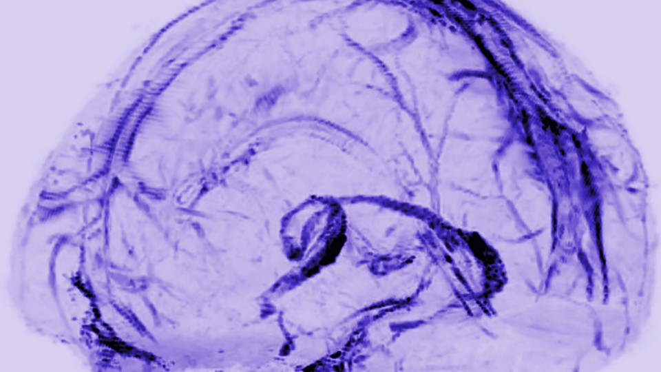
A transparent model of the brain with a network of vessels filled in
Daniel Reich / National Institute of Neurological Disorders and Stroke

You are now among the first people to see the brain’s lymphatic system. The vessels in the photo above transport fluid that is likely crucial to metabolic and inflammatory processes. Until now, no one knew for sure that they existed.

Doctors practicing today have been taught that there are no lymphatic vessels inside the skull. Those deep-purple vessels were seen for the first time in images published this week by researchers at the U.S. National Institute of Neurological Disorders and Stroke.

In the rest of the body, the lymphatic system collects and drains the fluid that bathes our cells, in the process exporting their waste. It also serves as a conduit for immune cells, which go out into the body looking for adversaries and learning how to distinguish self from other, and then travel back to lymph nodes and organs through lymphatic vessels.

So how was it even conceivable that this process wasn’t happening in our brains?

Reich (Daniel Reich, senior investigator) started his search in 2015, after a major study in Nature reported a similar conduit for lymph in mice. The University of Virginia team wrote at the time, “The discovery of the central-nervous-system lymphatic system may call for a reassessment of basic assumptions in neuroimmunology.” The study was regarded as a potential breakthrough in understanding how neurodegenerative disease is associated with the immune system.

Around the same time, researchers discovered fluid in the brains of mice and humans that would become known as the “glymphatic system.” [emphasis mine] It was described by a team at the University of Rochester in 2015 as not just the brain’s “waste-clearance system,” but as potentially helping fuel the brain by transporting glucose, lipids, amino acids, and neurotransmitters. Although since “the central nervous system completely lacks conventional lymphatic vessels,” the researchers wrote at the time, it remained unclear how this fluid communicated with the rest of the body.

There are occasional references to the idea of a lymphatic system in the brain in historic literature. Two centuries ago, the anatomist Paolo Mascagni made full-body models of the lymphatic system that included the brain, though this was dismissed as an error. [emphases mine]  A historical account in The Lancet in 2003 read: “Mascagni was probably so impressed with the lymphatic system that he saw lymph vessels even where they did not exist—in the brain.”

I couldn’t resist the reference to someone whose work had been dismissed summarily being proved right, eventually, and with the help of mouse brains. Do read Hamblin’s article in its entirety if you have time as these excerpts don’t do it justice.

Getting back to Princeton’s research, here’s their research paper,

Digital museum of retinal ganglion cells with dense anatomy and physiology,” by Alexander Bae, Shang Mu, Jinseop Kim, Nicholas Turner, Ignacio Tartavull, Nico Kemnitz, Chris Jordan, Alex Norton, William Silversmith, Rachel Prentki, Marissa Sorek, Celia David, Devon Jones, Doug Bland, Amy Sterling, Jungman Park, Kevin Briggman, Sebastian Seung and the Eyewirers, was published May 17 in the journal Cell with DOI 10.1016/j.cell.2018.04.040.

The research was supported by the Gatsby Charitable Foundation, National Institute of Health-National Institute of Neurological Disorders and Stroke (U01NS090562 and 5R01NS076467), Defense Advanced Research Projects Agency (HR0011-14-2- 0004), Army Research Office (W911NF-12-1-0594), Intelligence Advanced Research Projects Activity (D16PC00005), KT Corporation, Amazon Web Services Research Grants, Korea Brain Research Institute (2231-415) and Korea National Research Foundation Brain Research Program (2017M3C7A1048086).

This paper is behind a paywall. For the players amongst us, here’s the Eyewire website. Go forth,  play, and, maybe, discover new neurons!

World Science Festival May 29 – June 3, 2018 in New York City

I haven’t featured the festival since 2014 having forgotten all about it but I received (via email) an April 30, 2018 news release announcing the latest iteration,

ANNOUNCING WORLD SCIENCE FESTIVAL NEW YORK CITY

MAY 29 THROUGH JUNE 3, 2018

OVER 70 INSPIRING SCIENCE-THEMED EVENTS EXPLORE THE VERY EDGE OF
KNOWLEDGE

Over six extraordinary days in New York City, from May 29 through June
3, 2018; the world’s leading scientists will explore the very edge of
knowledge and share their insights with the public.  Festival goers of
all ages can experience vibrant discussions and debates, evocative
performances and films, world-changing research updates,
thought-provoking town hall gatherings and fireside chats, hands-on
experiments and interactive outdoor explorations.  It’s an action
adventure for your mind!

See the full list of programs here:
https://www.worldsciencefestival.com/festival/world-science-festival-2018/

This year will highlight some of the incredible achievements of Women in
Science, celebrating and exploring their impact on the history and
future of scientific discovery. Perennial favorites will also return in
full force, including WSF main stage Big Ideas programs, the Flame
Challenge, Cool Jobs, and FREE outdoor events.

The World Science Festival makes the esoteric understandable and the
familiar fascinating. It has drawn more than 2.5 million participants
since its launch in 2008, with millions more experiencing the programs
online.

THE 2018 WORLD SCIENCE FESTIVAL IS NOT TO BE MISSED, SO MARK YOUR
CALENDAR AND SAVE THE DATES!

Here are a few items from the 2018 Festival’s program page,

Thursday, May 31, 2018

6:00 pm – 9:00 pm

American Museum of Natural History

Host: Faith Salie

How deep is the ocean? Why do whales sing? How far is 20,000 leagues—and what is a league anyway? Raise a glass and take a deep dive into the foamy waters of oceanic arcana under the blue whale in the Museum’s Hall of Ocean Life. Comedian and journalist Faith Salie will regale you with a pub-style night of trivia questions, physical challenges, and hilarity to celebrate the Museum’s newest temporary exhibition, Unseen Oceans. Don’t worry. When the going gets tough, we won’t let you drown. Teams of top scientists—and even a surprise guest or two—will be standing by to assist you. Program includes one free drink and private access to the special exhibition Unseen Oceans. Special exhibition access is available to ticket holders beginning one hour before the program, from 6–7pm.

Learn More

Buy Tickets

Thursday, May 31, 2018

8:00 pm – 9:30 pm

Gerald W. Lynch Theater at John Jay College

Participants: Alvaro Pascual-Leone, Nim Tottenham, Carla Shatz, And Others

What if your brain at 77 were as plastic as it was at 7? What if you could learn Mandarin with the ease of a toddler or play Rachmaninoff without breaking a sweat? A growing understanding of neuroplasticity suggests these fantasies could one day become reality. Neuroplasticity may also be the key to solving diseases like Alzheimer’s, depression, and autism. This program will guide you through the intricate neural pathways inside our skulls, as leading neuroscientists discuss their most recent findings and both the tantalizing possibilities and pitfalls for our future cognitive selves.

The Big Ideas Series is supported in part by the John Templeton Foundation. 

Learn More

Buy Tickets

Friday, June 1, 2018

8:00 pm – 9:30 pm

NYU Skirball Center for the Performing Arts

Participants: Yann LeCun, Susan Schneider, Max Tegmark, And Others

“Success in creating effective A.I.,” said the late Stephen Hawking, “could be the biggest event in the history of our civilization. Or the worst. We just don’t know.” Elon Musk called A.I. “a fundamental risk to the existence of civilization.” Are we creating the instruments of our own destruction or exciting tools for our future survival? Once we teach a machine to learn on its own—as the programmers behind AlphaGo have done, to wondrous results—where do we draw moral and computational lines? Leading specialists in A.I, neuroscience, and philosophy will tackle the very questions that may define the future of humanity.

The Big Ideas Series is supported in part by the John Templeton Foundation. 

Learn More

Buy Tickets

Friday, June 1, 2018

8:00 pm – 9:30 pm

Gerald W. Lynch Theater at John Jay College

Participants Marcela Carena, Janet Conrad, Michael Doser, Hitoshi Murayama, Neil Turok

“If I had a world of my own,” said the Mad Hatter, “nothing would be what it is, because everything would be what it isn’t. And contrary wise, what is, it wouldn’t be.” Nonsensical as this may sound, it comes close to describing an interesting paradox: You exist. You shouldn’t. Stars and galaxies and planets exist. They shouldn’t. The nascent universe contained equal parts matter and antimatter that should have instantly obliterated each other, turning the Big Bang into the Big Fizzle. And yet, here we are: flesh, blood, stars, moons, sky. Why? Come join us as we dive deep down the rabbit hole of solving the mystery of the missing antimatter.

The Big Ideas Series is supported in part by the John Templeton Foundation.

Learn More

Buy Tickets

Saturday, June 2, 2018

10:00 am – 11:00 am

Museum of the City of New York

ParticipantsKubi Ackerman

What makes a city a city? How do you build buildings, plan streets, and design parks with humans and their needs in mind? Join architect and Future Lab Project Director, Kubi Ackerman, on an exploration in which you’ll venture outside to examine New York City anew, seeing it through the eyes of a visionary museum architect, and then head to the Future City Lab’s awesome interactive space where you will design your own park. This is a student-only program for kids currently enrolled in the 4th grade – 8th grade. Parents/Guardians should drop off their children for this event.

Supported by the Bezos Family Foundation.

Learn More

Buy Tickets

Saturday, June 2, 2018

11:00 am – 12:30 pm

NYU Global Center, Grand Hall

Kerouac called it “the only truth.” Shakespeare called it “the food of love.” Maya Angelou called it “my refuge.” And now scientists are finally discovering what these thinkers, musicians, or even any of us with a Spotify account and a set of headphones could have told you on instinct: music lights up multiple corners of the brain, strengthening our neural networks, firing up memory and emotion, and showing us what it means to be human. In fact, music is as essential to being human as language and may even predate it. Can music also repair broken networks, restore memory, and strengthen the brain? Join us as we speak with neuroscientists and other experts in the fields of music and the brain as we pluck the notes of these fascinating phenomenon.

The Big Ideas Series is supported in part by the John Templeton Foundation.

Learn More

Buy Tickets

Saturday, June 2, 2018

3:00 pm – 4:00 pm

NYU Skirball Center for the Performing Arts

Moderator“Science Bob” Pflugfelder

Participants William Clark, Matt Lanier, Michael Meacham, Casie Parish Fisher, Mike Ressler

Most people think of scientists as people who work in funny-smelling labs filled with strange equipment. But there are lots of scientists whose jobs often take them out of the lab, into the world, and beyond. Come join some of the coolest of them in Cool Jobs. You’ll get to meet a forensic scientist, a venomous snake-loving herpetologist, a NASA engineer who lands spacecrafts on Mars, and inventors who are changing the future of sports.

Learn More

Buy Tickets

Saturday, June 2, 2018

4:00 pm – 5:30 pm

NYU Global Center, Grand Hall

“We can rebuild him. We have the technology,” began the opening sequence of the hugely popular 70’s TV show, “The Six Million Dollar Man.” Forty-five years later, how close are we, in reality, to that sci-fi fantasy? More thornily, now that artificial intelligence may soon pass human intelligence, and the merging of human with machine is potentially on the table, what will it then mean to “be human”? Join us for an important discussion with scientists, technologists and ethicists about the path toward superhumanism and the quest for immortality.

The Big Ideas Series is supported in part by the John Templeton Foundation.

Learn More

Buy Tickets

Saturday, June 2, 2018

4:00 pm – 5:30 pm

Gerald W. Lynch Theater at John Jay College

Participants Brett Frischmann, Tim Hwang, Aviv Ovadya, Meredith Whittaker

“Move fast and break things,” went the Silicon Valley rallying cry, and for a long time we cheered along. Born in dorm rooms and garages, implemented by iconoclasts in hoodies, Big Tech, in its infancy, spouted noble goals of bringing us closer. But now, in its adolescence, it threatens to tear us apart. Some worry about an “Infocalypse”: a dystopian disruption so deep and dire we will no longer trust anything we see, hear, or read. Is this pessimistic vision of the future real or hyperbole? Is it time for tech to slow down, grow up, and stop breaking things? Big names in Big Tech will offer big thoughts on this massive societal shift, its terrifying pitfalls, and practical solutions both for ourselves and for future generations.

The Big Ideas Series is supported in part by the John Templeton Foundation.

Learn More

Buy Tickets

This looks like an exciting lineup and there’s a lot more for you to see on the 2018 Festival’s program page. You may also want to take a look at the list of participants which features some expected specialty speakers, an architect, a mathematician, a neuroscientist and some unexpected names such Kareem Abdul-Jabbar who I know as a basketball player and currently, a contestant on Dancing with the Stars. Bringing to mind that Walt Whitman quote, “I am large, I contain multitudes.” (from Whitman’s Song of Myself Wikipedia entry).

If you’re going, there are free events and note a few of the event are already sold out.