Tag Archives: hydrogel

Ouchies no more! Not from bandages, anyway.

An adhesive that US and Chinese scientists have developed shows great promise not just for bandages but wearable robotics too. From a December 14, 2018 news item on Nanowerk,

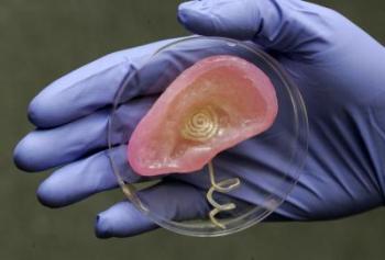
Researchers from the Harvard John A. Paulson School of Engineering and Applied Sciences (SEAS) and Xi’an Jiaotong University in China have developed a new type of adhesive that can strongly adhere wet materials — such as hydrogel and living tissue — and be easily detached with a specific frequency of light.

The adhesives could be used to attach and painlessly detach wound dressings, transdermal drug delivery devices, and wearable robotics.

A December 18, 2018 SEAS news release by Leah Burrows (also on EurekAlert but published Dec. 14, 2018), which originated the news item, delves further,

“Strong adhesion usually requires covalent bonds, physical interactions, or a combination of both,” said Yang Gao, first author of the paper and researcher at Xi’an Jiaotong University. “Adhesion through covalent bonds is hard to remove and adhesion through physical interactions usually requires solvents, which can be time-consuming and environmentally harmful. Our method of using light to trigger detachment is non-invasive and painless.”

The adhesive uses an aqueous solution of polymer chains spread between two, non-sticky materials — like jam between two slices of bread. On their own, the two materials adhere poorly together but the polymer chains act as a molecular suture, stitching the two materials together by forming a network with the two preexisting polymer networks. This process is known as topological entanglement.

When exposed to ultra-violet light, the network of stitches dissolves, separating the two materials.

The researchers, led by Zhigang Suo, the Allen E. and Marilyn M. Puckett Professor of Mechanics and Materials at SEAS, tested adhesion and detachment on a range of materials, sticking together hydrogels; hydrogels and organic tissue; elastomers; hydrogels and elastomers; and hydrogels and inorganic solids.

“Our strategy works across a range of materials and may enable broad applications,” said Kangling Wu, co-lead author and researcher at Xi’an Jiaotong University in China.
While the researchers focused on using UV light to trigger detachment, their work suggests the possibility that the stitching polymer could detach with near-infrared light, a feature which could be applied to a range of new medical procedures.

“In nature, wet materials don’t like to adhere together,” said Suo. “We have discovered a general approach to overcome this challenge. Our molecular sutures can strongly adhere wet materials together. Furthermore, the strong adhesion can be made permanent, transient, or detachable on demand, in response to a cue. So, as we see it, nature is full of loopholes, waiting to be stitched.”

Here’s a link to and  a citation for the paper,

Photodetachable Adhesion by Yang Gao, Kangling Wu, Zhigang Suo. https://doi.org/10.1002/adma.201806948 First published: 14 December 2018

This paper is behind a paywall.

Spider glue

Caption: An orb spider, glue-maker extraordinaire, at work on a web. Credit: The University of Akron

Scientists are taking inspiration from spiders in their quest to develop better adhesives. (Are they abandoning the gecko? Usually when scientists study adhesiveness, there’s talk of geckos. From a June 5, 2018 news item on ScienceDaily,

Ever wonder why paint peels off the wall during summer’s high humidity? It’s the same reason that bandages separate from skin when we bathe or swim.

Interfacial water, as it’s known, forms a slippery and non-adhesive layer between the glue and the surface to which it is meant to stick, interfering with the formation of adhesive bonds between the two.

Overcoming the effects of interfacial water is one of the challenges facing developers of commercial adhesives.

To find a solution, researchers at The University of Akron (UA) are looking to one of the strongest materials found in nature: spider silk.

The sticky glue that coats the silk threads of spider webs is a hydrogel, meaning it is full of water. One would think, then, that spiders would have difficulty catching prey, especially in humid conditions — but they do not. In fact, their sticky glue, which has been a subject of intensive research for years, is one of the most effective biological glues in all of nature.

A June 4, 2018 University of Akron news release (also on EurekAlert published on June 5, 2018), which originated the news item, provides more detail,

So how is spider glue able to stick in highly humid conditions?

That question was the subject of investigation by UA graduate students Saranshu Singla, Gaurav Amarpuri and Nishad Dhopatkar, who have been working with Dr. Ali Dhinojwala, interim dean of the College of Polymer Science and Polymer Engineering, and Dr. Todd Blackledge, professor of biology in the Integrated Bioscience program. Both professors are principal investigators in UA’s Biomimicry Research Innovation Center [BRIC], which specializes in emulating biological forms, processes, patterns and systems to solve technical challenges.

The team’s findings, which may provide the clue to developing stronger commercial adhesives, can be read in a paper recently published in the journal Nature Communications.

Singla and her colleagues set out to examine the secret behind the success of the common orb spider (Larinioides cornutus) glue and uncover how it overcomes the primary obstacle of achieving good adhesion in the humid conditions where water could be present between the glue and the target surface.

To investigate the processes involved, the team took orb spider glue, set it on sapphire substrate, then examined it using a combination of interface-sensitive spectroscopy and infrared spectroscopy.

Spider glue is made of three elements: two specialized glycoproteins, a collection of low molecular mass organic and inorganic compounds (LMMCs), and water. The LMMCs are hygroscopic (water-attracting), which keeps the glue soft and tacky to stick.

Singla and her team discovered that these glycoproteins act as primary binding agents to the surface. Glycoprotein-based glues have been identified in several other biological glues, such as fungi, algae, diatoms, sea stars, sticklebacks and English ivy.

But why doesn’t the water present in the spider glue interfere with the adhesive contact the way it does with most synthetic adhesives?

The LMMCs, the team concluded, perform a previously unknown function of sequestering interfacial water, preventing adhesive failure.

Singla and colleagues determined that it is the interaction of glycoproteins and LMMCs that governs the adhesive quality of the glue produced, with the respective proportions varying across species, thus optimizing adhesive strength to match the relative humidity of spider habitat.

“The hygroscopic compounds – known as water-absorbers – in spider glue play a previously unknown role in moving water away from the boundary, thereby preventing failure of spider glue at high humidity,” explained Singla.

The ability of the spider glue to overcome the problem of interfacial water by effectively absorbing it is the key finding of the research, and the one with perhaps the strongest prospect for commercial development.

“Imagine a paint that is guaranteed for life, come rain or shine,” Singla remarked.

All thanks to your friendly neighborhood spider glue.

Here’s a link to and a citation for the paper,

Hygroscopic compounds in spider aggregate glue remove interfacial water to maintain adhesion in humid conditions by Saranshu Singla, Gaurav Amarpuri, Nishad Dhopatkar, Todd A. Blackledge, & Ali Dhinojwala. Nature Communicationsvolume 9, Article number: 1890 (2018) Published 22 May 2018 DOI: https://doi.org/10.1038/s41467-018-04263-z

This paper is open access.

A cheaper way to make artificial organs

In the quest to develop artificial organs, the University of British Columbia (UBC) is the not the first research institution that comes to my mind. It seems I may need to reevaluate now that UBC (Okanagan) has announced some work on bio-inks and artificial organs in a Sept. 12, 2017 news  release (also on EurekAlert) by Patty Wellborn,,

A new bio-ink that may support a more efficient and inexpensive fabrication of human tissues and organs has been created by researchers at UBC’s Okanagan campus.

Keekyoung Kim, an assistant professor at UBC Okanagan’s School of Engineering, says this development can accelerate advances in regenerative medicine.

Using techniques like 3D printing, scientists are creating bio-material products that function alongside living cells. These products are made using a number of biomaterials including gelatin methacrylate (GelMA), a hydrogel that can serve as a building block in bio-printing. This type of bio-material—called bio-ink—are made of living cells, but can be printed and molded into specific organ or tissue shapes.

The UBC team analyzed the physical and biological properties of three different GelMA hydrogels—porcine skin, cold-water fish skin and cold-soluble gelatin. They found that hydrogel made from cold-soluble gelatin (gelatin which dissolves without heat) was by far the best performer and a strong candidate for future 3D organ printing.

“A big drawback of conventional hydrogel is its thermal instability. Even small changes in temperature cause significant changes in its viscosity or thickness,” says Kim. “This makes it problematic for many room temperature bio-fabrication systems, which are compatible with only a narrow range of hydrogel viscosities and which must generate products that are as uniform as possible if they are to function properly.”

Kim’s team created two new hydrogels—one from fish skin, and one from cold-soluble gelatin—and compared their properties to those of porcine skin GelMA. Although fish skin GelMA had some benefits, cold-soluble GelMA was the top overall performer. Not only could it form healthy tissue scaffolds, allowing cells to successfully grow and adhere to it, but it was also thermally stable at room temperature.

The UBC team also demonstrated that cold-soluble GelMA produces consistently uniform droplets at temperatures, thus making it an excellent choice for use in 3D bio-printing.

“We hope this new bio-ink will help researchers create improved artificial organs and lead to the development of better drugs, tissue engineering and regenerative therapies,” Kim says. “The next step is to investigate whether or not cold-soluble GelMA-based tissue scaffolds are can be used long-term both in the laboratory and in real-world transplants.”

Three times cheaper than porcine skin gelatin, cold-soluble gelatin is used primarily in culinary applications.

Here’s a link to and a citation for the paper,

Comparative study of gelatin methacrylate hydrogels from different sources for biofabrication applications by Zongjie Wang, Zhenlin Tian, Fredric Menard, and Keekyoung Kim. Biofabrication, Volume 9, Number 4 Special issue on Bioinks https://doi.org/10.1088/1758-5090/aa83cf Published 21 August 2017

© 2017 IOP Publishing Ltd

This paper is behind a paywall.

Accurate spatial orientation for regenerating cells

German scientists have developed a system that helps guide nerve cells into proper spatial alignmentswhen they are regenerating according to an April 5, 2017 news item on Nanowerk,

In many tissues of the human body, such as nerve tissue, the spatial organization of cells plays an important role. Nerve cells and their long protrusions assemble into nerve tracts and transport information throughout the body. When such a tissue is injured, an accurate spatial orientation of the cells facilitates the healing process. Scientists from the DWI – Leibniz Institute for Interactive Materials in Aachen developed an injectable gel, which can act as a guidance system for nerve cells.

An April 5, 2017 Leibniz Institute press release, which originated the news item, explains more about the research,

Inside the body, an extracellular matrix surrounds the cells. It provides mechanical support and promotes spatial tissue organization. In order to regenerate damaged tissue, an artificial matrix can temporally replace the natural extracellular matrix. This matrix needs to mimic the natural cell environment in order to efficiently stimulate the regenerative potential of the surrounding tissue. Solid implants, however, may impair remaining healthy tissue whereas soft, injectable materials allow for a minimal invasive therapy, which is particularly beneficial for sensitive tissues, such as the spinal cord. Unfortunately, up to now, artificial soft materials did not yet reproduce the complex structures and spatial properties of natural tissues.

A team of scientists, headed by Dr. Laura De Laporte from the DWI – Leibniz Institute for Interactive Materials, developed a new, minimal invasive material termed ‘Anisogel’. “If you aim to enhance the regeneration of damaged spinal cord tissue, you need to come up with a new material concept,” says Jonas Rose. He is a PhD student working on the Anisogel project. “We use micrometer-sized building blocks and assemble them into 3D hierarchically organized structures.” Anisogel consists of two gel components. Many, microscopically small, soft rod-shaped gels, incorporated with a low amount of magnetic nanoparticles, are the first component. Using a weak magnetic field, scientists can orient the gel rods, after which a very soft surrounding gel matrix is crosslinked, forming the structural guidance system. The gel rods, being stabilized by the gel matrix, maintain their orientation, even after removal of the magnetic field. Using cell culture experiments, the researchers demonstrate that cells can easily migrate through this gel matrix, and that nerve cells and fibroblasts orient along the paths provided by this guidance system.  A low amount of one percent gel rods inside the entire Anisogel volume is proven to be sufficient to induce linear nerve growth. The material, developed by the Aachen-based scientists, is the first injectable biomaterial, which assembles into a controlled oriented structure after injection and provides a functional guidance system for cells. “To meet the complex requirements of this approach, the project team includes researchers with very different areas of expertise,” says Laura De Laporte, whose research is supported by a Starting Grant of the European Research Council. “This interdisciplinary work is what makes this project so fascinating.”

“Although our cell culture experiments were successful, we are prepared to go a long way to translate our Anisogel into a medical therapy. In collaboration with the Uniklinik RWTH Aachen, we currently plan pre-clinical studies to further test and optimize this material,” Laura De Laporte explains.

Here’s a link to and a citation for the paper,

Nerve Cells Decide to Orient inside an Injectable Hydrogel with Minimal Structural Guidance  by Jonas C. Rose, María Cámara-Torres, Khosrow Rahimi, Jens Köhler, Martin Möller, and Laura De Laporte. Nano Lett., Article ASAP DOI: 10.1021/acs.nanolett.7b01123 Publication Date (Web): March 22, 2017

Copyright © 2017 American Chemical Society

This paper is behind a paywall.

Regrowing bone

The ability to grow bone or bone-like material could change life substantially for people with certain kinds of injuries. Scientists at Northwestern University and the University of Chicago have been able to regrow bone in a skull (according to a March 8, 2017 Northwestern University news release (also on EurekAlert),

A team of researchers repaired a hole in a mouse’s skull by regrowing “quality bone,” a breakthrough that could drastically improve the care of people who suffer severe trauma to the skull or face.

The work by a joint team of Northwestern Engineering and University of Chicago researchers was a resounding success, showing that a potent combination of technologies was able to regenerate the skull bone with supporting blood vessels in just the discrete area needed without developing scar tissue — and more rapidly than with previous methods.

“The results are very exciting,” said Guillermo Ameer, professor of biomedical engineering at Northwestern’s McCormick School of Engineering, and professor of surgery at Feinberg School of Medicine.

Supported by the China Scholarship Council, National Institute of Dental and Craniofacial Research, Chicago Community Trust, and National Center for Advancing Translational Sciences, the research was published last week in the journal PLOS One. Russell Reid, associate professor of surgery at the University of Chicago Medical Center, is the article’s corresponding author. Reid, his long-time collaborator Dr. Tong-Chuan He, and colleagues in Hyde Park brought the surgical and biological knowledge and skills. Zari P. Dumanian, affiliated with the medical center’s surgery department, was the paper’s first author.

“This project was a true collaborative team effort in which our Regenerative Engineering Laboratory provided the biomaterials expertise,” Ameer said.

Injuries or defects in the skull or facial bones are very challenging to treat, often requiring the surgeon to graft bone from the patient’s pelvis, ribs, or elsewhere, a painful procedure in itself. Difficulties increase if the injury area is large or if the graft needs to be contoured to the angle of the jaw or the cranial curve.

But if all goes well with this new approach, it may make painful bone grafting obsolete.

In the experiment, the researchers harvested skull cells from the mouse and engineered them to produce a potent protein to promote bone growth. They then used Ameer’s hydrogel, which acted like a temporary scaffolding, to deliver and contain these cells to the affected area. It was the combination of all three technologies that proved so successful, Ameer said.

Using calvaria or skull cells from the subject meant the body didn’t reject those cells.

The protein, BMP9, has been shown to promote bone cell growth more rapidly than other types of BMPs. Importantly, BMP9 also appeared to improve the creation of blood vessels in the area. Being able to safely deliver skull cells that are capable of rapidly regrowing bone in the affected site, in vivo as opposed to using them to grow bone in the laboratory, which would take a very long time, promises a therapy that might be more “surgeon friendly, if you will, and not too complicated to scale up for the patients,” Ameer said.

The scaffolding developed in Ameer’s laboratory, which is a material based on citric acid and called PPCN-g, is a liquid that when warmed to body temperature becomes a gel-like elastic material. “When applied, the liquid, which contains cells capable of producing bone, will conform to the shape of the bone defect to make a perfect fit,” Ameer said. “It then stays in place as a gel, localizing the cells to the site for the duration of the repair.” As the bone regrows, the PPCN-g is reabsorbed by the body.

“What we found is that these cells make natural-looking bone in the presence of the PPCN-g,” Ameer said. “The new bone is very similar to normal bone in that location.”

In fact, the three-part method was successful on a number of fronts: The regenerated bone was better quality, the bone growth was contained to the area defined by the scaffolding, the area healed much more quickly, and the new and old bone were continuous with no scar tissue.

The potential, if the procedure can be adapted to treat people that suffered trauma from car accidents or aggressive cancers that have affected the skull or face, would be huge, and give surgeons a much-sought-after option.

“The reconstruction procedure is a lot easier when you can harvest a few cells, make them produce the BMP9 protein, mix them in the PPCN-g solution, and apply it to the bone defect site to jump-start the new bone growth process where you want it.” Ameer said.

Ameer cautioned that the technology is years away to being used in humans, but added, “We did show proof of concept that we can heal large defects in the skull that would normally not heal on their own using a protein, cells and a new material that come together in a completely new way. Our team is very excited about these findings and the future of reconstructive surgery.”

Here’s a link and a citation for the paper,

Repair of critical sized cranial defects with BMP9-transduced calvarial cells delivered in a thermoresponsive scaffold by Zari P. Dumanian, Viktor Tollemar, Jixing Ye, Minpeng Lu, Yunxiao Zhu, Junyi Liao, Guillermo A. Ameer, Tong-Chuan He, Russell R. Reid. PLOS http://dx.doi.org/10.1371/journal.pone.0172327 Published: March 1, 2017

This is an open access paper.

Better technique for growing organoids taking them from the lab to the clinic

A Nov. 16, 2016 École Polytechnique Fédérale de Lausanne (EPFL) press release (also on EurekAlert) describes a new material for growing organoids,

Organoids are miniature organs that can be grown in the lab from a person’s stem cells. They can be used to model diseases, and in the future could be used to test drugs or even replace damaged tissue in patients. But currently organoids are very difficult to grow in a standardized and controlled way, which is key to designing and using them. EPFL scientists have now solved the problem by developing a patent-pending “hydrogel” that provides a fully controllable and tunable way to grow organoids. …

Organoids need a 3D scaffold

Growing organoids begins with stem cells — immature cells that can grow into any cell type of the human body and that play key roles in tissue function and regeneration. To form an organoid, the stem cells are grown inside three-dimensional gels that contain a mix of biomolecules that promote stem cell renewal and differentiation.

The role of these gels is to mimic the natural environment of the stem cells, which provides them with a protein- and sugar-rich scaffold called the “extracellular matrix”, upon which the stem cells build specific body tissues. The stem cells stick to the extracellular matrix gel, and then “self-organize” into miniature organs like retinas, kidneys, or the gut. These tiny organs retain key aspects of their real-life biology, and can be used to study diseases or test drugs before moving on to human trials.

But the current gels used for organoid growth are derived from mice, and have problems. First, it is impossible to control their makeup from batch to batch, which can cause stem cells to behave inconsistently. Second, their biochemical complexity makes them very difficult to fine-tune for studying the effect of different parameters (e.g. biological molecules, mechanical properties, etc.) on the growth of organoids. Finally, the gels can carry pathogens or immunogens, which means that they are not suitable for growing organoids to be used in the clinic.

A hydrogel solution

The lab of Matthias Lütolf at EPFL’s Institute of Bioengineering has developed a synthetic “hydrogel” that eschews the limitations of conventional, naturally derived gels. The patent-pending gel is made of water and polyethylene glycol, a substance used widely today in various forms, from skin creams and toothpastes to industrial applications and, as in this case, bioengineering.

Nikolce Gjorevski, the first author of the study, and his colleagues used the hydrogel to grow stem cells of the gut into a miniature intestine. The functional hydrogel was not only a goal in and of itself, but also a means to identify the factors that influence the stem cells’ ability to expand and form organoids. By carefully tweaking the hydrogel’s properties, they discovered that separate stages of the organoid formation process require different mechanical environments and biological components.

One such factor is a protein called fibronectin, which helps the stem cells attach to the hydrogel. Lütolf’s lab found that this attachment itself is immensely important for growing organoids, as it triggers a whole host of signals to the stem cell that tell it to grow and build an intestine-like structure. The researchers also discovered an essential role for the mechanical properties, i.e. the physical stiffness, of the gel in regulating intestinal stem cell behavior, shedding light on how cells are able to sense, process and respond to physical stimuli. This insight is particularly valuable – while the influence of biochemical signals on stem cells is well-understood, the effect of physical factors has been more mysterious.

Because the hydrogel is man-made, it is easy to control its chemical composition and key properties, and ensure consistency from batch to batch. And because it is artificial, it does not carry any risk of infection or triggering immune responses. As such, it provides a means of moving organoids from basic research to actual pharmaceutical and clinical applications in the future.

Lütolf’s lab is now researching other types of stem cells in order to extend the capacities of their hydrogel into other tissues.

Here’s a link to and a citation for the paper,

Designer matrices for intestinal stem cell and organoid culture by Nikolce Gjorevski, Norman Sachs, Andrea Manfrin, Sonja Giger, Maiia E. Bragina, Paloma Ordóñez-Morán, Hans Clevers, & Matthias P. Lutolf.  Nature (2016) doi:10.1038/nature20168 Published online 16 November 2016

This paper is behind a paywall.

Encapsulation of proteins in nanoparticles no longer necessary for time release?

A team of researchers at the University of Toronto (Canada) have developed a technique for the therapeutic use of proteins that doesn’t require ‘nanoencapsulation’ although nanoparticles are still used according to a May 27, 2016 news item on ScienceDaily,

A U of T [University of Toronto] Engineering team has designed a simpler way to keep therapeutic proteins where they are needed for long periods of time. The discovery is a potential game-changer for the treatment of chronic illnesses or injuries that often require multiple injections or daily pills.

For decades, biomedical engineers have been painstakingly encapsulating proteins in nanoparticles to control their release. Now, a research team led by University Professor Molly Shoichet has shown that proteins can be released over several weeks, even months, without ever being encapsulated. In this case the team looked specifically at therapeutic proteins relevant to tissue regeneration after stroke and spinal cord injury.

“It was such a surprising and unexpected discovery,” said co-lead author Dr. Irja Elliott Donaghue, who first found that the therapeutic protein NT3, a factor that promotes the growth of nerve cells, was slowly released when just mixed into a Jello-like substance that also contained nanoparticles. “Our first thought was, ‘What could be happening to cause this?'”

A May 27, 2016 University of Toronto news release (also on EurekAlert) by Marit Mitchell, which originated the news item, provides more in depth explanation,

Proteins hold enormous promise to treat chronic conditions and irreversible injuries — for example, human growth hormone is encapsulated in these tiny polymeric particles, and used to treat children with stunted growth. In order to avoid repeated injections or daily pills, researchers use complicated strategies both to deliver proteins to their site of action, and to ensure they’re released over a long enough period of time to have a beneficial effect.

This has long been a major challenge for protein-based therapies, especially because proteins are large and often fragile molecules. Until now, investigators have been treating proteins the same way as small drug molecules and encapsulating them in polymeric nanoparticles, often made of a material called poly(lactic-co-glycolic acid) or PLGA.

As the nanoparticles break down, the drug molecules escape. The same process is true for proteins; however, the encapsulating process itself often damages or denatures some of the encapsulated proteins, rendering them useless for treatment. Skipping encapsulation altogether means fewer denatured proteins, making for more consistent protein therapeutics that are easier to make and store.

“This is really exciting from a translational perspective,” said PhD candidate Jaclyn Obermeyer. “Having a simpler, more reliable fabrication process leaves less room for complications with scale-up for clinical use.”

The three lead authors, Elliott Donoghue, Obermeyer and Dr. Malgosia Pakulska have shown that to get the desired controlled release, proteins only need to be alongside the PLGA nanoparticles, not inside them. …

“We think that this could speed up the path for protein-based drugs to get to the clinic,” said Elliott Donaghue.

The mechanism for this encapsulation-free controlled release is surprisingly elegant. Shoichet’s group mixes the proteins and nanoparticles in a Jello-like substance called a hydrogel, which keeps them localized when injected at the site of injury. The positively charged proteins and negatively charged nanoparticles naturally stick together. As the nanoparticles break down they make the solution more acidic, weakening the attraction and letting the proteins break free.

“We are particularly excited to show long-term, controlled protein release by simply controlling the electrostatic interactions between proteins and polymeric nanobeads,” said Shoichet. “By manipulating the pH of the solution, the size and number of nanoparticles, we can control release of bioactive proteins. This has already changed and simplified the protein release strategies that we are pursuing in pre-clinical models of disease in the brain and spinal cord.”

“We’ve learned how to control this simple phenomena,” Pakulska said. “Our next question is whether we can do the opposite—design a similar release system for positively charged nanoparticles and negatively charged proteins.”

Here’s a link to and a citation for the paper,

Encapsulation-free controlled release: Electrostatic adsorption eliminates the need for protein encapsulation in PLGA nanoparticles by Malgosia M. Pakulska, Irja Elliott Donaghue, Jaclyn M. Obermeyer, Anup Tuladhar, Christopher K. McLaughlin, Tyler N. Shendruk, and Molly S. Shoichet. Science Advances  27 May 2016: Vol. 2, no. 5, e1600519 DOI: 10.1126/sciadv.1600519

This paper appears to be open access.

Dr. Molly Shoichet was featured here in a May 11, 2015 posting about the launch of her Canada-wide science communication project Research2.Reality.

Controlling water with ‘stick-slip surfaces’

Controlling water could lead to better designed microfluidic devices such as ‘lab-on-a-chip’. A July 27, 2015 news item on Nanowerk announces a new technique for controlling water,

Coating the inside of glass microtubes with a polymer hydrogel material dramatically alters the way capillary forces draw water into the tiny structures, researchers have found. The discovery could provide a new way to control microfluidic systems, including popular lab-on-a-chip devices.

Capillary action draws water and other liquids into confined spaces such as tubes, straws, wicks and paper towels, and the flow rate can be predicted using a simple hydrodynamic analysis. But a chance observation by researchers at the Georgia Institute of Technology [US] will cause a recalculation of those predictions for conditions in which hydrogel films line the tubes carrying water-based liquids.

“Rather than moving according to conventional expectations, water-based liquids slip to a new location in the tube, get stuck, then slip again – and the process repeats over and over again,” explained Andrei Fedorov, a professor in the George W. Woodruff School of Mechanical Engineering at Georgia Tech. “Instead of filling the tube with a rate of liquid penetration that slows with time, the water propagates at a nearly constant speed into the hydrogel-coated capillary. This was very different from what we had expected.”

A July 27, 2015 Georgia Institute of Technology (Georgia Tech) news release (also on EurekAlert) by John Toon, which originated the news item, describes the work in more detail,

When the opening of a thin glass tube is exposed to a droplet of water, the liquid begins to flow into the tube, pulled by a combination of surface tension in the liquid and adhesion between the liquid and the walls of the tube. Leading the way is a meniscus, a curved surface of the water at the leading edge of the water column. An ordinary borosilicate glass tube fills by capillary action at a gradually decreasing rate with the speed of meniscus propagation slowing as a square root of time.

But when the inside of a tube is coated with a very thin layer of poly(N-isopropylacrylamide), a so-called “smart” polymer (PNIPAM), everything changes. Water entering a tube coated on the inside with a dry hydrogel film must first wet the film and allow it to swell before it can proceed farther into the tube. The wetting and swelling take place not continuously, but with discrete steps in which the water meniscus first sticks and its motion remains arrested while the polymer layer locally deforms. The meniscus then rapidly slides for a short distance before the process repeats. This “stick-slip” process forces the water to move into the tube in a step-by-step motion.

The flow rate measured by the researchers in the coated tube is three orders of magnitude less than the flow rate in an uncoated tube. A linear equation describes the time dependence of the filling process instead of a classical quadratic equation which describes filling of an uncoated tube.

“Instead of filling the capillary in a hundredth of a second, it might take tens of seconds to fill the same capillary,” said Fedorov. “Though there is some swelling of the hydrogel upon contact with water, the change in the tube diameter is negligible due to the small thickness of the hydrogel layer. This is why we were so surprised when we first observed such a dramatic slow-down of the filing process in our experiments.”

The researchers – who included graduate students James Silva, Drew Loney and Ren Geryak and senior research engineer Peter Kottke – tried the experiment again using glycerol, a liquid that is not absorbed by the hydrogel. With glycerol, the capillary action proceeded through the hydrogel-coated microtube as with an uncoated tube in agreement with conventional theory. After using high-resolution optical visualization to study the meniscus propagation while the polymer swelled, the researchers realized they could put this previously-unknown behavior to good use.

Water absorption by the hydrogels occurs only when the materials remain below a specific transition temperature. When heated above that temperature, the materials no longer absorb water, eliminating the “stick-slip” phenomenon in the microtubes and allowing them to behave like ordinary tubes.

This ability to turn the stick-slip behavior on and off with temperature could provide a new way to control the flow of water-based liquid in microfluidic devices, including labs-on-a-chip. The transition temperature can be controlled by varying the chemical composition of the hydrogel.

“By locally heating or cooling the polymer inside a microfluidic chamber, you can either speed up the filling process or slow it down,” Fedorov said. “The time it takes for the liquid to travel the same distance can be varied up to three orders of magnitude. That would allow precise control of fluid flow on demand using external stimuli to change polymer film behavior.”

The heating or cooling could be done locally with lasers, tiny heaters, or thermoelectric devices placed at specific locations in the microfluidic devices.

That could allow precise timing of reactions in microfluidic devices by controlling the rate of reactant delivery and product removal, or allow a sequence of fast and slow reactions to occur. Another important application could be controlled drug release in which the desired rate of molecule delivery could be dynamically tuned over time to achieve the optimal therapeutic outcome.

In future work, Fedorov and his team hope to learn more about the physics of the hydrogel-modified capillaries and study capillary flow using partially-transparent microtubes. They also want to explore other “smart” polymers which change the flow rate in response to different stimuli, including the changing pH of the liquid, exposure to electromagnetic radiation, or the induction of mechanical stress – all of which can change the properties of a particular hydrogel designed to be responsive to those triggers.

“These experimental and theoretical results provide a new conceptual framework for liquid motion confined by soft, dynamically evolving polymer interfaces in which the system creates an energy barrier to further motion through elasto-capillary deformation, and then lowers the barrier through diffusive softening,” the paper’s authors wrote. “This insight has implications for optimal design of microfluidic and lab-on-a-chip devices based on stimuli-responsive smart polymers.”

In addition to those already mentioned, the research team included Professor Vladimir Tsukruk from the Georgia Tech School of Materials Science and Engineering and Rajesh Naik, Biotechnology Lead and Tech Advisor of the Nanostructured and Biological Materials Branch of the Air Force Research Laboratory (AFRL).

Here’s a link to and a citation for the paper,

Stick–slip water penetration into capillaries coated with swelling hydrogel by J. E. Silva, R. Geryak, D. A. Loney, P. A. Kottke, R. R. Naik, V. V. Tsukruk, and A. G. Fedorov. Soft Matter, 2015,11, 5933-5939 DOI: 10.1039/C5SM00660K First published online 23 Jun 2015

This paper is behind a paywall.

Assembly-line 3-D tissue engineering

It looks as if the researchers at Singapore’s Institute of Bioengineering and Nanotechnology (IBN), have developed a template for producing complex tissues such as those in liver and in fat, from an Aug. 20, 2013 news item on ScienceDaily,

Researchers at the Institute of Bioengineering and Nanotechnology (IBN) have developed a simple method of organizing cells and their microenvironments in hydrogel fibers. Their unique technology provides a feasible template for assembling complex structures, such as liver and fat tissues, as described in their recent publication in Nature Communications.

According to IBN Executive Director Professor Jackie Y. Ying, “Our tissue engineering approach gives researchers great control and flexibility over the arrangement of individual cell types, making it possible to engineer prevascularized tissue constructs easily. This innovation brings us a step closer toward developing viable tissue or organ replacements.”

The Aug. 19, 2013 A*STAR’s (Singapore’s Agency for Science and Technology Research) IBN  press release, which originated the news item, offers a detailed explanation of how this discovery could make tissue and organ replacements much easier,

IBN Team Leader and Principal Research Scientist, Dr Andrew Wan, elaborated, “Critical to the success of an implant is its ability to rapidly integrate with the patient’s circulatory system. This is essential for the survival of cells within the implant, as it would ensure timely access to oxygen and essential nutrients, as well as the removal of metabolic waste products. Integration would also facilitate signaling between the cells and blood vessels, which is important for tissue development.”

Tissues designed with pre-formed vascular networks are known to promote rapid vascular integration with the host. Generally, prevascularization has been achieved by seeding or encapsulating endothelial cells, which line the interior surfaces of blood vessels, with other cell types. In many of these approaches, the eventual distribution of vessels within a thick structure is reliant on in vitro cellular infiltration and self-organization of the cell mixture. These are slow processes, often leading to a non-uniform network of vessels within the tissue. As vascular self-assembly requires a large concentration of endothelial cells, this method also severely restricts the number of other cells that may be co-cultured.

Alternatively, scientists have attempted to direct the distribution of newly formed vessels via three-dimensional (3D) co-patterning of endothelial cells with other cell types in a hydrogel. This approach allows large concentrations of endothelial cells to be positioned in specific regions within the tissue, leaving the rest of the construct available for other cell types. The hydrogel also acts as a reservoir of nutrients for the encapsulated cells. However, co-patterning multiple cell types within a hydrogel is not easy. Conventional techniques, such as micromolding and organ printing, are limited by slow cell assembly, large volumes of cell suspension, complicated multi-step processes and expensive instruments. These factors also make it difficult to scale up the production of implantable 3D cell-patterned constructs. To date, these approaches have been unsuccessful in achieving vascularization and mass transport through thick engineered tissues.

To overcome these limitations, IBN researchers have used interfacial polyelectrolyte complexation (IPC) fiber assembly, a unique cell patterning technology patented by IBN, to produce cell-laden hydrogel fibers under aqueous conditions at room temperature. Unlike other methods, IBN’s novel technique allows researchers to incorporate different cell types separately into different fibers, and these cell-laden fibers may then be assembled into more complex constructs with hierarchical tissue structures. In addition, IBN researchers are able to tailor the microenvironment for each cell type for optimal functionality by incorporating the appropriate factors, e.g. proteins, into the fibers. Using IPC fiber assembly, the researchers have engineered an endothelial vessel network, as well as cell-patterned fat and liver tissue constructs, which have successfully integrated with the host circulatory system in a mouse model and produced vascularized tissues.

Here’s a citation for and link to the published paper,

Patterned prevascularised tissue constructs by assembly of polyelectrolyte hydrogel fibres by Meng Fatt Leong,    Jerry K. C. Toh, Chan Du, Karthikeyan Narayanan, Hong Fang Lu, Tze Chiun Lim, Andrew C. A. Wan, & Jackie Y. Ying. Nature Communications 4, Article number: 2353 doi:10.1038/ncomms3353 Published 19 August 2013

This article is behind a paywall although you can preview it with ReadingCube access.

More than human—a bionic ear that extends hearing beyond the usual frequencies

It’s now possible to print a bionic ear in 3D that can hear beyond the human range and all you need is off-the-shelf printing equipment—and technical expertise. A May 2, 2013 news item on Azonano provides more detail,

Scientists at Princeton University used off-the-shelf printing tools to create a functional ear that can “hear” radio frequencies far beyond the range of normal human capability.

“In general, there are mechanical and thermal challenges with interfacing electronic materials with biological materials,” said Michael McAlpine, an assistant professor of mechanical and aerospace engineering at Princeton and the lead researcher. “Previously, researchers have suggested some strategies to tailor the electronics so that this merger is less awkward. That typically happens between a 2D sheet of electronics and a surface of the tissue. However, our work suggests a new approach — to build and grow the biology up with the electronics synergistically and in a 3D interwoven format.”

McAlpine’s team has made several advances in recent years involving the use of small-scale medical sensors and antenna. Last year, a research effort led by McAlpine and Naveen Verma, an assistant professor of electrical engineering, and Fio Omenetto of Tufts University, resulted in the development of a “tattoo” made up of a biological sensor and antenna that can be affixed to the surface of a tooth.

The tooth tattoo is mentioned in my Nov. 9, 2012 posting; I focused more on Tufts University than Princeton in that piece. As for the ear (from the news item on Azonano),

The finished ear consists of a coiled antenna inside a cartilage structure. Two wires lead from the base of the ear and wind around a helical “cochlea” – the part of the ear that senses sound – which can connect to electrodes. Although McAlpine cautions that further work and extensive testing would need to be done before the technology could be used on a patient, he said the ear in principle could be used to restore or enhance human hearing. He said electrical signals produced by the ear could be connected to a patient’s nerve endings, similar to a hearing aid. The current system receives radio waves, but he said the research team plans to incorporate other materials, such as pressure-sensitive electronic sensors, to enable the ear to register acoustic sounds.

Here’s the technique the researchers used to create their bionic ear (from the news item),

Standard tissue engineering involves seeding types of cells, such as those that form ear cartilage, onto a scaffold of a polymer material called a hydrogel. However, the researchers said that this technique has problems replicating complicated three dimensional biological structures. Ear reconstruction “remains one of the most difficult problems in the field of plastic and reconstructive surgery,” they wrote.

To solve the problem, the team turned to a manufacturing approach called 3D printing. These printers use computer-assisted design to conceive of objects as arrays of thin slices. The printer then deposits layers of a variety of materials – ranging from plastic to cells – to build up a finished product. Proponents say additive manufacturing promises to revolutionize home industries by allowing small teams or individuals to create work that could previously only be done by factories.

Creating organs using 3D printers is a recent advance; several groups have reported using the technology for this purpose in the past few months. But this is the first time that researchers have demonstrated that 3D printing is a convenient strategy to interweave tissue with electronics.

The technique allowed the researchers to combine the antenna electronics with tissue within the highly complex topology of a human ear. The researchers used an ordinary 3D printer to combine a matrix of hydrogel and calf cells with silver nanoparticles that form an antenna. The calf cells later develop into cartilage.

Here’s an image of the ear,

Scientists used 3-D printing to merge tissue and an antenna capable of receiving radio signals. Credit: Photo by Frank Wojciechowski

Scientists used 3-D printing to merge tissue and an antenna capable of receiving radio signals. Credit: Photo by Frank Wojciechowski

For interested parties,a link to and a citation for the published research,

A 3D Printed Bionic Ear by Manu S Mannoor , Ziwen Jiang , Teena James , Yong Lin Kong , Karen A Malatesta , Winston Soboyejo , Naveen Verma , David H Gracias , and Michael C. McAlpine. Nano Lett., Just Accepted Manuscript DOI: 10.1021/nl4007744 Publication Date (Web): May 1, 2013

Copyright © 2013 American Chemical Society

This article is behind a paywall.

At this point, the ear is strictly for use in the laboratory they have not run any ‘in vivo’ experiments, which would be one of the next steps and a prerequisite before  human clinical trials are considered.

I have written about human enhancement before, notably in my Aug. 30, 2011 posting where I highlighted this excerpt from an article by Paul Hochman,

“I don’t think I would have said this if it had never happened,” says Bailey, referring to the accident that tore off his pinkie, ring, and middle fingers. “But I told Touch Bionics I’d cut the rest of my hand off if I could make all five of my fingers robotic.” [originally excerpted from Paul Hochman’s Feb. 1, 2010 article, Bionic Legs, i-Limbs, and Other Super Human Prostheses You’ll Envy for Fast Company]

The Bailey quote stimulated this question for me, what would you choose if you could get an ear that hears beyond the human range?