Tag Archives: Imperial College London

2023 Nobel prizes (medicine, physics, and chemistry)

For the first time in the 15 years this blog has been around, the Nobel prizes awarded in medicine, physics, and chemistry all are in areas discussed here at one or another. As usual where people are concerned, some of these scientists had a tortuous journey to this prestigious outcome.

Medicine

Two people (Katalin Karikó and Drew Weissman) were awarded the prize in medicine according to the October 2, 2023 Nobel Prize press release, Note: Links have been removed,

The Nobel Assembly at Karolinska Institutet [Sweden]

has today decided to award

the 2023 Nobel Prize in Physiology or Medicine

jointly to

Katalin Karikó and Drew Weissman

for their discoveries concerning nucleoside base modifications that enabled the development of effective mRNA vaccines against COVID-19

The discoveries by the two Nobel Laureates were critical for developing effective mRNA vaccines against COVID-19 during the pandemic that began in early 2020. Through their groundbreaking findings, which have fundamentally changed our understanding of how mRNA interacts with our immune system, the laureates contributed to the unprecedented rate of vaccine development during one of the greatest threats to human health in modern times.

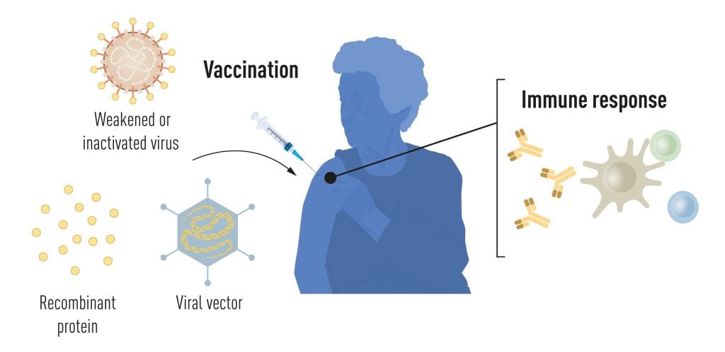
Vaccines before the pandemic

Vaccination stimulates the formation of an immune response to a particular pathogen. This gives the body a head start in the fight against disease in the event of a later exposure. Vaccines based on killed or weakened viruses have long been available, exemplified by the vaccines against polio, measles, and yellow fever. In 1951, Max Theiler was awarded the Nobel Prize in Physiology or Medicine for developing the yellow fever vaccine.

Thanks to the progress in molecular biology in recent decades, vaccines based on individual viral components, rather than whole viruses, have been developed. Parts of the viral genetic code, usually encoding proteins found on the virus surface, are used to make proteins that stimulate the formation of virus-blocking antibodies. Examples are the vaccines against the hepatitis B virus and human papillomavirus. Alternatively, parts of the viral genetic code can be moved to a harmless carrier virus, a “vector.” This method is used in vaccines against the Ebola virus. When vector vaccines are injected, the selected viral protein is produced in our cells, stimulating an immune response against the targeted virus.

Producing whole virus-, protein- and vector-based vaccines requires large-scale cell culture. This resource-intensive process limits the possibilities for rapid vaccine production in response to outbreaks and pandemics. Therefore, researchers have long attempted to develop vaccine technologies independent of cell culture, but this proved challenging.

Illustration of methods for vaccine production before the COVID-19 pandemic.
Figure 1. Methods for vaccine production before the COVID-19 pandemic. © The Nobel Committee for Physiology or Medicine. Ill. Mattias Karlén

mRNA vaccines: A promising idea

In our cells, genetic information encoded in DNA is transferred to messenger RNA (mRNA), which is used as a template for protein production. During the 1980s, efficient methods for producing mRNA without cell culture were introduced, called in vitro transcription. This decisive step accelerated the development of molecular biology applications in several fields. Ideas of using mRNA technologies for vaccine and therapeutic purposes also took off, but roadblocks lay ahead. In vitro transcribed mRNA was considered unstable and challenging to deliver, requiring the development of sophisticated carrier lipid systems to encapsulate the mRNA. Moreover, in vitro-produced mRNA gave rise to inflammatory reactions. Enthusiasm for developing the mRNA technology for clinical purposes was, therefore, initially limited.

These obstacles did not discourage the Hungarian biochemist Katalin Karikó, who was devoted to developing methods to use mRNA for therapy. During the early 1990s, when she was an assistant professor at the University of Pennsylvania, she remained true to her vision of realizing mRNA as a therapeutic despite encountering difficulties in convincing research funders of the significance of her project. A new colleague of Karikó at her university was the immunologist Drew Weissman. He was interested in dendritic cells, which have important functions in immune surveillance and the activation of vaccine-induced immune responses. Spurred by new ideas, a fruitful collaboration between the two soon began, focusing on how different RNA types interact with the immune system.

The breakthrough

Karikó and Weissman noticed that dendritic cells recognize in vitro transcribed mRNA as a foreign substance, which leads to their activation and the release of inflammatory signaling molecules. They wondered why the in vitro transcribed mRNA was recognized as foreign while mRNA from mammalian cells did not give rise to the same reaction. Karikó and Weissman realized that some critical properties must distinguish the different types of mRNA.

RNA contains four bases, abbreviated A, U, G, and C, corresponding to A, T, G, and C in DNA, the letters of the genetic code. Karikó and Weissman knew that bases in RNA from mammalian cells are frequently chemically modified, while in vitro transcribed mRNA is not. They wondered if the absence of altered bases in the in vitro transcribed RNA could explain the unwanted inflammatory reaction. To investigate this, they produced different variants of mRNA, each with unique chemical alterations in their bases, which they delivered to dendritic cells. The results were striking: The inflammatory response was almost abolished when base modifications were included in the mRNA. This was a paradigm change in our understanding of how cells recognize and respond to different forms of mRNA. Karikó and Weissman immediately understood that their discovery had profound significance for using mRNA as therapy. These seminal results were published in 2005, fifteen years before the COVID-19 pandemic.

Illustration of the four different bases mRNA contains.
Figure 2. mRNA contains four different bases, abbreviated A, U, G, and C. The Nobel Laureates discovered that base-modified mRNA can be used to block activation of inflammatory reactions (secretion of signaling molecules) and increase protein production when mRNA is delivered to cells.  © The Nobel Committee for Physiology or Medicine. Ill. Mattias Karlén

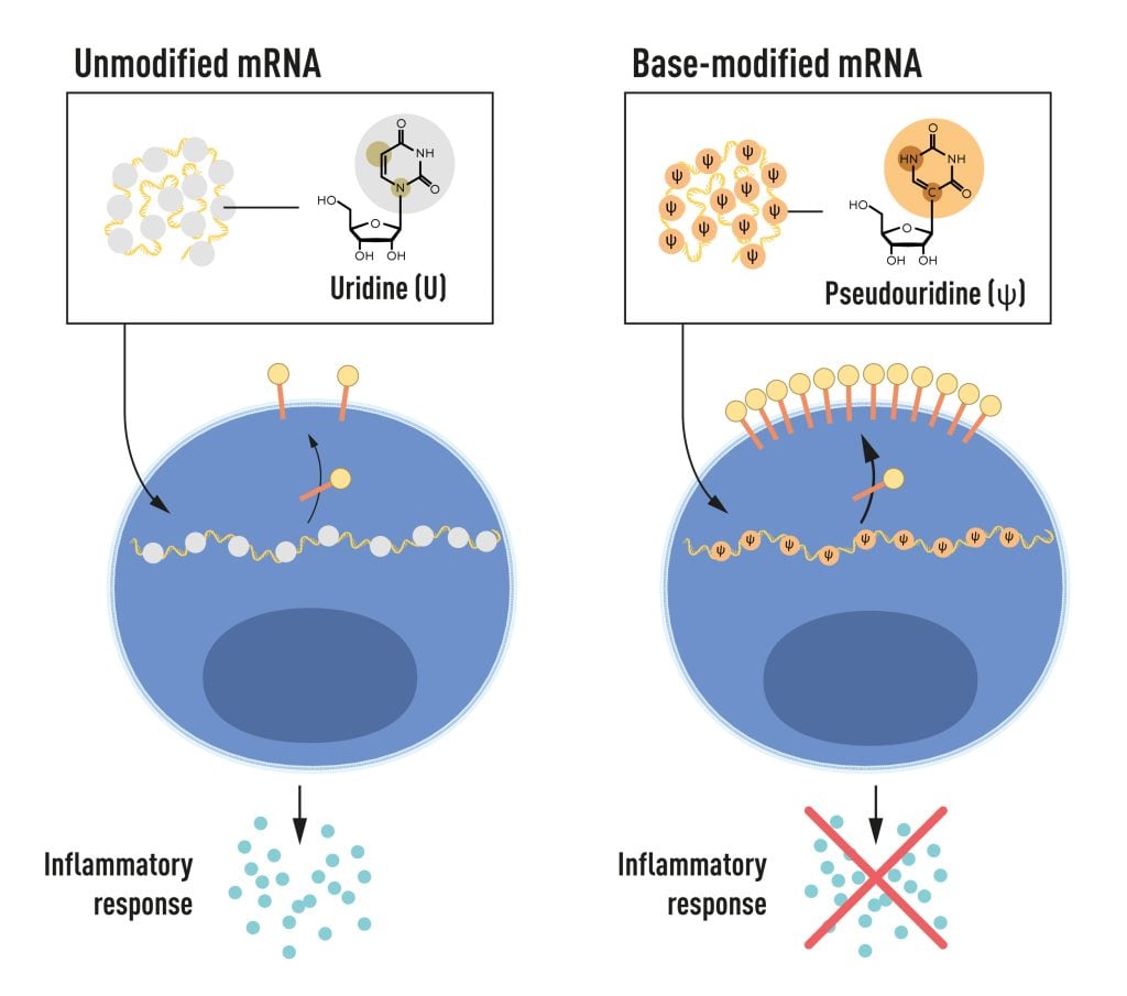
In further studies published in 2008 and 2010, Karikó and Weissman showed that the delivery of mRNA generated with base modifications markedly increased protein production compared to unmodified mRNA. The effect was due to the reduced activation of an enzyme that regulates protein production. Through their discoveries that base modifications both reduced inflammatory responses and increased protein production, Karikó and Weissman had eliminated critical obstacles on the way to clinical applications of mRNA.

mRNA vaccines realized their potential

Interest in mRNA technology began to pick up, and in 2010, several companies were working on developing the method. Vaccines against Zika virus and MERS-CoV were pursued; the latter is closely related to SARS-CoV-2. After the outbreak of the COVID-19 pandemic, two base-modified mRNA vaccines encoding the SARS-CoV-2 surface protein were developed at record speed. Protective effects of around 95% were reported, and both vaccines were approved as early as December 2020.

The impressive flexibility and speed with which mRNA vaccines can be developed pave the way for using the new platform also for vaccines against other infectious diseases. In the future, the technology may also be used to deliver therapeutic proteins and treat some cancer types.

Several other vaccines against SARS-CoV-2, based on different methodologies, were also rapidly introduced, and together, more than 13 billion COVID-19 vaccine doses have been given globally. The vaccines have saved millions of lives and prevented severe disease in many more, allowing societies to open and return to normal conditions. Through their fundamental discoveries of the importance of base modifications in mRNA, this year’s Nobel laureates critically contributed to this transformative development during one of the biggest health crises of our time.

Read more about this year’s prize

Scientific background: Discoveries concerning nucleoside base modifications that enabled the development of effective mRNA vaccines against COVID-19

Katalin Karikó was born in 1955 in Szolnok, Hungary. She received her PhD from Szeged’s University in 1982 and performed postdoctoral research at the Hungarian Academy of Sciences in Szeged until 1985. She then conducted postdoctoral research at Temple University, Philadelphia, and the University of Health Science, Bethesda. In 1989, she was appointed Assistant Professor at the University of Pennsylvania, where she remained until 2013. After that, she became vice president and later senior vice president at BioNTech RNA Pharmaceuticals. Since 2021, she has been a Professor at Szeged University and an Adjunct Professor at Perelman School of Medicine at the University of Pennsylvania.

Drew Weissman was born in 1959 in Lexington, Massachusetts, USA. He received his MD, PhD degrees from Boston University in 1987. He did his clinical training at Beth Israel Deaconess Medical Center at Harvard Medical School and postdoctoral research at the National Institutes of Health. In 1997, Weissman established his research group at the Perelman School of Medicine at the University of Pennsylvania. He is the Roberts Family Professor in Vaccine Research and Director of the Penn Institute for RNA Innovations.

The University of Pennsylvania October 2, 2023 news release is a very interesting announcement (more about why it’s interesting afterwards), Note: Links have been removed,

The University of Pennsylvania messenger RNA pioneers whose years of scientific partnership unlocked understanding of how to modify mRNA to make it an effective therapeutic—enabling a platform used to rapidly develop lifesaving vaccines amid the global COVID-19 pandemic—have been named winners of the 2023 Nobel Prize in Physiology or Medicine. They become the 28th and 29th Nobel laureates affiliated with Penn, and join nine previous Nobel laureates with ties to the University of Pennsylvania who have won the Nobel Prize in Medicine.

Nearly three years after the rollout of mRNA vaccines across the world, Katalin Karikó, PhD, an adjunct professor of Neurosurgery in Penn’s Perelman School of Medicine, and Drew Weissman, MD, PhD, the Roberts Family Professor of Vaccine Research in the Perelman School of Medicine, are recipients of the prize announced this morning by the Nobel Assembly in Solna, Sweden.

After a chance meeting in the late 1990s while photocopying research papers, Karikó and Weissman began investigating mRNA as a potential therapeutic. In 2005, they published a key discovery: mRNA could be altered and delivered effectively into the body to activate the body’s protective immune system. The mRNA-based vaccines elicited a robust immune response, including high levels of antibodies that attack a specific infectious disease that has not previously been encountered. Unlike other vaccines, a live or attenuated virus is not injected or required at any point.

When the COVID-19 pandemic struck, the true value of the pair’s lab work was revealed in the most timely of ways, as companies worked to quickly develop and deploy vaccines to protect people from the virus. Both Pfizer/BioNTech and Moderna utilized Karikó and Weissman’s technology to build their highly effective vaccines to protect against severe illness and death from the virus. In the United States alone, mRNA vaccines make up more than 655 million total doses of SARS-CoV-2 vaccines that have been administered since they became available in December 2020.

Editor’s Note: The Pfizer/BioNTech and Moderna COVID-19 mRNA vaccines both use licensed University of Pennsylvania technology. As a result of these licensing relationships, Penn, Karikó and Weissman have received and may continue to receive significant financial benefits in the future based on the sale of these products. BioNTech provides funding for Weissman’s research into the development of additional infectious disease vaccines.

Science can be brutal

Now for the interesting bit: it’s in my March 5, 2021 posting (mRNA, COVID-19 vaccines, treating genetic diseases before birth, and the scientist who started it all),

Before messenger RNA was a multibillion-dollar idea, it was a scientific backwater. And for the Hungarian-born scientist behind a key mRNA discovery, it was a career dead-end.

Katalin Karikó spent the 1990s collecting rejections. Her work, attempting to harness the power of mRNA to fight disease, was too far-fetched for government grants, corporate funding, and even support from her own colleagues.

“Every night I was working: grant, grant, grant,” Karikó remembered, referring to her efforts to obtain funding. “And it came back always no, no, no.”

By 1995, after six years on the faculty at the University of Pennsylvania, Karikó got demoted. [emphasis mine] She had been on the path to full professorship, but with no money coming in to support her work on mRNA, her bosses saw no point in pressing on.

She was back to the lower rungs of the scientific academy.

“Usually, at that point, people just say goodbye and leave because it’s so horrible,” Karikó said.

There’s no opportune time for demotion, but 1995 had already been uncommonly difficult. Karikó had recently endured a cancer scare, and her husband was stuck in Hungary sorting out a visa issue. Now the work to which she’d devoted countless hours was slipping through her fingers.

In time, those better experiments came together. After a decade of trial and error, Karikó and her longtime collaborator at Penn — Drew Weissman [emphasis mine], an immunologist with a medical degree and Ph.D. from Boston University — discovered a remedy for mRNA’s Achilles’ heel.

You can get the whole story from my March 5, 2021 posting, scroll down to the “mRNA—it’s in the details, plus, the loneliness of pioneer researchers, a demotion, and squabbles” subhead. If you are very curious about mRNA and the rough and tumble of the world of science, there’s my August 20, 2021 posting “Getting erased from the mRNA/COVID-19 story” where Ian MacLachlan is featured as a researcher who got erased and where Karikó credits his work.

‘Rowing Mom Wins Nobel’ (credit: rowing website Row 2K)

Karikó’s daughter is a two-time gold medal Olympic athlete as the Canadian Broadcasting Corporation’s (CBC) radio programme, As It Happens, notes in an interview with the daughter (Susan Francia). From an October 4, 2023 As It Happens article (with embedded audio programme excerpt) by Sheena Goodyear,

Olympic gold medallist Susan Francia is coming to terms with the fact that she’s no longer the most famous person in her family.

That’s because the retired U.S. rower’s mother, Katalin Karikó, just won a Nobel Prize in Medicine. The biochemist was awarded alongside her colleague, vaccine researcher Drew Weissman, for their groundbreaking work that led to the development of COVID-19 vaccines. 

“Now I’m like, ‘Shoot! All right, I’ve got to work harder,'” Francia said with a laugh during an interview with As It Happens host Nil Köksal. 

But in all seriousness, Francia says she’s immensely proud of her mother’s accomplishments. In fact, it was Karikó’s fierce dedication to science that inspired Francia to win Olympic gold medals in 2008 and 2012.

“Sport is a lot like science in that, you know, you have a passion for something and you just go and you train, attain your goal, whether it be making this discovery that you truly believe in, or for me, it was trying to be the best in the world,” Francia said.

“It’s a grind and, honestly, I love that grind. And my mother did too.”

… one of her [Karikó] favourite headlines so far comes from a little blurb on the rowing website Row 2K: “Rowing Mom Wins Nobel.”

Nowadays, scientists are trying to harness the power of mRNA to fight cancer, malaria, influenza and rabies. But when Karikó first began her work, it was a fringe concept. For decades, she toiled in relative obscurity, struggling to secure funding for her research.

“That’s also that same passion that I took into my rowing,” Francia said.

But even as Karikó struggled to make a name for herself, she says her own mother, Zsuzsanna, always believed she would earn a Nobel Prize one day.

Every year, as the Nobel Prize announcement approached, she would tell Karikó she’d be watching for her name. 

“I was laughing [and saying] that, ‘Mom, I am not getting anything,'” she said. 

But her mother, who died a few years ago, ultimately proved correct. 

Congratulations to both Katalin Karikó and Drew Weissman and thank you both for persisting!

Physics

This prize is for physics at the attoscale.

Aaron W. Harrison (Assistant Professor of Chemistry, Austin College, Texas, US) attempts an explanation of an attosecond in his October 3, 2023 essay (in English “What is an attosecond? A physical chemist explains the tiny time scale behind Nobel Prize-winning research” and in French “Nobel de physique : qu’est-ce qu’une attoseconde?”) for The Conversation, Note: Links have been removed,

“Atto” is the scientific notation prefix that represents 10-18, which is a decimal point followed by 17 zeroes and a 1. So a flash of light lasting an attosecond, or 0.000000000000000001 of a second, is an extremely short pulse of light.

In fact, there are approximately as many attoseconds in one second as there are seconds in the age of the universe.

Previously, scientists could study the motion of heavier and slower-moving atomic nuclei with femtosecond (10-15) light pulses. One thousand attoseconds are in 1 femtosecond. But researchers couldn’t see movement on the electron scale until they could generate attosecond light pulses – electrons move too fast for scientists to parse exactly what they are up to at the femtosecond level.

Harrison does a very good job of explaining something that requires a leap of imagination. He also explains why scientists engage in attosecond research. h/t October 4, 2023 news item on phys.org

Amelle Zaïr (Imperial College London) offers a more technical explanation in her October 4, 2023 essay about the 2023 prize winners for The Conversation. h/t October 4, 2023 news item on phys.org

Main event

Here’s the October 3, 2023 Nobel Prize press release, Note: A link has been removed,

The Royal Swedish Academy of Sciences has decided to award the Nobel Prize in Physics 2023 to

Pierre Agostini
The Ohio State University, Columbus, USA

Ferenc Krausz
Max Planck Institute of Quantum Optics, Garching and Ludwig-Maximilians-Universität München, Germany

Anne L’Huillier
Lund University, Sweden

“for experimental methods that generate attosecond pulses of light for the study of electron dynamics in matter”

Experiments with light capture the shortest of moments

The three Nobel Laureates in Physics 2023 are being recognised for their experiments, which have given humanity new tools for exploring the world of electrons inside atoms and molecules. Pierre Agostini, Ferenc Krausz and Anne L’Huillier have demonstrated a way to create extremely short pulses of light that can be used to measure the rapid processes in which electrons move or change energy.

Fast-moving events flow into each other when perceived by humans, just like a film that consists of still images is perceived as continual movement. If we want to investigate really brief events, we need special technology. In the world of electrons, changes occur in a few tenths of an attosecond – an attosecond is so short that there are as many in one second as there have been seconds since the birth of the universe.

The laureates’ experiments have produced pulses of light so short that they are measured in attoseconds, thus demonstrating that these pulses can be used to provide images of processes inside atoms and molecules.

In 1987, Anne L’Huillier discovered that many different overtones of light arose when she transmitted infrared laser light through a noble gas. Each overtone is a light wave with a given number of cycles for each cycle in the laser light. They are caused by the laser light interacting with atoms in the gas; it gives some electrons extra energy that is then emitted as light. Anne L’Huillier has continued to explore this phenomenon, laying the ground for subsequent breakthroughs.

In 2001, Pierre Agostini succeeded in producing and investigating a series of consecutive light pulses, in which each pulse lasted just 250 attoseconds. At the same time, Ferenc Krausz was working with another type of experiment, one that made it possible to isolate a single light pulse that lasted 650 attoseconds.

The laureates’ contributions have enabled the investigation of processes that are so rapid they were previously impossible to follow.

“We can now open the door to the world of electrons. Attosecond physics gives us the opportunity to understand mechanisms that are governed by electrons. The next step will be utilising them,” says Eva Olsson, Chair of the Nobel Committee for Physics.

There are potential applications in many different areas. In electronics, for example, it is important to understand and control how electrons behave in a material. Attosecond pulses can also be used to identify different molecules, such as in medical diagnostics.

Read more about this year’s prize

Popular science background: Electrons in pulses of light (pdf)
Scientific background: “For experimental methods that generate attosecond pulses of light for the study of electron dynamics in matter” (pdf)

Pierre Agostini. PhD 1968 from Aix-Marseille University, France. Professor at The Ohio State University, Columbus, USA.

Ferenc Krausz, born 1962 in Mór, Hungary. PhD 1991 from Vienna University of Technology, Austria. Director at Max Planck Institute of Quantum Optics, Garching and Professor at Ludwig-Maximilians-Universität München, Germany.

Anne L’Huillier, born 1958 in Paris, France. PhD 1986 from University Pierre and Marie Curie, Paris, France. Professor at Lund University, Sweden.

A Canadian connection?

An October 3, 2023 CBC online news item from the Associated Press reveals a Canadian connection of sorts ,

Three scientists have won the Nobel Prize in physics Tuesday for giving us the first split-second glimpse into the superfast world of spinning electrons, a field that could one day lead to better electronics or disease diagnoses.

The award went to French-Swedish physicist Anne L’Huillier, French scientist Pierre Agostini and Hungarian-born Ferenc Krausz for their work with the tiny part of each atom that races around the centre, and that is fundamental to virtually everything: chemistry, physics, our bodies and our gadgets.

Electrons move around so fast that they have been out of reach of human efforts to isolate them. But by looking at the tiniest fraction of a second possible, scientists now have a “blurry” glimpse of them, and that opens up whole new sciences, experts said.

“The electrons are very fast, and the electrons are really the workforce in everywhere,” Nobel Committee member Mats Larsson said. “Once you can control and understand electrons, you have taken a very big step forward.”

L’Huillier is the fifth woman to receive a Nobel in Physics.

L’Huillier was teaching basic engineering physics to about 100 undergraduates at Lund when she got the call that she had won, but her phone was on silent and she didn’t pick up. She checked it during a break and called the Nobel Committee.

Then she went back to teaching.

Agostini, an emeritus professor at Ohio State University, was in Paris and could not be reached by the Nobel Committee before it announced his win to the world

Here’s the Canadian connection (from the October 3, 2023 CBC online news item),

Krausz, of the Max Planck Institute of Quantum Optics and Ludwig Maximilian University of Munich, told reporters that he was bewildered.

“I have been trying to figure out since 11 a.m. whether I’m in reality or it’s just a long dream,” the 61-year-old said.

Last year, Krausz and L’Huillier won the prestigious Wolf prize in physics for their work, sharing it with University of Ottawa scientist Paul Corkum [emphasis mine]. Nobel prizes are limited to only three winners and Krausz said it was a shame that it could not include Corkum.

Corkum was key to how the split-second laser flashes could be measured [emphasis mine], which was crucial, Krausz said.

Congratulations to Pierre Agostini, Ferenc Krausz and Anne L’Huillier and a bow to Paul Corkum!

For those who are curious. a ‘Paul Corkum’ search should bring up a few postings on this blog but I missed this piece of news, a May 4, 2023 University of Ottawa news release about Corkum and the 2022 Wolf Prize, which he shared with Krausz and L’Huillier,

Chemistry

There was a little drama where this prize was concerned, It was announced too early according to an October 4, 2023 news item on phys.org and, again, in another October 4, 2023 news item on phys.org (from the Oct. 4, 2023 news item by Karl Ritter for the Associated Press),

Oops! Nobel chemistry winners are announced early in a rare slip-up

The most prestigious and secretive prize in science ran headfirst into the digital era Wednesday when Swedish media got an emailed press release revealing the winners of the Nobel Prize in chemistry and the news prematurely went public.

Here’s the fully sanctioned October 4, 2023 Nobel Prize press release, Note: A link has been removed,

The Royal Swedish Academy of Sciences has decided to award the Nobel Prize in Chemistry 2023 to

Moungi G. Bawendi
Massachusetts Institute of Technology (MIT), Cambridge, MA, USA

Louis E. Brus
Columbia University, New York, NY, USA

Alexei I. Ekimov
Nanocrystals Technology Inc., New York, NY, USA

“for the discovery and synthesis of quantum dots”

They planted an important seed for nanotechnology

The Nobel Prize in Chemistry 2023 rewards the discovery and development of quantum dots, nanoparticles so tiny that their size determines their properties. These smallest components of nanotechnology now spread their light from televisions and LED lamps, and can also guide surgeons when they remove tumour tissue, among many other things.

Everyone who studies chemistry learns that an element’s properties are governed by how many electrons it has. However, when matter shrinks to nano-dimensions quantum phenomena arise; these are governed by the size of the matter. The Nobel Laureates in Chemistry 2023 have succeeded in producing particles so small that their properties are determined by quantum phenomena. The particles, which are called quantum dots, are now of great importance in nanotechnology.

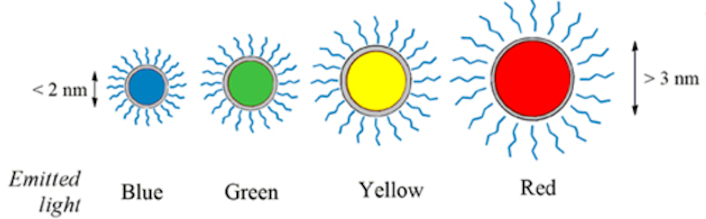
“Quantum dots have many fascinating and unusual properties. Importantly, they have different colours depending on their size,” says Johan Åqvist, Chair of the Nobel Committee for Chemistry.

Physicists had long known that in theory size-dependent quantum effects could arise in nanoparticles, but at that time it was almost impossible to sculpt in nanodimensions. Therefore, few people believed that this knowledge would be put to practical use.

However, in the early 1980s, Alexei Ekimov succeeded in creating size-dependent quantum effects in coloured glass. The colour came from nanoparticles of copper chloride and Ekimov demonstrated that the particle size affected the colour of the glass via quantum effects.

A few years later, Louis Brus was the first scientist in the world to prove size-dependent quantum effects in particles floating freely in a fluid.

In 1993, Moungi Bawendi revolutionised the chemical production of quantum dots, resulting in almost perfect particles. This high quality was necessary for them to be utilised in applications.

Quantum dots now illuminate computer monitors and television screens based on QLED technology. They also add nuance to the light of some LED lamps, and biochemists and doctors use them to map biological tissue.

Quantum dots are thus bringing the greatest benefit to humankind. Researchers believe that in the future they could contribute to flexible electronics, tiny sensors, thinner solar cells and encrypted quantum communication – so we have just started exploring the potential of these tiny particles.

Read more about this year’s prize

Popular science background: They added colour to nanotechnology (pdf)
Scientific background: Quantum dots – seeds of nanoscience (pdf)

Moungi G. Bawendi, born 1961 in Paris, France. PhD 1988 from University of Chicago, IL, USA. Professor at Massachusetts Institute of Technology (MIT), Cambridge, MA, USA.

Louis E. Brus, born 1943 in Cleveland, OH, USA. PhD 1969 from Columbia University, New York, NY, USA. Professor at Columbia University, New York, NY, USA.

Alexei I. Ekimov, born 1945 in the former USSR. PhD 1974 from Ioffe Physical-Technical Institute, Saint Petersburg, Russia. Formerly Chief Scientist at Nanocrystals Technology Inc., New York, NY, USA.


The most recent ‘quantum dot’ (a particular type of nanoparticle) story here is a January 5, 2023 posting, “Can I have a beer with those carbon quantum dots?

Proving yet again that scientists can have a bumpy trip to a Nobel prize, an October 4, 2023 news item on phys.org describes how one of the winners flunked his first undergraduate chemistry test, Note: Links have been removed,

Talk about bouncing back. MIT professor Moungi Bawendi is a co-winner of this year’s Nobel chemistry prize for helping develop “quantum dots”—nanoparticles that are now found in next generation TV screens and help illuminate tumors within the body.

But as an undergraduate, he flunked his very first chemistry exam, recalling that the experience nearly “destroyed” him.

The 62-year-old of Tunisian and French heritage excelled at science throughout high school, without ever having to break a sweat.

But when he arrived at Harvard University as an undergraduate in the late 1970s, he was in for a rude awakening.

You can find more about the winners and quantum dots in an October 4, 2023 news item on Nanowerk and in Dr. Andrew Maynard’s (Professor of Advanced Technology Transitions, Arizona State University) October 4, 2023 essay for The Conversation (h/t October 4, 2023 news item on phys.org), Note: Links have been removed,

This year’s prize recognizes Moungi Bawendi, Louis Brus and Alexei Ekimov for the discovery and development of quantum dots. For many years, these precisely constructed nanometer-sized particles – just a few hundred thousandths the width of a human hair in diameter – were the darlings of nanotechnology pitches and presentations. As a researcher and adviser on nanotechnology [emphasis mine], I’ve [Dr. Andrew Maynard] even used them myself when talking with developers, policymakers, advocacy groups and others about the promise and perils of the technology.

The origins of nanotechnology predate Bawendi, Brus and Ekimov’s work on quantum dots – the physicist Richard Feynman speculated on what could be possible through nanoscale engineering as early as 1959, and engineers like Erik Drexler were speculating about the possibilities of atomically precise manufacturing in the the 1980s. However, this year’s trio of Nobel laureates were part of the earliest wave of modern nanotechnology where researchers began putting breakthroughs in material science to practical use.

Quantum dots brilliantly fluoresce: They absorb one color of light and reemit it nearly instantaneously as another color. A vial of quantum dots, when illuminated with broad spectrum light, shines with a single vivid color. What makes them special, though, is that their color is determined by how large or small they are. Make them small and you get an intense blue. Make them larger, though still nanoscale, and the color shifts to red.

The wavelength of light a quantum dot emits depends on its size. Maysinger, Ji, Hutter, Cooper, CC BY

There’s also an October 4, 2023 overview article by Tekla S. Perry and Margo Anderson for the IEEE Spectrum about the magazine’s almost twenty-five years of reporting on quantum dots

Red blue and green dots mass in rows, with some dots moving away

Image credit: Brandon Palacio/IEEE Spectrum

Your Guide to the Newest Nobel Prize: Quantum Dots

What you need to know—and what we’ve reported—about this year’s Chemistry award

It’s not a long article and it has a heavy focus on the IEEEE’s (Institute of Electrical and Electtronics Engineers) the road quantum dots have taken to become applications and being commercialized.

Congratulations to Moungi Bawendi, Louis Brus, and Alexei Ekimov!

Tapping into wound healing by harnessing the natural healing process

If you’re imagining an enhanced chakra balancing experience or more efficient digestion of your vitamin supplements, you will be a little disappointed in this latest news from the Imperial College of London (ICL). On the other hand, if you have damaged tissue, this discovery could make your recovery much easier. From a January 7, 2019 news item on phys.org,

Materials are widely used to help heal wounds: Collagen sponges help treat burns and pressure sores, and scaffold-like implants are used to repair bones. However, the process of tissue repair changes over time, so scientists are developing biomaterials that interact with tissues as healing takes place

Now, Dr. Ben Almquist and his team at Imperial College London have created a new molecule that could change the way traditional materials work with the body. Known as traction force-activated payloads (TrAPs), their method lets materials talk to the body’s natural repair systems to drive healing.

The researchers say incorporating TrAPs into existing medical materials could revolutionise the way injuries are treated. Dr. Almquist, from Imperial’s Department of Bioengineering, said: “Our technology could help launch a new generation of materials that actively work with tissues to drive healing.”

A January 7, 2019 ICL press release (also on EurekAlert) by Caroline Brogan, which originated the news item, expands on the theme,

After an injury, cells ‘crawl’ through the collagen ‘scaffolds’ found in wounds, like spiders navigating webs. As they move, they pull on the scaffold, which activates hidden healing proteins that begin to repair injured tissue.

The researchers in the study designed TrAPs as a way to recreate this natural healing method. They folded the DNA segments into three-dimensional shapes known as aptamers that cling tightly to proteins. Then, they attached a customisable ‘handle’ that cells can grab onto on one end, before attaching the opposite end to a scaffold such as collagen.

During laboratory testing of their technique, they found that cells pulled on the TrAPs as they crawled through the collagen scaffolds. The pulling made the TrAPs unravel like shoelaces to reveal and activate the healing proteins. These proteins instruct the healing cells to grow and multiply

The researchers also found that by changing the cellular ‘handle’, they can change which type of cell can grab hold and pull, letting them tailor TrAPs to release specific therapeutic proteins based on which cells are present at a given point in time. In doing so, the TrAPs produce materials that can smartly interact with the correct type of cell at the correct time during wound repair.

This is the first time scientists have activated healing proteins using differing cell types in man-made materials. The technique mimics healing methods found in nature. Dr Almquist said: “Creatures from sea sponges to humans use cell movement to activate healing. Our approach mimics this by using the different cell varieties in wounds to drive healing.””

From lab to humans

This approach is adaptable to different cell types, so could be used in a variety of injuries such as fractured bones, scar tissue after heart attacks, and damaged nerves. New techniques are also desperately needed for patients whose wounds won’t heal despite current interventions, like diabetic foot ulcers, which are the leading cause of non-traumatic lower leg amputations.

TrAPs are relatively straightforward to create and are fully man-made, meaning they are easily recreated in different labs and can be scaled up to industrial quantities. Their adaptability also means they could help scientists create new methods for laboratory studies of diseases, stem cells, and tissue development.

Aptamers are currently used as drugs, meaning they are already proven safe and optimised for clinical use. Because TrAPs take advantage of aptamers that are safe for humans, they may be able to take a shorter path to the clinic than methods that start from ground zero.

Dr Almquist said: “TrAPs provide a flexible method of actively communicating with wounds, as well as key instructions when and where they are needed. This intelligent healing is useful during every phase of the healing process, has the potential to increase the body’s chance to recover, and has far-reaching uses on many different types of wounds. This technology could serve as a conductor of wound repair, orchestrating different cells over time to work together to heal damaged tissues.”

The researchers have made available an image and a video abstract illustrating their work,

TrAPs could harness the body’s natural healing powers to repair bone. Courtesy: Imperial College of London

By the way, the video was produced by www.animateyour.science (based in Adelaide, Australia) and they have a very interesting About page,

Our story

Your research is brilliant and novel. I’m sure of it. You might even be a pioneer in your field. But ask yourself honestly, is it enough? Is it truly enough to make a difference in the world?
 
My name is Tullio Rossi, and I founded Animate Your Science on my quest to make a positive impact on society through science.
 
During my Ph.D., I found that my peer-reviewed paper alone wasn’t cutting it. If I wanted to reach my peers, let alone the general public, I needed to communicate my findings in a fun and imaginative way.
 
This realization changed everything and inspired me to create “Lost at Sea,” an award-winning video that reached the hearts and minds of thousands of people.
 
The success of this first video blew my mind. And I got to thinking, maybe other scientists are lost at sea, so to speak. Maybe others want to reach the masses with their research, but just don’t know where to start.
 
This was the day Animate Your Science was born.

Why we do it

What we really want to do is bring science into society. That’s the true value of this company and the reason we believe in it.

We love science but we believe that, if not communicated properly, science is of limited use to society.​
 
As scientists, it’s our privilege and duty to unearth these revelations and package them in a way that appeals to our peers as well as the general public.

Getting back to TrAPS, here’s a link to and a citation for the paper,

Biologically Inspired, Cell‐Selective Release of Aptamer‐Trapped Growth Factors by Traction Forces by Anna Stejskalová, Nuria Oliva, Frances J. England, Benjamin D. Almquist. Advanced Materials DOI: https://doi.org/10.1002/adma.201806380 First published: 07 January 2019

This paper is open access.

Donna Strickland, first female Nobel Prize winner in 55 years and one of only three (so far) in history

It’s been quite the fascinating week in the world of physics culminating with Donna Strickland’s shiny new Nobel Prize in physics.

For my purposes, this week in physics started on Friday, September 28, 2018 with Allesanndro Strumia’s presentation at CERN’s (European Particle Physics Laboratory) “1st workshop on high energy theory and gender” where he claimed and proved ‘scientifically’ that physics has become “sexist against men.” I’ll get back to Strumia in a moment but, first, let’s celebrate Donna Strickland and her achievements.

Only three women, including Strickland, in the history (117 years) of the Nobel Prize for Physics have won it, Marie Curie in 1903, Maria Goeppert Mayer in 1963, and, now, Strickland in 2018.

The University of Waterloo (Ontario, Canada) had this to say in an October 2, 2018 news release,

Donna Strickland wins Nobel Prize in Physics

Tuesday, October 2, 2018

Dr. Donna Strickland

Donna Strickland, a University of Waterloo professor who helped revolutionize laser physics, has been named a winner of this year’s Nobel Prize in Physics.

Strickland, an associate professor in the Department of Physics and Astronomy, shares half the $1.4 million prize with French laser physicist Gérard Mourou. The other half was awarded to U.S. physicist Arthur Ashkin.

The Royal Swedish Academy of Sciences stated that Mourou and Strickland paved the way toward the shortest and most intense laser pulses created by mankind. Their revolutionary article was published in 1985 and was the foundation of Strickland’s doctoral thesis.

Strickand conducted her Nobel-winning research while a PhD student under Mourou in 1989 at the University of Rochester in New York. The team’s research has a number of applications in industry and medicine.

It was great to have had the opportunity to work with one of the pioneers of ultrafast lasers, Gerard Mourou,” said Strickland. “It was a small community back then. It was a new, burgeoning field. I got to be part of that. It was very exciting.”

A Nobel committee member said billions of people make daily use of laser printers and optical scanners and millions undergo laser surgery.

“This is a tremendous day for Professor Strickland and needless to say a tremendous day for the University of Waterloo,” said Feridun Hamdullahpur, president and vice-chancellor of the University of Waterloo. “This is Waterloo’s first Nobel laureate and the first woman to receive the Nobel Prize in Physics in 55 years.”

During an interview, Strickland told the Globe and Mail [national newspaper]: “We need to celebrate women physicists because we’re out there, and hopefully in time it’ll start to move forward at a faster rate.”

Charmaine Dean, vice-president research at the University of Waterloo said: “Donna Strickland exemplifies research excellence at Waterloo. Her groundbreaking work is a testament to the importance of fundamental research as it has established the foundation for laser-based technologies that we see today from micromachining to laser eye surgery.”

An October 2, 2018 news item on Nanowerk focuses on the three winners,

Arthur Ashkin, an American physicist has been awarded half the prize for his invention of optical tweezers and their application to biological systems. His amazing tool has helped to reach the old dream of grabing [sic] particles, atoms, viruses and other living cells. The optical tweezers work with the radiation pressure of light to hold and move tiny object and are widely used to study the machinery of life.

French physicist Gérard Mourou and Canadian physicist Donna Strickland share the other half for their method of generating ultra-short and very intense optical pulses. Ultra-sharp laser beams have made possible to cut or drill holes in various materials extremely precisely – even in living matter. The technique this duo pioneered is called chirped pulse amplification or CPA and it has led to corrective eye surgeries for millions of people.

An Oct. 2, 2018 article by Marina Koren for The Atlantic is my favourite of the ones focusing on Strickland. One of Koren’s major focal points is Strickland’s new Wikipedia page (Note: Links have been removed),

It was about five in the morning in Ontario, Canada, when Donna Strickland’s phone rang. The Nobel Prize committee was on the line in Stockholm, calling to tell her she had won the prize in physics.

“We wondered if it was a prank,” Strickland said Tuesday [October 2 ,2018], in an interview with a Nobel official after the call. She had been asleep when the call arrived. “But then I knew it was the right day, and it would have been a cruel prank.”

Lasers, focused beams of light particles, were invented in the 1960s. Scientists immediately started tinkering with them, looking for ways to harness and manipulate these powerful devices.

Strickland and [Gérard] Mourou] found a way to stretch and compress lasers to produce short, intense pulses that are now used, among other things, in delicate surgeries to fix vision problems. [Arthur] Ashkin figured out a way to maneuver laser light so that it could push small particles toward the center of the beam, hold them in place, and even move them around. This technique became the delightfully named “optical tweezer.” It allowed Ashkin to use the power of light to capture and hold living bacteria and viruses without harming the organisms.

Unlike her fellow winners, Strickland did not have a Wikipedia page at the time of the announcement. A Wikipedia user tried to set up a page in May, but it was denied by a moderator with the message: “This submission’s references do not show that the subject qualifies for a Wikipedia article.” Strickland, it was determined, had not received enough dedicated coverage elsewhere on the internet to warrant a page.

On Tuesday, a newly created page flooded with edits: “Added in her title.” “Add Nobel-winning paper.” “Added names of other women Nobelists [sic] in physics.”

The construction of the Wikipedia page feels like a metaphor for a historic award process that has long been criticized for neglecting women in its selection, and for the shortage of women’s stories in the sciences at large. To scroll through the “history” tab of Strickland’s page, where all edits are recorded and tracked, is to witness in real time the recognition of a scientist whose story likely deserved attention long before the Nobel Prize committee called.

Strickland’s historic win comes a day after CERN, the European organization that operates the world’s most powerful particle accelerator, suspended a senior scientist for saying that physics was “invented and built by men.” Alessandro Strumia, a professor at the University of Pisa, made the statement during a recent speech at a seminar on gender issues in physics that was attended by mostly female physicists. Strumia said “men prefer working with things and women prefer working with people,” and that between men and women there is a “difference even in children before any social influence.” His remarks were widely circulated online and prompted fierce backlash.

The remarks don’t faze Strickland, who very publicly proved them wrong on Tuesday. In an interview with the BBC on Tuesday, she called Strumia’s claims “silly.”

For anyone curious about the Strumia situation, there’s an October 2, 2018 CBC Radio (As It Happens) online news article. Note: Links have been removed,

Not only was Alessandro Strumia being offensive when he said that physics “was invented and built by men” — he was also wrong, says physicist Jess Wade.

“Actually, women have contributed hugely to physics throughout the whole of history, but for an incredibly long time we haven’t documented or told those stories,” Wade told As It Happens host Carol Off.

And she would know. The Imperial College London research associate has made it her mission to write hundreds of Wikipedia entries about women in science and engineering.

Wade was in the room on Friday when Strumia, a physicist at Pisa University, made the inflammatory remarks during a gender workshop in Geneva, organized by the European nuclear research centre CERN.

CERN cut ties with Strumia after the BBC reported the content of his presentation.

This article includes some of the slides in Strumia’s now infamous presentation.

Tommaso Dorigo in an October 1, 2018 posting on the Science 2.0 blog offers another analysis,

The world of particle physics is in turmoil because of a presentation by Alessandro Strumia, an Italian phenomenologist, at CERN’s “1st workshop on high energy theory and gender”, and its aftermath.

By now the story has been echoed by many major newscasters around the world, and discussed in public and private forums, blogs, twitter feeds. I wanted to stay away from it here, mainly because it is a sensitive issue and the situation is still evolving, but after all, why not offer to you my personal pitch on the matter? Strumia, by the way, has been an occasional commenter to this blog – you can find some of his comments signed as “AS” in threads of past articles. Usually he makes good points here, as long as physics is the subject.

Anyway, first of all let me give you a quick recall of the events. The three-day workshop, which took place on September 26-28, was meant to”focus on recent developments in theoretical high-energy physics and cosmology, and discuss issues of gender and equal opportunities in the field“; it followed three previous events which combined string theory and gender issues. Strumia’s presentation was titled “Experimental tests of a new global symmetry“, a physicist’s way of describing the issue of man-woman equality. It is important to note that the talk was not an invited one – its author had asked the organizers for a slot as he said he would be talking of bibliometrics, and indeed his contribution was listed in the agenda of September 28 with the innocuous title “Bibliometrics data about gender issues in fundamental theory“.

Strumia’s slides contain a collection of half-baked claims, coming from his analysis of InSpire data from citations and authorship of articles in theoretical physics. I consider his talk offensive on many levels. It starts by casting the woman discrimination issue in scientific academia as a test of hypothesis of whether the “man-woman” symmetry is explicitly broken (i.e. there is no symmetry) or spontaneously broken (by a difference of treatment) – something that could even raise a smile in a geeky physicist; but the fun ends there.

Dorigo offers a detailed ‘takedown’ of Strumia’s assertions. I found the post intriguing for the insight it offers into physics. Never in a million years would I have thought this title, “Experimental tests of a new global symmetry,” would indicate a discussion on gender balance in the field of physics.

As I said in the opening, it has been quite the week in physics. On a final note, Brava to Doctor Donna Strickland!

Tractor beams for artificial cells

This particular piece has videos of cells moving around. I won’t be including all of them but they are weirdly fascinating. First, a May 14, 2018 news item on Nanowerk announces the latest in tractor beam news from the Imperial College London (ICL; UK),

Researchers have used lasers to connect, arrange and merge artificial cells, paving the way for networks of artificial cells that act like tissues.

The team say that by altering artificial cell membranes they can now get the cells to stick together like ‘stickle bricks’ – allowing them to be arranged into whole new structures.

Biological cells can perform complex functions, but are difficult to controllably engineer.

Artificial cells, however, can in principle be made to order. Now, researchers from Imperial College London and Loughborough University have demonstrated a new level of complexity with artificial cells by arranging them into basic tissue structures with different types of connectivity.

These structures could be used to perform functions like initiating chemical reactions or moving chemicals around networks of artificial and biological cells. This could be useful in carrying out chemical reactions in ultra-small volumes, in studying the mechanisms through which cells communicate with one another, and in the development of a new generation of smart biomaterials.

A May 14, 2018 ICL press release by Hayley Dunning , which originated the news item, provides more detail,

Cells are the basic units of biology, which are capable of working together as a collective when arranged into tissues. In order to do this, cells must be connected and be capable of exchanging materials with one another.

The team were able to link up artificial cells into a range of new architectures, the results of which are published today in Nature Communications.

The artificial cells have a membrane-like layer as their shell, which the researchers engineered to ‘stick’ to each other. In order to get the cells to come close enough, the team first had to manipulate the cells with ‘optical tweezers’ that act like mini ‘tractor beams’ dragging and dropping cells into any position. Once connected in this way the cells can be moved as one unit.

Lead researcher Dr Yuval Elani, an EPSRC Research Fellow from the Department of Chemistry at Imperial, said: “Artificial cell membranes usually bounce off each other like rubber balls. By altering the biophysics of the membranes in our cells, we got them instead to stick to each other like stickle bricks.

“With this, we were able to form networks of cells connected by ‘biojunctions’. By reinserting biological components such as proteins in the membrane, we could get the cells to communicate and exchange material with one another. This mimics what is seen in nature, so it’s a great step forward in creating biological-like artificial cell tissues.”

Building up complexity

The team were also able to engineer a ‘tether’ between two cells. Here the membranes are not stuck together, but a tendril of membrane material links them so that they can be moved together.

Once they had perfected the cell-sticking process, the team were able to build up more complex arrangements. These include lines of cells, 2D shapes like squares, and 3D shapes like pyramids. Once the cells are stuck together, they can be rearranged, and also pulled by the laser beam as an ensemble

Finally, the team were also able to connect two cells, and then make them merge into one larger cell. This was achieved by coating the membranes with gold nanoparticles.

When the laser beam at the heart of the ‘optical tweezer’ technology was concentrated at the junction between the two cells, the nanoparticles resonated, breaking the membranes at that point. The membrane then reforms as a whole.

Merging cells in this way allowed whatever chemicals they were carrying to mix within the new, larger cell, kicking off chemical reactions. This could be useful, for example, for delivering materials such as drugs into cells, and in changing the composition of cells in real time, getting them to adopt new functions.

Professor Oscar Ces, also from the Department of Chemistry at Imperial, said: “Connecting artificial cells together is a valuable technology in the wider toolkit we are assembling for creating these biological systems using bottom-up approaches.

“We can now start to scale up basic cell technologies into larger tissue-scale networks, with precise control over the kind of architecture we create.”

Here’s one of the videos that has been embedded with ICL press release,

You can see the whole series if you go to the May 14, 2018 ICL press release.

Here’s a link to and a citation for the paper,

Sculpting and fusing biomimetic vesicle networks using optical tweezers by Guido Bolognesi, Mark S. Friddin, Ali Salehi-Reyhani, Nathan E. Barlow, Nicholas J. Brooks, Oscar Ces, & Yuval Elani. Nature Communicationsvolume 9, Article number: 1882 (2018) doi:10.1038/s41467-018-04282-w Published: 14 May 2018

This paper is open access.

Edible water bottles by Ooho!

Courtesy: Skipping Rocks Lab

As far as I’m concerned, that looks more like a breast implant than a water bottle, which, from a psycho-social perspective, could lead to some interesting research papers. It is, in fact a new type of water bottle.  From an April 10, 2017 article by Adele Peters for Fast Company (Note: Links have been removed),

If you run in a race in London in the near future and pass a hydration station, you may be handed a small, bubble-like sphere of water instead of a bottle. The gelatinous packaging, called the Ooho, is compostable–or even edible, if you want to swallow it. And after two years of development, its designers are ready to bring it to market.

Three London-based design students first created a prototype of the edible bottle in 2014 as an alternative to plastic bottles. The idea gained internet hype (though also some scorn for a hilarious video that made the early prototypes look fairly impossible to use without soaking yourself).
The problem it was designed to solve–the number of disposable bottles in landfills–keeps growing. In the U.K. alone, around 16 million are trashed each day; another 19 million are recycled, but still have the environmental footprint of a product made from oil. In the U.S., recycling rates are even lower. …

The new packaging is based on the culinary technique of spherification, which is also used to make fake caviar and the tiny juice balls added to boba tea [bubble tea?]. Dip a ball of ice in calcium chloride and brown algae extract, and you can form a spherical membrane that keeps holding the ice as it melts and returns to room temperature.

An April 25, 2014 article by Kashmira Gander for Independent.co.uk describes the technology and some of the problems that had to be solved before bringing this product to market,

To make the bottle [Ooho!], students at the Imperial College London gave a frozen ball of water a gelatinous layer by dipping it into a calcium chloride solution.

They then soaked the ball in another solution made from brown algae extract to encapsulate the ice in a second membrane, and reinforce the structure.

However, Ooho still has teething problems, as the membrane is only as thick as a fruit skin, and therefore makes transporting the object more difficult than a regular bottle of water.

“This is a problem we’re trying to address with a double container,” Rodrigo García González, who created Ooho with fellow students Pierre Paslier and Guillaume Couche, explained to the Smithsonian. “The idea is that we can pack several individual edible Oohos into a bigger Ooho container [to make] a thicker and more resistant membrane.”

According to Peters’ Fast Company article, the issues have been resolved,

Because the membrane is made from food ingredients, you can eat it instead of throwing it away. The Jell-O-like packaging doesn’t have a natural taste, but it’s possible to add flavors to make it more appetizing.

The package doesn’t have to be eaten every time, since it’s also compostable. “When people try it for the first time, they want to eat it because it’s part of the experience,” says Pierre Paslier, cofounder of Skipping Rocks Lab, the startup developing the packaging. “Then it will be just like the peel of a fruit. You’re not expected to eat the peel of your orange or banana. We are trying to follow the example set by nature for packaging.”

The outer layer of the package is always meant to be peeled like fruit–one thin outer layer of the membrane peels away to keep the inner layer clean and can then be composted. (While compostable cups are an alternative solution, many can only be composted in industrial facilities; the Ooho can be tossed on a simple home compost pile, where it will decompose within weeks).

The company is targeting both outdoor events and cafes. “Where we see a lot of potential for Ooho is outdoor events–festivals, marathons, places where basically there are a lot of people consuming packaging over a very short amount of time,” says Paslier.

I encourage you to read Peters’ article in its entirety if you have the time. You can also find more information on the Skipping Rocks Lab website and on the company’s crowdfunding campaign on CrowdCube.

New form of light could lead to circuits that run on photons instead of electrons

If circuits are running on photons instead of electrons, does that mean there will be no more electricity and electronics?  Apparently, the answer is not exactly. First, an Aug. 5, 2016 news item on ScienceDaily makes the announcement about photons and circuits,

New research suggests that it is possible to create a new form of light by binding light to a single electron, combining the properties of both.

According to the scientists behind the study, from Imperial College London, the coupled light and electron would have properties that could lead to circuits that work with packages of light — photons — instead of electrons.

It would also allow researchers to study quantum physical phenomena, which govern particles smaller than atoms, on a visible scale.

An Aug. 5, 2016 Imperial College of London (ICL) press release, which originated the news item, describes the research further (Note: A link has been removed),

In normal materials, light interacts with a whole host of electrons present on the surface and within the material. But by using theoretical physics to model the behaviour of light and a recently-discovered class of materials known as topological insulators, Imperial researchers have found that it could interact with just one electron on the surface.

This would create a coupling that merges some of the properties of the light and the electron. Normally, light travels in a straight line, but when bound to the electron it would instead follow its path, tracing the surface of the material.

Improved electronics

In the study, published today in Nature Communications, Dr Vincenzo Giannini and colleagues modelled this interaction around a nanoparticle – a small sphere below 0.00000001 metres in diameter – made of a topological insulator.

Their models showed that as well as the light taking the property of the electron and circulating the particle, the electron would also take on some of the properties of the light. [emphasis mine]

Normally, as electrons are travelling along materials, such as electrical circuits, they will stop when faced with a defect. However, Dr Giannini’s team discovered that even if there were imperfections in the surface of the nanoparticle, the electron would still be able to travel onwards with the aid of the light.

If this could be adapted into photonic circuits, they would be more robust and less vulnerable to disruption and physical imperfections.

Quantum experiments

Dr Giannini said: “The results of this research will have a huge impact on the way we conceive light. Topological insulators were only discovered in the last decade, but are already providing us with new phenomena to study and new ways to explore important concepts in physics.”

Dr Giannini added that it should be possible to observe the phenomena he has modelled in experiments using current technology, and the team is working with experimental physicists to make this a reality.

He believes that the process that leads to the creation of this new form of light could be scaled up so that the phenomena could observed much more easily.

Currently, quantum phenomena can only be seen when looking at very small objects or objects that have been super-cooled, but this could allow scientists to study these kinds of behaviour at room temperature.

An electron that takes on the properties of light? I find that fascinating.

Artistic image of light trapped on the surface of a nanoparticle topological insulator. Credit: Vincenzo Giannini

Artistic image of light trapped on the surface of a nanoparticle topological insulator. Credit: Vincenzo Giannini

For those who’d like more information, here’s a link to and a citation for the paper,

Single-electron induced surface plasmons on a topological nanoparticle by G. Siroki, D.K.K. Lee, P. D. Haynes,V. Giannini. Nature Communications 7, Article number: 12375  doi:10.1038/ncomms12375 Published 05 August 2016

This paper is open access.

Happy International Women’s Day March 8, 2016!

The UK’s Medical Research Council’s Clinical Science Centre and  Imperial College have found an interesting way to celebrate   International Women’s Day 2016 according to a March 8, 2016 posting by Stuart Clark for the Guardian (Note: Links have been removed),

Tonight [March 8, 2016] at the Royal Society, London, around a dozen women will be presented with Suffrage Science awards. Organised by the Medical Research Council’s Clinical Science Centre, Imperial College, they honour women’s contributions to science and are timing to coincide with International Women’s Day.

One of today’s awardees is Pippa Goldschmidt. She is being honoured for her work in science communication. With a PhD in astronomy, …

Her latest project is editing the short story collection I Am Because You Are. These stories all take their inspiration from Albert Einstein’s General Theory of Relativity, which is currently celebrating its 100th anniversary.

What can fiction bring to science?

Science is too often a closed book for many people, they study it at school and are bored by it, or find it difficult or irrelevant to their lives. But fiction has this incredible ability to reflect and examine all aspects of the real world, and writing fiction about science is a great way of opening it up to new audiences, and helping to demystify it.

Science is also heavily reliant on literary concepts, such as metaphors, to get its points across; we often hear the phrases ‘the Universe is like an expanding balloon’, or ‘DNA is like an alphabet’. So I think fiction and science have more in common with each other than may first appear.

Should you be able to attend, I’d be delighted to hear more about the event.

Next, I have a March 8, 2016 article by Lauren J. Young on Inverse.com (Note: Links have been removed),

Women have achieved a lot throughout history. That’s why today, on March 8, thousands of events are taking place in more than 40 countries across the world to celebrate International Women’s Day. This year’s theme is Planet 50-50 by 2030: Step it up for Gender Equality, alluding to the United Nations’ Sustainable Development Goals — a 15-year plan for growth and development in all countries including gender equality and education for all.

International Women’s Day dates back to February 28, 1909, when the Socialist Party of America observed it for the first time in the United States, and two years later, the leader of the Women’s Office for Germany’s Social Democratic Party, Clara Zetkin, expanded the idea internationally. It gained support by the United Nations in 1975, which strengthened the movement.

International Women’s Day is also a day to celebrate science: The United Nations created an interactive timeline documenting some of the most significant contributions made by women. Here are the three:

In Ancient Greece, Agnodice was one of the first female gynecologists. She risked her life to practice medicine even though women who were caught were sentenced to death.

You can find the UN timeline here.

Finally, the UN has a separate International Day of Women and Girls in Science celebrated on Feb. 11 (presumably of each year).

The evolution of molecules as observed with femtosecond stimulated Raman spectroscopy

A July 3, 2014 news item on Azonano features some recent research from the Université de Montréal (amongst other institutions),

Scientists don’t fully understand how ‘plastic’ solar panels work, which complicates the improvement of their cost efficiency, thereby blocking the wider use of the technology. However, researchers at the University of Montreal, the Science and Technology Facilities Council, Imperial College London and the University of Cyprus have determined how light beams excite the chemicals in solar panels, enabling them to produce charge.

A July 2, 2014 University of Montreal news release, which originated the news item, provides a fascinating description of the ultrafast laser process used to make the observations,

 “We used femtosecond stimulated Raman spectroscopy,” explained Tony Parker of the Science and Technology Facilities Council’s Central Laser Facility. “Femtosecond stimulated Raman spectroscopy is an advanced ultrafast laser technique that provides details on how chemical bonds change during extremely fast chemical reactions. The laser provides information on the vibration of the molecules as they interact with the pulses of laser light.” Extremely complicated calculations on these vibrations enabled the scientists to ascertain how the molecules were evolving. Firstly, they found that after the electron moves away from the positive centre, the rapid molecular rearrangement must be prompt and resemble the final products within around 300 femtoseconds (0.0000000000003 s). A femtosecond is a quadrillionth of a second – a femtosecond is to a second as a second is to 3.7 million years. This promptness and speed enhances and helps maintain charge separation.  Secondly, the researchers noted that any ongoing relaxation and molecular reorganisation processes following this initial charge separation, as visualised using the FSRS method, should be extremely small.

As for why the researchers’ curiosity was stimulated (from the news release),

The researchers have been investigating the fundamental beginnings of the reactions that take place that underpin solar energy conversion devices, studying the new brand of photovoltaic diodes that are based on blends of polymeric semiconductors and fullerene derivatives. Polymers are large molecules made up of many smaller molecules of the same kind – consisting of so-called ‘organic’ building blocks because they are composed of atoms that also compose molecules for life (carbon, nitrogen, sulphur). A fullerene is a molecule in the shape of a football, made of carbon. “In these and other devices, the absorption of light fuels the formation of an electron and a positive charged species. To ultimately provide electricity, these two attractive species must separate and the electron must move away. If the electron is not able to move away fast enough then the positive and negative charges simple recombine and effectively nothing changes. The overall efficiency of solar devices compares how much recombines and how much separates,” explained Sophia Hayes of the University of Cyprus, last author of the study.

… “Our findings open avenues for future research into understanding the differences between material systems that actually produce efficient solar cells and systems that should as efficient but in fact do not perform as well. A greater understanding of what works and what doesn’t will obviously enable better solar panels to be designed in the future,” said the University of Montreal’s Carlos Silva, who was senior author of the study.

Here’s a link to and a citation for the paper,

Direct observation of ultrafast long-range charge separation at polymer–fullerene heterojunctions by Françoise Provencher, Nicolas Bérubé, Anthony W. Parker, Gregory M. Greetham, Michael Towrie, Christoph Hellmann, Michel Côté, Natalie Stingelin, Carlos Silva & Sophia C. Hayes. Nature Communications 5, Article number: 4288 doi:10.1038/ncomms5288 Published 01 July 2014

This article is behind a paywall but there is a free preview available vie ReadCube Access.

Controlling crystal growth for plastic electronics

A July 4, 2013 news item on Nanowerk highlights research into plastic electronics taking place at Imperial College London (ICL), Note: A link has been removed,

Scientists have discovered a way to better exploit a process that could revolutionise the way that electronic products are made.

The scientists from Imperial College London say improving the industrial process, which is called crystallisation, could revolutionise the way we produce electronic products, leading to advances across a whole range of fields; including reducing the cost and improving the design of plastic solar cells.

The process of making many well-known products from plastics involves controlling the way that microscopic crystals are formed within the material. By controlling the way that these crystals are grown engineers can determine the properties they want such as transparency and toughness. Controlling the growth of these crystals involves engineers adding small amounts of chemical additives to plastic formulations. This approach is used in making food boxes and other transparent plastic containers, but up until now it has not been used in the electronics industry.

The team from Imperial have now demonstrated that these additives can also be used to improve how an advanced type of flexible circuitry called plastic electronics is made.

The team found that when the additives were included in the formulation of plastic electronic circuitry they could be printed more reliably and over larger areas, which would reduce fabrication costs in the industry.

The team reported their findings this month in the journal Nature Materials (“Microstructure formation in molecular and polymer semiconductors assisted by nucleation agents”).

The June 7, 2013 Imperial College London news release by Joshua Howgego, which originated the news item, describes the researchers and the process in more detail,

Dr Natalie Stingelin, the leader of the study from the Department of Materials and Centre of Plastic Electronics at Imperial, says:

“Essentially, we have demonstrated a simple way to gain control over how crystals grow in electrically conducting ‘plastic’ semiconductors. Not only will this help industry fabricate plastic electronic devices like solar cells and sensors more efficiently. I believe it will also help scientists experimenting in other areas, such as protein crystallisation, an important part of the drug development process.”

Dr Stingelin and research associate Neil Treat looked at two additives, sold under the names IrgaclearÒ XT 386 and MilladÒ 3988, which are commonly used in industry. These chemicals are, for example, some of the ingredients used to improve the transparency of plastic drinking bottles. The researchers experimented with adding tiny amounts of these chemicals to the formulas of several different electrically conducting plastics, which are used in technologies such as security key cards, solar cells and displays.

The researchers found the additives gave them precise control over where crystals would form, meaning they could also control which parts of the printed material would conduct electricity. In addition, the crystallisations happened faster than normal. Usually plastic electronics are exposed to high temperatures to speed up the crystallisation process, but this can degrade the materials. This heat treatment treatment is no longer necessary if the additives are used.

Another industrially important advantage of using small amounts of the additives was that the crystallisation process happened more uniformly throughout the plastics, giving a consistent distribution of crystals.  The team say this could enable circuits in plastic electronics to be produced quickly and easily with roll-to-roll printing procedures similar to those used in the newspaper industry. This has been very challenging to achieve previously.

Dr Treat says: “Our work clearly shows that these additives are really good at controlling how materials crystallise. We have shown that printed electronics can be fabricated more reliably using this strategy. But what’s particularly exciting about all this is that the additives showed fantastic performance in many different types of conducting plastics. So I’m excited about the possibilities that this strategy could have in a wide range of materials.”

Dr Stingelin and Dr Treat collaborated with scientists from the University of California Santa Barbara (UCSB), and the National Renewable Energy Laboratory in Golden, US, and the Swiss Federal Institute of Technology on this study. The team are planning to continue working together to see if subtle chemical changes to the additives improve their effects – and design new additives.

There are some big plans for this discovery, from the news release,

They [the multinational team from ICL, UCSB, National Renewable Energy Laboratory, and Swiss Federal Institute of Technology]  will be working with the new Engineering and Physical Sciences Research Council (EPSRC)-funded Centre for Innovative Manufacturing in Large Area Electronics in order to drive the industrial exploitation of their process. The £5.6 million of funding for this centre, to be led by researchers from Cambridge University, was announced earlier this year [2013]. They are also exploring collaborations with printing companies with a view to further developing their circuit printing technique.

For the curious, here’s a link to and a citation for the published paper,

Microstructure formation in molecular and polymer semiconductors assisted by nucleation agents by Neil D. Treat, Jennifer A. Nekuda Malik, Obadiah Reid, Liyang Yu, Christopher G. Shuttle, Garry Rumbles, Craig J. Hawker, Michael L. Chabinyc, Paul Smith, & Natalie Stingelin. Nature Materials 12, 628–633 (2013) doi:10.1038/nmat3655 Published online 02 June 2013

This article is open access (at least for now).

e-Gnosis chip (nanopore sensor) competition on Marblar—winning money and developing a reputation for brilliance

It’s probably best to explain Marblar, a creative ‘playground’ or, as it could be called, a ‘wisdom of the crowd initiative’, before describing the e-Gnosis chip project.

Basically, Marblar is inviting people to participate in an online game/conversation where competitors make suggestions to ‘host’ inventors about how to best commercialize their inventions. Anyone can register to join in; there are two types of incentives for ‘game players’. First, they can accumulate marbles/points by voting and/or contributing ideas. Second, they can win cash prizes. Here’s how the Marblar community describes itself, from the About page,

Marblar is a creative playground that takes over-looked technology and unleashes a crowd of multi-disciplined, brilliant Marblars to discover new applications.

It is like a big game where many minds work together to realise the promise of science. Working with tech holders, we find the best technology deserving of a second look and transform these into challenges for the crowd of Marblars. The best ideas win points, kudos, and prizes. Best yet anyone can tackle any challenge. We don’t care what your background is…we care about your applied brilliance.

There’s a very interesting list of organizations backing this initiative, heavily weighted towards UK institutions but with a solid international presence, from the Partners page,

University of Oxford
Oxford, England

MRC Laboratory of Molecular Biology
Cambridge, England

Svaya Nanotechnologies
California, USA

Imperial Innovations
London, England

Edinburgh Research and Innovation
Edinburgh, Scotland

King’s College London
London, UK

Exploit Technologies
Singapore

Virginia Tech
Virginia, USA

Getting back to the game, for the hosted competitions, participants get to brainstorm ideas for a fixed period of time. These ideas are then refined over another fixed period of time with the inventor finally choosing a winner.

Now on to the specific game/project, the e-Gnosis chip (nanopore sensor). The inventor, Peter Kollensperger of the Imperial College London, has created a portable diagnostic device. There are many such diagnostic devices being developed all over the world, many of them designed for medical use. Kollensperger wants to find another market niche for his e-Gnosis chip device,

The vast majority of biosensors today are based on some form of optical readout to get the  results you want. You usually have a choice between inexpensive (but non-quantitative) methods such as lateral flow tests (e.g. pregnancy tests), which just show you a blue line if positive, or more sensitive tests that can tell you how much of the analyte is present using specialised optical equipment. These quantitative tests generally require several extra wash steps and additional reagents and are carried out by labs or on specialised microfluidic or robotic platforms. We wanted to develop a sensitive, quantitative technology that doesn’t require expensive platforms but instead:

  • Could be read using a low-cost smartphone or laptop accessory (<$20);
  • Works with a small amount of sample (~10 microlitre, such as a tiny drop of blood, urine or saliva)
  • Requires no (or just one) washing steps.
  • Runs several different tests on the same sample simultaneously.
  • Is as easy to use as a pregnancy test.

Here’s what the inventor is looking for (from the e-Gnosis chip page),

We’ve been looking at the field of medical diagnostics for a while, but the point-of-care market is highly competitive, fragmented into relatively small markets, with high entry barriers in the form of FDA [US Food and Drug Administration]/EMA [European Medicines Agency] approval. So for any medical diagnostic we’d need a large market, where our device’s unique features (multiplexing, rapid & simple point-of-care use without sample prep) offer a very significant competitive advantage, and can justify the high barrier costs for approval.

We’d be very interested to hear ideas about a consumer market to prove the device commercially, keeping in mind:

  • While the chip-manufacturing part of the process is cheap, the cost/test is unlikely to ever fall below $6-8 due to functionalization and assembly. We need an application where customers would pay enough to allow a reasonable profit margin.
  • Need a high-volume application to justify setup costs of chip-manufacture (>$300k). What’s your market size?
  • What would be the market entry route? Who’d be our commercial partners? What are the competing devices and their price? How would distinguish ourselves against these?

Here’s a little more about Kollensperger (from the e-Gnosis chip page),

I’m Peter Kollensperger and I’m working with Prof. Green in the Optical and Semiconductor Devices Group of the Electrical and Electronic Engineering Department at Imperial College London.

My research to date has focused on the use of nanotechnology for biosensing applications, but my overarching interest is in making diagnostic/sensing technologies more accessible both to doctors and the general public.

The combination of scalable nanotechnology and the hugely parallel processing of semiconductor foundries holds great promise for the area of biosensors and we are looking for applications where the end-user wants to get results on the go without spending a large upfront amount on a reader. This can be in medical diagnostics, but ideally would be in an underserved consumer market where the combination of properties of our chip can make a real difference.

The Marblar community offers video services for the inventors hosting competitions and this is Kollensperger’s

Diagnostics Array from Marblar on Vimeo.

There’s still time (20 days) to enter the competition. Good luck!

By the way, I owe a big thank you to Daniel Bayley for contacting me about the project and about Marblar.