Tag Archives: Inserm

Changing synaptic connectivity with a memristor

The French have announced some research into memristive devices that mimic both short-term and long-term neural plasticity according to a Dec. 6, 2016 news item on Nanowerk,

Leti researchers have demonstrated that memristive devices are excellent candidates to emulate synaptic plasticity, the capability of synapses to enhance or diminish their connectivity between neurons, which is widely believed to be the cellular basis for learning and memory.

The breakthrough was presented today [Dec. 6, 2016] at IEDM [International Electron Devices Meeting] 2016 in San Francisco in the paper, “Experimental Demonstration of Short and Long Term Synaptic Plasticity Using OxRAM Multi k-bit Arrays for Reliable Detection in Highly Noisy Input Data”.

Neural systems such as the human brain exhibit various types and time periods of plasticity, e.g. synaptic modifications can last anywhere from seconds to days or months. However, prior research in utilizing synaptic plasticity using memristive devices relied primarily on simplified rules for plasticity and learning.

The project team, which includes researchers from Leti’s sister institute at CEA Tech, List, along with INSERM and Clinatec, proposed an architecture that implements both short- and long-term plasticity (STP and LTP) using RRAM devices.

A Dec. 6, 2016 Laboratoire d’électronique des technologies de l’information (LETI) press release, which originated the news item, elaborates,

“While implementing a learning rule for permanent modifications – LTP, based on spike-timing-dependent plasticity – we also incorporated the possibility of short-term modifications with STP, based on the Tsodyks/Markram model,” said Elisa Vianello, Leti non-volatile memories and cognitive computing specialist/research engineer. “We showed the benefits of utilizing both kinds of plasticity with visual pattern extraction and decoding of neural signals. LTP allows our artificial neural networks to learn patterns, and STP makes the learning process very robust against environmental noise.”

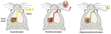
Resistive random-access memory (RRAM) devices coupled with a spike-coding scheme are key to implementing unsupervised learning with minimal hardware footprint and low power consumption. Embedding neuromorphic learning into low-power devices could enable design of autonomous systems, such as a brain-machine interface that makes decisions based on real-time, on-line processing of in-vivo recorded biological signals. Biological data are intrinsically highly noisy and the proposed combined LTP and STP learning rule is a powerful technique to improve the detection/recognition rate. This approach may enable the design of autonomous implantable devices for rehabilitation purposes

Leti, which has worked on RRAM to develop hardware neuromorphic architectures since 2010, is the coordinator of the H2020 [Horizon 2020] European project NeuRAM3. That project is working on fabricating a chip with architecture that supports state-of-the-art machine-learning algorithms and spike-based learning mechanisms.

That’s it folks.

Improving the quality of sight in artificial retinas

Researchers at France’s Centre national de la recherche scientifique (CNRS) and elsewhere have taken a step forward to improving sight derived from artificial retinas according to an Aug. 25, 2016 news item on Nanowerk (Note: A link has been removed),

A major therapeutic challenge, the retinal prostheses that have been under development during the past ten years can enable some blind subjects to perceive light signals, but the image thus restored is still far from being clear. By comparing in rodents the activity of the visual cortex generated artificially by implants against that produced by “natural sight”, scientists from CNRS, CEA [Commissariat à l’énergie atomique et aux énergies alternatives is the French Alternative Energies and Atomic Energy Commission], INSERM [Institut national de la santé et de la recherche médicale is the French National Institute of Health and Medical Research], AP-HM [Assistance Publique – Hôpitaux de Marseille] and Aix-Marseille Université identified two factors that limit the resolution of prostheses.

Based on these findings, they were able to improve the precision of prosthetic activation. These multidisciplinary efforts, published on 23 August 2016 in eLife (“Probing the functional impact of sub-retinal prosthesis”), thus open the way towards further advances in retinal prostheses that will enhance the quality of life of implanted patients.

An Aug. 24, 2015 CNRS press release, which originated the news item, expands on the theme,

A retinal prosthesis comprises three elements: a camera (inserted in the patient’s spectacles), an electronic microcircuit (which transforms data from the camera into an electrical signal) and a matrix of microscopic electrodes (implanted in the eye in contact with the retina). This prosthesis replaces the photoreceptor cells of the retina: like them, it converts visual information into electrical signals which are then transmitted to the brain via the optic nerve. It can treat blindness caused by a degeneration of retinal photoreceptors, on condition that the optical nerve has remained functional1. Equipped with these implants, patients who were totally blind can recover visual perceptions in the form of light spots, or phosphenes.  Unfortunately, at present, the light signals perceived are not clear enough to recognize faces, read or move about independently.

To understand the resolution limits of the image generated by the prosthesis, and to find ways of optimizing the system, the scientists carried out a large-scale experiment on rodents.  By combining their skills in ophthalmology and the physiology of vision, they compared the response of the visual system of rodents to both natural visual stimuli and those generated by the prosthesis.

Their work showed that the prosthesis activated the visual cortex of the rodent in the correct position and at ranges comparable to those obtained under natural conditions.  However, the extent of the activation was much too great, and its shape was much too elongated.  This deformation was due to two separate phenomena observed at the level of the electrode matrix. Firstly, the scientists observed excessive electrical diffusion: the thin layer of liquid situated between the electrode and the retina passively diffused the electrical stimulus to neighboring nerve cells. And secondly, they detected the unwanted activation of retinal fibers situated close to the cells targeted for stimulation.

Armed with these findings, the scientists were able to improve the properties of the interface between the prosthesis and retina, with the help of specialists in interface physics.  Together, they were able to generate less diffuse currents and significantly improve artificial activation, and hence the performance of the prosthesis.

This lengthy study, because of the range of parameters covered (to study the different positions, types and intensities of signals) and the surgical problems encountered (in inserting the implant and recording the images generated in the animal’s brain) has nevertheless opened the way towards making promising improvements to retinal prostheses for humans.

This work was carried out by scientists from the Institut de Neurosciences de la Timone (CNRS/AMU) and AP-HM, in collaboration with CEA-Leti and the Institut de la Vision (CNRS/Inserm/UPMC).

Artificial retinas


© F. Chavane & S. Roux.

Activation (colored circles at the level of the visual cortex) of the visual system by prosthetic stimulation (in the middle, in red, the insert shows an image of an implanted prosthesis) is greater and more elongated than the activation achieved under natural stimulation (on the left, in yellow). Using a protocol to adapt stimulation (on the right, in green), the size and shape of the activation can be controlled and are more similar to natural visual activation (yellow).


Here’s a link to and a citation for the paper,

Probing the functional impact of sub-retinal prosthesis by Sébastien Roux, Frédéric Matonti, Florent Dupont, Louis Hoffart, Sylvain Takerkart, Serge Picaud, Pascale Pham, and Frédéric Chavane. eLife 2016;5:e12687 DOI: http://dx.doi.org/10.7554/eLife.12687 Published August 23, 2016

This paper appears to be open access.

The French glue wounds shut with aqueous solutions of nanoparticles

An April 21, 2014 news item on Azonano, features a new treatment for gluing wounds shut,

 A significant breakthrough could revolutionize surgical practice and regenerative medicine. A team led by Ludwik Leibler from the Laboratoire Matière Molle et Chimie (CNRS/ESPCI Paris Tech) and Didier Letourneur from the Laboratoire Recherche Vasculaire Translationnelle (INSERM/Universités Paris Diderot and Paris 13), has just demonstrated that the principle of adhesion by aqueous solutions of nanoparticles can be used in vivo to repair soft-tissue organs and tissues.

This easy-to-use gluing method has been tested on rats. When applied to skin, it closes deep wounds in a few seconds and provides aesthetic, high quality healing. It has also been shown to successfully repair organs that are difficult to suture, such as the liver. Finally, this solution has made it possible to attach a medical device to a beating heart, demonstrating the method’s potential for delivering drugs and strengthening tissues. This work has just been published on the website of the journal Angewandte Chemie.

An April 17, 2014 Inserm (Institut national de la santé et de la recherche médicale) press release, which originated the news item, describes the work leading up to this latest announcement (Note: Links to footnotes have been disabled),

In an issue of Nature published in December last year, a team led by Ludwik Leibler [1] presented a novel concept for gluing gels and biological tissues using nanoparticles [2]. The principle is simple: nanoparticles contained in a solution spread out on surfaces to be glued bind to the gel’s (or tissue’s) molecular network. This phenomenon is called adsorption. At the same time the gel (or tissue) binds the particles together. Accordingly, myriad connections form between the two surfaces. This adhesion process, which involves no chemical reaction, only takes a few seconds. In their latest, newly published study, the researchers used experiments performed on rats to show that this method, applied in vivo , has the potential to revolutionize clinical practice.

The press release goes on to describe some of the experiments,

In a first experiment, the researchers compared two methods for skin closure in a deep wound: traditional sutures, and the application of the aqueous nanoparticle solution with a brush. The latter is easy to use and closes skin rapidly until it heals completely, without inflammation or necrosis. The resulting scar is almost invisible.

Phase 1 Skin injury
Phase 2 Application of the solution
Phase 3 Using pressure to hold the edges together
Phase 4 Skin closure

Schéma plaie cutanée

Illustration of the first experiment conducted by the resear chers on rats: a deep wound is repaired by applying the aqueous nanoparticle solution. The wound closes in thirty seconds.
© “Matière Molle et Chimie” Laboratory (CNRS/ESPCI Paris Tech)

In a second experiment, still on rats, the researchers applied this solution to soft-tissue organs such as the liver, lungs or spleen that are difficult to suture because they tear when the needle passes through them. At present, no glue is sufficiently strong as well as harmless for the organism. Confronted with a deep gash in the liver with severe bleeding, the researchers closed the wound by spreading the aqueous nanoparticle solution and pressing the two edges of the wound toget her. The bleeding stopped. To repair a sectioned liver lobe, the researchers also used nanoparticles: they glued a film coated with nanoparticles onto the wound, and stopped the bleeding. In both situations, organ function was unaffected and the animals survived.

“Gluing a film to stop leakage” is only one example of the possibilities opened up by adhesion brought by nanoparticles. In an entirely different field, the researchers have succeeded in using anoparticles to attach a biodegradable membrane used for cardiac cell therapy, and to achieve this despite the substantial mechanical constraints due to its beating. They thus showed that it would be possible to attach various medical devices to organs and tissues for therapeutic, repair or mechanical strengthening purposes.

This adhesion method is exceptional because of its potential spectrum of clinical applications. It is simple, easy to use and the nanoparticles employed (silica, iron oxides) can be metabolized by the organism. It can easily be integrated into ongoing research on healing and tissue regeneration and contribute to the development of regenerative medicine.

Here’s a link to and a citation for the paper,

Organ Repair, Hemostasis, and In Vivo Bonding of Medical Devices by Aqueous Solutions of Nanoparticles by Anne Meddahi-Pellé, Aurélie Legrand, Alba Marcellan, Liliane Louedec, Didier Letourneur, Ludwik Leibler. Angewandte Chemie. Published online April 16, 2014. DOI: 10.1002/anie.201401043

This article is open access.