Tag Archives: Japan Riken research institute

An emotional android child

Caption: The six emotional expressions assessed in the second experiment. See if you can identify them! Note: this is a video made by filming Nikola’s head as it sat on a desk – it is not a computer animated graphic. Credit: RIKEN

This work comes from Japan according to a February 16, 2022 news item on ScienceDaily,

Researchers from the RIKEN Guardian Robot Project in Japan have made an android child named Nikola that successfully conveys six basic emotions. The new study, published in Frontiers in Psychology, tested how well people could identify six facial expressions — happiness, sadness, fear, anger, surprise, and disgust — which were generated by moving “muscles” in Nikola’s face. This is the first time that the quality of android-expressed emotion has been tested and verified for these six emotions.

A February 11, 2022 RIKEN press release (also on EurekAlert but published February 15, 2022), which originated the news item, provides more detail about the work,

Rosie the robot maid was considered science fiction when she debuted on the Jetson’s cartoon over 50 years ago. Although the reality of the helpful robot is currently more science and less fiction, there are still many challenges that need to be met, including being able to detect and express emotions. The recent study led by Wataru Sato from the RIKEN Guardian Robot Project focused on building a humanoid robot, or android, that can use its face to express a variety of emotions. The result is Nikola, an android head that looks like a hairless boy.

Inside Nikola’s face are 29 pneumatic actuators that control the movements of artificial muscles. Another 6 actuators control head and eyeball movements. Pneumatic actuators are controlled by air pressure, which makes the movements silent and smooth. The team placed the actuators based on the Facial Action Coding System (FACS), which has been used extensively to study facial expressions. Past research has identified numerous facial action units—such as ‘cheek raiser’ and ‘lip pucker’—that comprise typical emotions such as happiness or disgust, and the researchers incorporated these action units in Nikola’s design.

Typically, studies of emotions, particularly how people react to emotions, have a problem. It is difficult to do a properly controlled experiment with live people interacting, but at the same time, looking at photos or videos of people is less natural, and reactions aren’t the same. “The hope is that with androids like Nikola, we can have our cake and eat it too,” says Sato. “We can control every aspect of Nikola’s behavior, and at the same time study live interactions.” The first step was to see if Nikola’s facial expressions were understandable.

A person certified in FACS [Facial Action Coding System] scoring was able to identify each facial action unit, indicating that Nikola’s facial movements accurately resemble those of a real human. A second test showed that everyday people could recognize the six prototypical emotions—happiness, sadness, fear, anger, surprise, and disgust—in Nikola’s face, albeit to varying accuracies. This is because Nikola’s silicone skin is less elastic than real human skin and cannot form wrinkles very well. Thus, emotions like disgust were harder to identify because the action unit for nose wrinkling could not be included.

“In the short term, androids like Nikola can be important research tools for social psychology or even social neuroscience,” says Sato. “Compared with human confederates, androids are good at controlling behaviors and can facilitate rigorous empirical investigation of human social interactions.” As an example, the researchers asked people to rate the naturalness of Nikola’s emotions as the speed of his facial movements was systematically controlled. They researchers found that the most natural speed was slower for some emotions like sadness than it was for others like surprise.

While Nikola still lacks a body, the ultimate goal of the Guardian Robot Project is to build an android that can assist people, particularly those which physical needs who might live alone. “Androids that can emotionally communicate with us will be useful in a wide range of real-life situations, such as caring for older people, and can promote human wellbeing,” says Sato.

Here’s a link to and a citation for the paper,

An Android for Emotional Interaction: Spatiotemporal Validation of Its Facial Expressions by Wataru Sato, Shushi Namba, Dongsheng Yang, Shin’ya Nishida, Carlos Ishi, and Takashi Minato. Front. Psychol., 04 February 2022 DOI: https://doi.org/10.3389/fpsyg.2021.800657

This paper is open access.

For anyone who’d like to investigate the worlds of robots, artificial intelligence, and emotions, I have my December 3, 2021 posting “True love with AI (artificial intelligence): The Nature of Things explores emotional and creative AI (long read)” and there’s also Hiroshi Ishiguro’s work, which I’ve mentioned a number of times here, most recently in a March 27, 2017 posting “Ishiguro’s robots and Swiss scientist question artificial intelligence at SXSW (South by Southwest) 2017.”
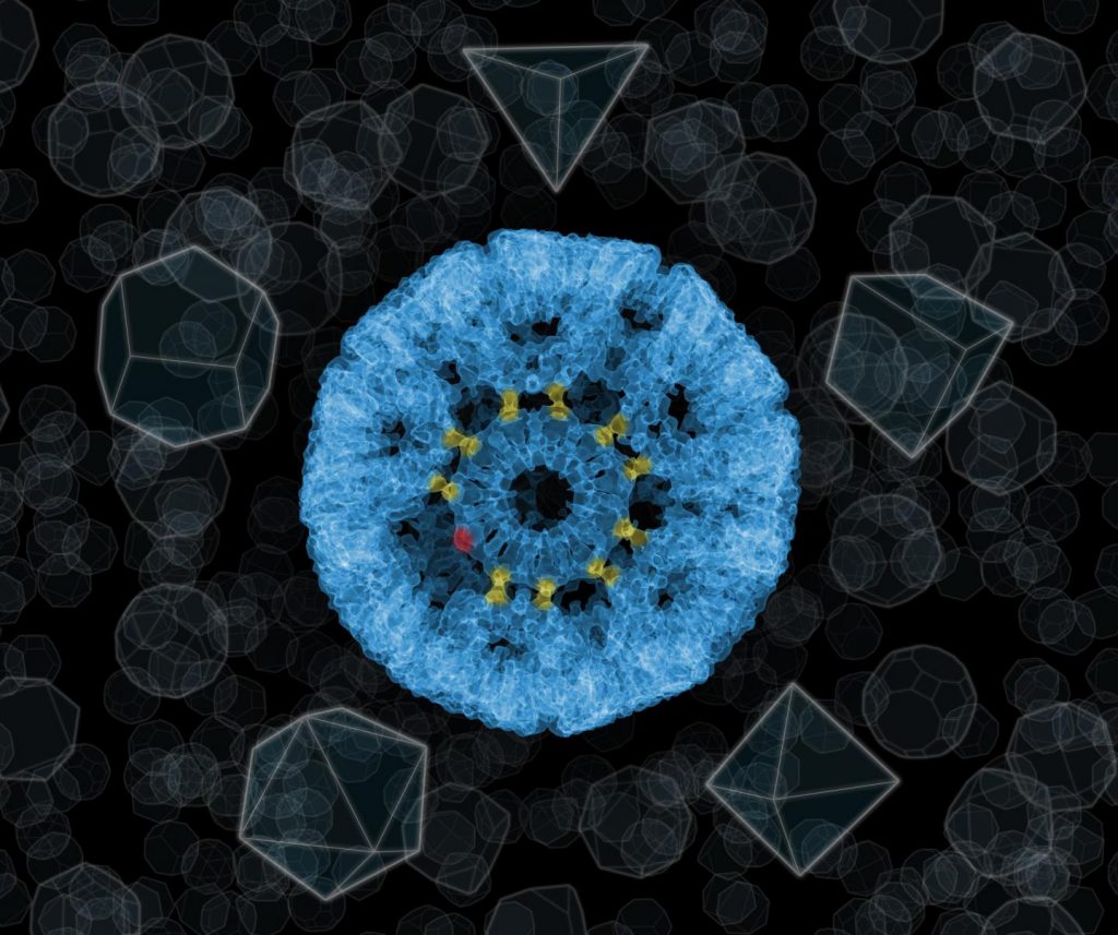
Gold glue?

If you’re hoping for gold flecks in your glue, this is not going to satisfy you, given that it’s all at the nanoscale. An August 7, 2019 news item on Nanowerk briefly describes this gold glue (Note: A link has been removed),

It has long been known that gold can be used to do things that philosophers have never even dreamed of. The Institute of Nuclear Physics of the Polish Academy of Sciences in Cracow has confirmed the existence of ‘gold glue’: bonds involving gold atoms, capable of permanently bonding protein rings. Skilfully used by an international team of scientists, the bonds have made it possible to construct molecular nanocages with a structure so far unparalleled in nature or even in mathematics (Nature, “An ultra-stable gold-coordinated protein cage displaying reversible assembly”).

Caption: The ‘impossible’ sphere, i.e. a molecular nanocage of 24 protein rings, each of which has an 11-sided structure. The rings are connected by bonds with the participation of gold atoms, here marked in yellow. Depending on their position in the structure, not all gold atoms have to be used to attach adjacent proteins (an unused gold atom is marked in red). Credit: Source: UJ, IFJ PAN

An August 6, 2019 Polish Academy of Sciences press release (also on EurekAlert but published August 7, 2019), which originated the news item, expands on the theme,

The world of science has been interested in molecular cages for years. Not without reason. Chemical molecules, including those that would under normal conditions enter into chemical reactions, can be enclosed within their empty interiors. The particles of the enclosed compound, separated by the walls of the cage from the environment, have nothing to bond with. These cages can be therefore be used, for example, to transport drugs safely into a cancer cell, only releasing the drug when they are inside it.

Molecular cages are polyhedra made up of smaller ‘bricks’, usually protein molecules. The bricks can’t be of any shape. For example, if we wanted to build a molecular polyhedron using only objects with the outline of an equilateral triangle, geometry would limit us to only three solid figures: a tetrahedron, an octahedron or an icosahedron. So far, there have been no other structural possibilities.

“Fortunately, Platonic idealism is not a dogma of the physical world. If you accept certain inaccuracies in the solid figure being constructed, you can create structures with shapes that are not found in nature, what’s more, with very interesting properties,” says Dr. Tomasz Wrobel from the Cracow Institute of Nuclear Physics of the Polish Academy of Sciences (IFJ PAN).

Dr. Wrobel is one of the members of an international team of researchers who have recently carried out the ‘impossible’: they built a cage similar in shape to a sphere out of eleven-walled proteins. The main authors of this spectacular success are scientists from the group of Prof. Jonathan Heddle from the Malopolska Biotechnology Centre of the Jagiellonian University in Cracow and the Japanese RIKEN Institute in Wako. The work described in Nature magazine took place with the participation of researchers from universities in Osaka and Tsukuba (Japan), Durham (Great Britain), Waterloo (Canada) and other research centres.

Each of the walls of the new nanocages was formed by a protein ring from which eleven cysteine molecules stuck out at regular intervals. It was to the sulphur atom found in each cysteine molecule that the ‘glue’, i.e. the gold atom, was planned to be attached. In the appropriate conditions, it could bind with one more sulphur atom, in the cysteine of a next ring. In this way a permanent chemical bond would be formed between the two rings. But would the gold atom under these conditions really be able to form a bond between the rings?

“In the Spectroscopic Imaging Laboratory of IFJ PAS we used Raman spectroscopy and X-ray photoelectron spectroscopy to show that in the samples provided to us with the test nanocages, the gold really did form bonds with sulphur atoms in cysteines. In other words, in a difficult, direct measurement, we proved that gold ‘glue’ for bonding protein rings in cages really does exist,” explains Dr. Wrobel.

Each gold atom can be treated as a stand-alone clip that makes it possible to attach another ring. The road to the ‘impossible’ begins when we realize that we don’t always have to use all of the clips! So, although all the rings of the new nanocages are physically the same, depending on their place in the structure they connect with their neighbours with a different number of gold atoms, and thus function as polygons with different numbers of vertices. 24 nanocage walls presented by the researchers were held together by 120 gold atoms. The outer diameter of the cages was 22 nanometres and the inner diameter was 16 nm.

Using gold atoms as a binder for nanocages is also important due to its possible applications. In earlier molecular structures, proteins were glued together using many weak chemical bonds. The complexity of the bonds and their similarity to the bonds responsible for the existence of the protein rings themselves did not allow for precise control over the decomposition of the cages. This is not the case in the new structures. On the one hand, gold-bonded nanocages are chemically and thermally stable (for example, they withstand hours of boiling in water). On the other hand, however, gold bonds are sensitive to an increase in acidity. By its increase, the nanocage can be decomposed in a controlled way and the contents can be released into the environment. Since the acidity within cells is greater than outside them, gold-bonded nanocages are ideal for biomedical applications.

The ‘impossible’ nanocage is the presentation of a qualitatively new approach to the construction of molecular cages, with gold atoms in the role of loose clips. The demonstrated flexibility of the gold bonds will make it possible in the future to create nanocages with sizes and features precisely tailored to specific needs.

Here’s a link to and a citation for the paper.

An ultra-stable gold-coordinated protein cage displaying reversible assembly by Ali D. Malay, Naoyuki Miyazaki, Artur Biela, Soumyananda Chakraborti, Karolina Majsterkiewicz, Izabela Stupka, Craig S. Kaplan, Agnieszka Kowalczyk, Bernard M. A. G. Piette, Georg K. A. Hochberg, Di Wu, Tomasz P. Wrobel, Adam Fineberg, Manish S. Kushwah, Mitja Kelemen, Primož Vavpetič, Primož Pelicon, Philipp Kukura, Justin L. P. Benesch, Kenji Iwasaki & Jonathan G. Heddle Nature volume 569, pages438–442 (2019) Issue Date: 16 May 2019 DOI: https://doi.org/10.1038/s41586-019-1185-4 Published online: 08 May 2019

This paper is behind a paywall.

Watching motor proteins at work

Researchers in the UK and in Japan have described these motor proteins as ‘swinging on monkey bars’,

A Sept. 14, 2015 news item on Nanowerk provides more information about the motor protein observations,

These proteins are vital to complex life, forming the transport infrastructure that allows different parts of cells to specialise in particular functions. Until now, the way they move has never been directly observed.

Researchers at the University of Leeds and in Japan used electron microscopes to capture images of the largest type of motor protein, called dynein, during the act of stepping along its molecular track.

A Sept 14, 2015 Leeds University press release, (also on EurekAlert*) which originated the news item, expands on the theme with what amounts to a transcript of sorts for the video (Note: Links have been removed),

Dr Stan Burgess, at the University of Leeds’ School of Molecular and Cellular Biology, who led the research team, said: “Dynein has two identical motors tied together and it moves along a molecular track called a microtubule. It drives itself along the track by alternately grabbing hold of a binding site, executing a power stroke, then letting go, like a person swinging on monkey bars.

“Previously, dynein movement had only been tracked by attaching fluorescent molecules to the proteins and observing the fluorescence using very powerful light microscopes. It was a bit like tracking vehicles from space with GPS. It told us where they were, their speed and for how long they ran, stopped and so on, but we couldn’t see the molecules in action themselves. These are the first images of these vital processes.”

An understanding of motor proteins is important to medical research because of their fundamental role in complex cellular life. Many viruses hijack motor proteins to hitch a ride to the nucleus for replication. Cell division is driven by motor proteins and so insights into their mechanics could be relevant to cancer research. Some motor neurone diseases are also associated with disruption of motor protein traffic.

The team at Leeds, working within the world-leading Astbury Centre for Structural Molecular Biology, combined purified microtubules with purified dynein motors and added the chemical fuel ATP (adenosine triphosphate) to power the motor.

Dr Hiroshi Imai, now Assistant Professor in the Department of Biological Sciences at Chuo University, Japan, carried out the experiments while working at the University of Leeds.

He explained: “We set the dyneins running along their tracks and then we froze them in ‘mid-stride’ by cooling them at about a million degrees a second, fast enough to prevent the water from forming ice crystals as it solidified. Then using a cryo-electron microscope we took many thousands of images of the motors caught during the act of stepping. By combining many images of individual motors, we were able to sharpen up our picture of the dynein and build up a dynamic idea of how it moved. It is a bit like figuring out how to swing along monkey bars by studying photographs of many people swinging on them.”

Dr Burgess said: “Our most striking discovery was the existence of a hinge between the long, thin stalk and the ‘grappling hook’, like the wrist between a human arm and hand. This allows a lot of variation in the angle of attachment of the motor to its track.

“Each of the two arms of a dynein motor protein is about 25 nanometres (0.000025 millimetre) long, while the binding sites it attaches to are only 8 nanometres apart. That means dynein can reach not only the next rung but the one after that and the one after that and appears to give it flexibility in how it moves along the ‘track’.”

Dynein is not only the biggest but also the most versatile of the motor proteins in living cells and, like all motor proteins, is vital to life. Motor proteins transport cargoes and hold many cellular components in position within the cell. For instance, dynein is responsible for carrying messages from the tips of active nerve cells back to the nucleus and these messages keep the nerve cells alive.

Co-author Peter Knight, Professor of Molecular Contractility in the University of Leeds’ School of Molecular and Cellular Biology, said: “If a cell is like a city, these are like the truckers on its road and rail networks. If you didn’t have a transport system, you couldn’t have specialised regions. Every part of the cell would be doing the same thing and that would mean you could not have complex life.”

“Dynein is the multi-purpose vehicle of cellular transport. Other motor proteins, called kinesins and myosins, are much smaller and have specific functions, but dynein can turn its hand to a lot of different of functions,” Professor Knight said.

For instance, in the motor neurone connecting the central nervous system to the big toe—which is a single cell a metre long— dynein provides the transport from the toe back to the nucleus. Another vital role is in the movement of cells.

Dr Burgess said: “During brain development, neurones must crawl into their correct position and dynein molecules in this instance grab hold of the nucleus and pull it along with the moving mass of the cell. If they didn’t, the nucleus would be left behind and the cytoplasm would crawl away.”

The study involved researchers from the University of Leeds and Japan’s Waseda and Osaka universities, as well as the Quantitative Biology Center at Japan’s Riken research institute and the Japan Science and Technology Agency (JST). The research was funded by the Human Frontiers Science Program and the Biotechnology and Biological Sciences Research Council (BBSRC).

Here’s a link to and a citation for the paper,

Direct observation shows superposition and large scale flexibility within cytoplasmic dynein motors moving along microtubules by Hiroshi Imai, Tomohiro Shima, Kazuo Sutoh, Matthew L. Walker, Peter J. Knight, Takahide Kon, & Stan A. Burgess. Nature Communications 6, Article number: 8179  doi:10.1038/ncomms9179 Published 14 September 2015

This paper is open access.

*The EurekAlert link added Sept. 15, 2015 at 1200 hours PST.