Tag Archives: Jared Sagoff

Nanoparticle snapshots with femtosecond photography

Caption: Here are "stills" from an X-ray "movie" of an exploding nanoparticle. The nanoparticle is superheated with an intense optical pulse and subsequently explodes (left). A series of ultrafast x-ray diffraction images (right) maps the process and contains information how the explosion starts with surface softening and proceeds from the outside in. Credit: Christoph Bostedt

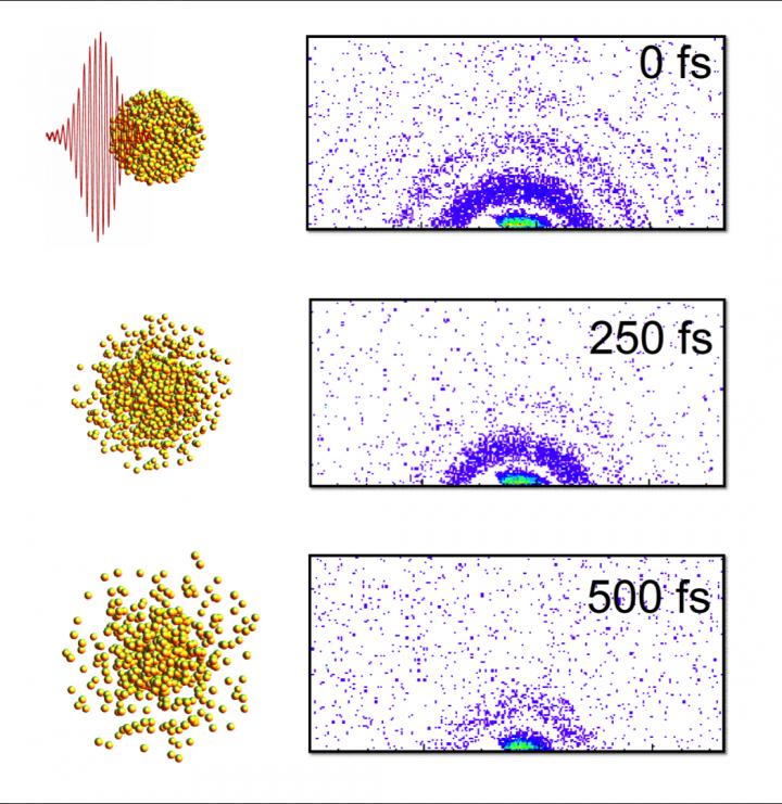
Caption: Here are “stills” from an X-ray “movie” of an exploding nanoparticle. The nanoparticle is superheated with an intense optical pulse and subsequently explodes (left). A series of ultrafast x-ray diffraction images (right) maps the process and contains information how the explosion starts with surface softening and proceeds from the outside in. Credit: Christoph Bostedt

A Feb. 10, 2016 news item on Nanotechnology Now provides more information about the ‘snapshots,

Just as a photographer needs a camera with a split-second shutter speed to capture rapid motion, scientists looking at the behavior of tiny materials need special instruments with the capacity to see changes that happen in the blink of an eye.

An international team of researchers led by X-ray scientist Christoph Bostedt of the U.S. Department of Energy’s (DOE) Argonne National Laboratory and Tais Gorkhover of DOE’s SLAC National Accelerator Laboratory used two special lasers to observe the dynamics of a small sample of xenon as it was heated to a plasma.

A Feb. 10, 2016 Argonne National Laboratory news release (also on EurekAlert) by Jared Sagoff, which originated the news item, provides more technical details,

Bostedt and Gorkhover were able to use the Linac Coherent Light Source (LCLS) at SLAC to make observations of the sample in time steps of approximately a hundred femtoseconds – a femtosecond being one millionth of a billionth of a second [emphasis mine]. The exposure time of the individual images was so short that the quickly moving particles in the gas phase appeared frozen. “The advantage of a machine like the LCLS is that it gives us the equivalent of high-speed flash photography as opposed to a pinhole camera,” Bostedt said. The LCLS is a DOE Office of Science User Facility.

The researchers used an optical laser to heat the sample cluster and an X-ray laser to probe the dynamics of the cluster as it changed over time. As the laser heated the cluster, the photons freed electrons initially bound to the atoms; however, these electrons still remained loosely bound to the cluster.

By imaging exploding nanoparticles, the team was able to make measurements of how they change over time in extreme environments. “Ultimately, we want to understand how the energy from the light affects the system,” Gorkhover said.

“There are really no other techniques that give us this good a resolution in both time and space simultaneously,” she added. “Other methods require us to take averages over many different ‘exposures,’ which can obscure relevant details. Additionally, techniques like electron microscopy involve a substrate material that can interfere with the behavior of the sample.”

According to Bostedt, the research could also impact the study of aerosols in the environment or in combustion, as the dual-laser “pump and probe” model could be adapted to study materials in the gas phase. “Although our material goes from solid to plasma very quickly, there are other types of materials you could study with this or a similar technique,” he said.

I marvel at how very brief the time intervals are at the femtoscale and for that matter, the other subatomic scales.

Here’s a link to and a citation for the paper,

Femtosecond and nanometre visualization of structural dynamics in superheated nanoparticles by Tais Gorkhover, Sebastian Schorb, Ryan Coffee, Marcus Adolph, Lutz Foucar, Daniela Rupp, Andrew Aquila, John D. Bozek, Sascha W. Epp, Benjamin Erk, Lars Gumprecht, Lotte Holmegaard, Andreas Hartmann, Robert Hartmann, Günter Hauser, Peter Holl, Andre Hömke, Per Johnsson, Nils Kimmel, Kai-Uwe Kühnel, Marc Messerschmidt, Christian Reich, Arnaud Rouzée, Benedikt Rudek, Carlo Schmidt et al. Nature Photonics 10, 93–97 (2016) doi:10.1038/nphoton.2015.264 Published online 25 January 2016

This paper is behind a paywall.

Dumbbells at the nanoscale according to researchers at the (US) Argonne National Laboratory

Researchers at the US Dept. of Energy’s Argonne National Laboratory are providing new insight into how nanoparticles ‘grow’. From a Dec. 5, 2014 news item on Nanowerk,

Like snowflakes, nanoparticles come in a wide variety of shapes and sizes. The geometry of a nanoparticle is often as influential as its chemical makeup in determining how it behaves, from its catalytic properties to its potential as a semiconductor component.

Thanks to a new study from the U.S. Department of Energy’s (DOE) Argonne National Laboratory, researchers are closer to understanding the process by which nanoparticles made of more than one material – called heterostructured nanoparticles – form. This process, known as heterogeneous nucleation, is the same mechanism by which beads of condensation form on a windowpane.

The scientists have provided an image which illustrates their findings,

This picture combines a transmission electron microscope image of a nanodumbbell with a gold domain oriented in direction. The seed and gold domains in the dumbbell in the image on the right are identified by geometric phase analysis. Image credit: Soon Gu Kwon.

This picture combines a transmission electron microscope image of a nanodumbbell with a gold domain oriented in direction. The seed and gold domains in the dumbbell in the image on the right are identified by geometric phase analysis. Image credit: Soon Gu Kwon.

A Dec. 4, 2014 Argonne National Laboratory news release by Jared Sagoff, which originated the news item, describes the structures being examined and the reason for doing so,

Heterostructured nanoparticles can be used as catalysts and in advanced energy conversion and storage systems. Typically, these nanoparticles are created from tiny “seeds” of one material, on top of which another material is grown.  In this study, the Argonne researchers noticed that the differences in the atomic arrangements of the two materials have a big impact on the shape of the resulting nanoparticle.

“Before we started this experiment, it wasn’t entirely clear what’s happening at the interface when one material grows on another,” said nanoscientist Elena Shevchenko of Argonne Center for Nanoscale Materials, a DOE Office of Science user facility.

In this study, the researchers observed the formation of a nanoparticle consisting of platinum and gold.  The researchers started with a platinum seed and grew gold around it. Initially, the gold covered the platinum seed’s surface uniformly, creating a type of nanoparticle known as “core-shell.” However, as more gold was deposited, it started to grow unevenly, creating a dumbbell-like structure.

Thanks to state-of-the-art X-ray analysis provided by Argonne’s Advanced Photon Source (APS), a DOE Office of Science user facility, the researchers identified the cause of the dumbbell formation as “lattice mismatch,” in which the spacing between the atoms in the two materials doesn’t align.

“Essentially, you can think of lattice mismatch as having a row of smaller boxes on the bottom layer and larger boxes on the top layer.  When you try to fit the larger boxes into the space for a smaller box, it creates an immense strain,” said Argonne physicist Byeongdu Lee.

While the lattice mismatch is only fractions of a nanometer, the effect accumulates as layer after layer of gold forms on the platinum. The mismatch can be handled by the first two layers of gold atoms – creating the core-shell effect – but afterwards it proves too much to overcome. “The arrangement of atoms is the same in the two materials, but the distance between atoms is different,” said Argonne postdoctoral researcher Soon Gu Kwon. “Eventually, this becomes unstable, and the growth of the gold becomes unevenly distributed.”

As the gold continues to accumulate on one side of the seed nanoparticle, small quantities “slide” down the side of the nanoparticle like grains of sand rolling down the side of a sand hill, creating the dumbbell shape.

The advantage of the Argonne study comes from the researchers’ ability to perform in situ observations of the material in realistic conditions using the APS. “This is the first time anyone has been able to study the kinetics of this heterogeneous nucleation process of nanoparticles in real-time under realistic conditions,” said Argonne physicist Byeongdu Lee. “The combination of two X-ray techniques gave us the ability to observe the material at both the atomic level and the nanoscale, which gave us a good view of how the nanoparticles form and transform.” All conclusions made based on the X-ray studies were further confirmed using atomic-resolution microscopy in the group of Professor Robert Klie of the University of Illinois at Chicago.

This analysis of nanoparticle formation will help to lay the groundwork for the formation of new materials with different and controllable properties, according to Shevchenko. “In order to design materials, you have to understand how these processes happen at a very basic level,” she said.

Here’s a link to and a citation for the paper,

Heterogeneous nucleation and shape transformation of multicomponent metallic nanostructures by Soon Gu Kwon, Galyna Krylova, Patrick J. Phillips, Robert F. Klie, Soma Chattopadhyay, Tomohiro Shibata, Emilio E. Bunel, Yuzi Liu, Vitali B. Prakapenka, Byeongdu Lee, & Elena V. Shevchenko. Nature Materials (2014) doi:10.1038/nmat4115 Published online 02 November 2014

This paper is behind a paywall but there is a free preview via ReadCube Access.

Studying corrosion from the other side

Corrosion can be beautiful as well as destructive,

Typically, the process of corrosion has been studied from the metal side of the equation - See more at: http://www.anl.gov/articles/core-corrosion#sthash.ZPqFF13I.dpuf. Courtesy of the Argonne National Laboratory

Typically, the process of corrosion has been studied from the metal side of the equation – See more at: http://www.anl.gov/articles/core-corrosion#sthash.ZPqFF13I.dpuf. Courtesy of the Argonne National Laboratory

A Feb. 18, 2014 news item on Nanowerk expands on the theme of corrosion as destruction (Note: Links have been removed),

Anyone who has ever owned a car in a snowy town – or a boat in a salty sea – can tell you just how expensive corrosion can be.

One of the world’s most common and costly chemical reactions, corrosion happens frequently at the boundaries between water and metal surfaces. In the past, the process of corrosion has mostly been studied from the metal side of the equation.

However, in a new study (“Chloride ions induce order-disorder transition at water-oxide interfaces”), scientists at the Center for Nanoscale Materials at the U.S. Department of Energy’s Argonne National Laboratory investigated the problem from the other side, looking at the dynamics of water containing dissolved ions located in the regions near a metal surface.

The Feb. 14, 2014 Argonne National Laboratory news release by Jared Sagoff, which originated the news item, describes how the scientists conducted their research,

A team of researchers led by Argonne materials scientist Subramanian Sankaranarayanan simulated the physical and chemical dynamics of dissolved ions in water at the atomic level as it corrodes metal oxide surfaces. “Water-based solutions behave quite differently near a metal or oxide surface than they do by themselves,” Sankaranarayanan said. “But just how the chemical ions in the water interact with a surface has been an area of intense debate.”

Under low-chlorine conditions, water tends to form two-dimensional ordered layers near solid interfaces because of the influence of its strong hydrogen bonds. However, the researchers found that increasing the proportion of chlorine ions above a certain threshold causes a change in which the solution loses its ordered nature near the surface and begins to act similar to water away from the surface. This transition, in turn, can increase the rate at which materials corrode as well as the freezing temperature of the solution.

This switch between an ordered and a disordered structure near the metal surface happens incredibly quickly, in just fractions of a nanosecond. The speed of the chemical reaction necessitates the use of high-performance computers like Argonne’s Blue/Gene Q supercomputer, Mira.

To further explore these electrochemical oxide interfaces with high-performance computers, Sankaranarayanan and his colleagues from Argonne, Harvard University and the University of Missouri have also been awarded 40 million processor-hours of time on Mira.

“Having the ability to look at these reactions in a more powerful simulation will give us the opportunity to make a more educated guess of the rates of corrosion for different scenarios,” Sankaranarayanan said. Such studies will open up for the first time fundamental studies of corrosion behavior and will allow scientists to tailor materials surfaces to improve the stability and lifetime of materials.

Here’s a link to and a citation for the paper,

Chloride ions induce order-disorder transition at water-oxide interfaces by Sanket Deshmukh, Ganesh Kamath, Shriram Ramanathan, and Subramanian K. R. S. Sankaranarayanan. Phys. Rev. E 88 (6), 062119 (2013) [5 pages]

This article is behind a paywall on both the primary site and the beta site (the American Physical Society is testing a new website for its publications).