Tag Archives: Jeffrey Hartgerink

Mussels to muscles for biocompatible fibres

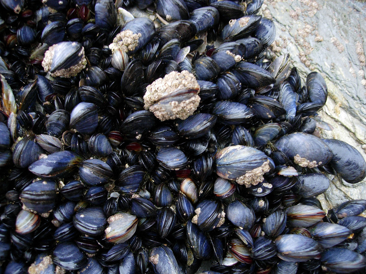
Mussels and barnacles in the intertidal near Newquay, Cornwall, England.
Wilson44691 at English Wikipedia – Photograph taken by Mark A. Wilson (Department of Geology, The College of Wooster

I love puns and word play so I was happy to see this in a June 9, 2017 news item on ScienceDaily,

Rice University chemists can thank the mussel for putting the muscle into their new macroscale scaffold fibers.

A June 9, 2017 Rice University news release (also on EurekAlert), which originated the news item, provides more details about the research,

The Rice lab of chemist Jeffrey Hartgerink had already figured out how to make biocompatible nanofibers out of synthetic peptides. In new work, the lab is using an amino acid found in the sticky feet of mussels to make those fibers line up into strong hydrogel strings.

Hartgerink and Rice graduate student I-Che Li introduced their room-temperature method this month in an open-access paper in the Journal of the American Chemical Society.

The hydrogel strings can be picked up and moved with tweezers, and Li said he expects they will help labs gain better control over the growth of cell cultures.

“Usually when cells grow on a surface, they spread randomly,” he said. “There are a lot of biomaterials we want to grow in a specific direction. With the hydrogel scaffold aligned, we can expect cells to grow the way we want them to. One example would be neuron cells, which we want to grow head-to-tail to aid nerve regeneration.

“Basically, this could allow us to direct cell growth from here to there,” he said. “That’s why this material is so exciting.”

In previous research Hartgerink’s lab had developed synthetic hydrogels that could be injected into the body to serve as scaffolds for tissue growth. The hydrogels contained hydrophobic peptides that self-assembled into fibers about 6 nanometers wide and up to several microns long. However, because the fibers did not interact with one other, they generally appeared in microscope images as a tangled mass.

Experiments showed the fibers could be coaxed into alignment with the application of shear forces, in the same way that playing cards are aligned during shuffling by pushing on both the top and bottom of the deck.

Hartgerink and Li decided to try pushing the fibers through a needle to force them into alignment, a process that would be easier if the material was water soluble. So they added a chain of amino acids known as DOPA to the sides of the fibers to allow them to remain water-soluble in the syringe, Li said.

DOPA — short for 3,4-dihydroxyphenylalanine — is the compound that lets mussels stick to just about anything. Hartgerink and Li found that the combination of DOPA and shear stress from passing through the needle prompted the fibers to form visible, rope-like bundles.

They also found that DOPA promoted chemical cross-linking reactions that helped the bundles hold their shape. “DOPA is really sensitive to oxidizing agents,” Li said. “Even exposing DOPA to air oxidizes it, and that aids in cross-linking the fibers.”

As a bonus, the aligned fibers also proved to have a curious and useful optical property called “uniform birefringence,” or double-refraction. Li said this could allow researchers to use polarized light to see exactly where the aligned fibers are, even if they’re covered by cells.

“This will be an important technique for us to make sure of the long-range order of fiber alignment when we are testing directed cell growth,” he said.

The researchers expect the aligned fibers can be used for macroscale medical applications but with nanoscale control over the structures.

“Self-assembly is basically the ability of a molecule to make ordered structure from chaos, and what I-Che has done is push this organization to a new level with his aligned strings,” said Hartgerink, a professor of chemistry and of bioengineering. “With this material, we are excited to see if we can impose this organization onto the growth of cells that interact with it.”

Here’s a link to and a citation for the paper,

Covalent Capture of Aligned Self-Assembling Nanofibers by I-Che Li and Jeffrey D. Hartgerink. J. Am. Chem. Soc., Article ASAP DOI: 10.1021/jacs.7b04655 Publication Date (Web): June 5, 2017

Copyright © 2017 American Chemical Society

This paper is open access.

Snake venom as a healing agent in hydrogels

The Brazilian lancehead is one of several South American pit vipers that produce venom that has proven to be a powerful blood coagulant. Scientists at Rice University have combined a derivative of the venom with their injectable hydrogels to create a material that can quickly stop bleeding and protect wounds, even in patients who take anti-coagulant medications. (Credit: Photo by Greg Hume via Wikipedia)

The Brazilian lancehead is one of several South American pit vipers that produce venom that has proven to be a powerful blood coagulant. Scientists at Rice University have combined a derivative of the venom with their injectable hydrogels to create a material that can quickly stop bleeding and protect wounds, even in patients who take anti-coagulant medications. (Credit: Photo by Greg Hume via Wikipedia)

Mesmerizing and beautiful in their way, venomous snakes are healers, as well as, killers. An Oct. 27, 2015 news item on Azonano describes a new healing use for their venom,

A nanofiber hydrogel infused with snake venom may be the best material to stop bleeding quickly, according to Rice University scientists.

The hydrogel called SB50 incorporates batroxobin, a venom produced by two species of South American pit viper. It can be injected as a liquid and quickly turns into a gel that conforms to the site of a wound, keeping it closed, and promotes clotting within seconds.

An Oct. 26, 2015 Rice University news release, which originated the news item, provides more details (Note: Links have been removed),

Rice chemist Jeffrey Hartgerink, lead author Vivek Kumar and their colleagues reported their discovery in the American Chemical Society journal ACS Biomaterials Science and Engineering. The hydrogel may be most useful for surgeries, particularly for patients who take anti-coagulant drugs to thin their blood.

“It’s interesting that you can take something so deadly and turn it into something that has the potential to save lives,” Hartgerink said.

Batroxobin was recognized for its properties as a coagulant – a substance that encourages blood to clot – in 1936. It has been used in various therapies as a way to remove excess fibrin proteins from the blood to treat thrombosis and as a topical hemostat. It has also been used as a diagnostic tool to determine blood-clotting time in the presence of heparin, an anti-coagulant drug.

“From a clinical perspective, that’s far and away the most important issue here,” Hartgerink said. “There’s a lot of different things that can trigger blood coagulation, but when you’re on heparin, most of them don’t work, or they work slowly or poorly. That obviously causes problems if you’re bleeding.

“Heparin blocks the function of thrombin, an enzyme that begins a cascade of reactions that lead to the clotting of blood,” he said. “Batroxobin is also an enzyme with similar function to thrombin, but its function is not blocked by heparin. This is important because surgical bleeding in patients taking heparin can be a serious problem. The use of batroxobin allows us to get around this problem because it can immediately start the clotting process, regardless of whether heparin is there or not.”

The batroxobin combined with the Rice lab’s hydrogels isn’t taken directly from snakes, Hartgerink said. The substance used for medicine is produced by genetically modified bacteria and then purified, avoiding the risk of other contaminant toxins.

The Rice researchers combined batroxobin with their synthetic, self-assembling nanofibers, which can be loaded into a syringe and injected at the site of a wound, where they reassemble themselves into a gel.

Tests showed the new material stopped a wound from bleeding in as little as six seconds, and further prodding of the wound minutes later did not reopen it. The researchers also tested several other options: the hydrogel without batroxobin, the batroxobin without the hydrogel, a current clinical hemostat known as GelFoam and an alternative self-assembling hemostat known as Puramatrix and found that none were as effective, especially in the presence of anti-coagulants.

The new work builds upon the Rice lab’s extensive development of injectable hydrogel scaffolds that help wounds heal and grow natural tissue. The synthetic scaffolds are built from the peptide sequences to mimic natural processes.

“To be clear, we did not discover nor do any of the initial investigations of batroxobin,” Hartgerink said. “Its properties have been well-known for many decades. What we did was combine it with the hydrogel we’ve been working on for a long time.

“We think SB50 has great potential to stop surgical bleeding, particularly in difficult cases in which the patient is taking heparin or other anti-coagulants,” he said. “SB50 takes the powerful clotting ability of this snake venom and makes it far more effective by delivering it in an easily localized hydrogel that prevents possible unwanted systemic effects from using batroxobin alone.”

SB50 will require FDA approval before clinical use, Hartgerink said. While batroxobin is already approved, the Rice lab’s hydrogel has not yet won approval, a process he expects will take several more years of testing.

Here’s a link to and a citation for the paper,

Nanofibrous Snake Venom Hemostat by Vivek A. Kumar, Navindee C. Wickremasinghe, Siyu Shi, and Jeffrey D. Hartgerink. ACS Biomater. Sci. Eng., Article ASAP
DOI: 10.1021/acsbiomaterials.5b00356 Publication Date (Web): October 22, 2015

Copyright © 2015 American Chemical Society

This paper is behind a paywall.