Tag Archives: Jeremy J. Baumberg

Colo(u)r-changing building surfaces thanks to gold nanoparticles

Gold, at the nanoscale, has different properties than it has at the macroscale and research at the University of Cambridge has found a new way to exploit gold’s unique properties at the nanoscale according to a May 13, 2019 news item item on ScienceDaily,

The smallest pixels yet created — a million times smaller than those in smartphones, made by trapping particles of light under tiny rocks of gold — could be used for new types of large-scale flexible displays, big enough to cover entire buildings.

The colour pixels, developed by a team of scientists led by the University of Cambridge, are compatible with roll-to-roll fabrication on flexible plastic films, dramatically reducing their production cost. The results are reported in the journal Science Advances [May 10, 2019].

A May 10,2019 University of Cambridge press release (also on EurekAlert), which originated the news item, delves further into the research,

It has been a long-held dream to mimic the colour-changing skin of octopus or squid, allowing people or objects to disappear into the natural background, but making large-area flexible display screens is still prohibitively expensive because they are constructed from highly precise multiple layers.

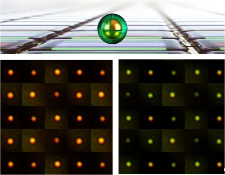
At the centre of the pixels developed by the Cambridge scientists is a tiny particle of gold a few billionths of a metre across. The grain sits on top of a reflective surface, trapping light in the gap in between. Surrounding each grain is a thin sticky coating which changes chemically when electrically switched, causing the pixel to change colour across the spectrum.

The team of scientists, from different disciplines including physics, chemistry and manufacturing, made the pixels by coating vats of golden grains with an active polymer called polyaniline and then spraying them onto flexible mirror-coated plastic, to dramatically drive down production cost.

The pixels are the smallest yet created, a million times smaller than typical smartphone pixels. They can be seen in bright sunlight and because they do not need constant power to keep their set colour, have an energy performance that makes large areas feasible and sustainable. “We started by washing them over aluminized food packets, but then found aerosol spraying is faster,” said co-lead author Hyeon-Ho Jeong from Cambridge’s Cavendish Laboratory.

“These are not the normal tools of nanotechnology, but this sort of radical approach is needed to make sustainable technologies feasible,” said Professor Jeremy J Baumberg of the NanoPhotonics Centre at Cambridge’s Cavendish Laboratory, who led the research. “The strange physics of light on the nanoscale allows it to be switched, even if less than a tenth of the film is coated with our active pixels. That’s because the apparent size of each pixel for light is many times larger than their physical area when using these resonant gold architectures.”

The pixels could enable a host of new application possibilities such as building-sized display screens, architecture which can switch off solar heat load, active camouflage clothing and coatings, as well as tiny indicators for coming internet-of-things devices.
The team are currently working at improving the colour range and are looking for partners to develop the technology further.

The research is funded as part of a UK Engineering and Physical Sciences Research Council (EPSRC) investment in the Cambridge NanoPhotonics Centre, as well as the European Research Council (ERC) and the China Scholarship Council.

This image accompanies the press release,

Caption: eNPoMs formed from gold nanoparticles (Au NPs) encapsulated in a conductive polymer shell. Credit: NanoPhotonics Cambridge/Hyeon-Ho Jeong, Jialong Peng Credit: NanoPhotonics Cambridge/Hyeon-Ho Jeong, Jialong Peng

Here’s a link to and a citation for the paper,

Scalable electrochromic nanopixels using plasmonics by Jialong Peng, Hyeon-Ho Jeong, Qianqi Lin, Sean Cormier, Hsin-Ling Liang, Michael F. L. De Volder, Silvia Vignolini, and Jeremy J. Baumberg. Science Advances Vol. 5, no. 5, eaaw2205 DOI: 10.1126/sciadv.aaw2205 Published: 01 May 2019

This paper appears to be open access.

Gold nanoparticles concentrate light so atomic bonds can be viewed

 Artist's impression light waves capable of revealing atomic bonds Credit: NanoPhotonics Cambridge/Bart deNijs

Artist’s impression light waves capable of revealing atomic bonds Credit: NanoPhotonics Cambridge/Bart deNijs

This research upends centuries of scientific thought according to a Nov. 10, 2016 news item on ScienceDaily,

For centuries, scientists believed that light, like all waves, couldn’t be focused down smaller than its wavelength, just under a millionth of a metre. Now, researchers led by the University of Cambridge have created the world’s smallest magnifying glass, which focuses light a billion times more tightly, down to the scale of single atoms.

If they’ve created is a ‘magnifying glass’ as they call it in the news item, then I suppose you could call the ‘pico-cavity’ mentioned in the following press release, a lens.

A Nov. 10, 2016 University of Cambridge press release (also on EurekAlert), which originated the news item, describes the research in more detail,

In collaboration with European colleagues, the team used highly conductive gold nanoparticles to make the world’s tiniest optical cavity, so small that only a single molecule can fit within it. The cavity – called a ‘pico-cavity’ by the researchers – consists of a bump in a gold nanostructure the size of a single atom, and confines light to less than a billionth of a metre. The results, reported in the journal Science, open up new ways to study the interaction of light and matter, including the possibility of making the molecules in the cavity undergo new sorts of chemical reactions, which could enable the development of entirely new types of sensors.

According to the researchers, building nanostructures with single atom control was extremely challenging. “We had to cool our samples to -260°C in order to freeze the scurrying gold atoms,” said Felix Benz, lead author of the study. The researchers shone laser light on the sample to build the pico-cavities, allowing them to watch single atom movement in real time.

“Our models suggested that individual atoms sticking out might act as tiny lightning rods, but focusing light instead of electricity,” said Professor Javier Aizpurua from the Center for Materials Physics in San Sebastian in Spain, who led the theoretical section of this work.

“Even single gold atoms behave just like tiny metallic ball bearings in our experiments, with conducting electrons roaming around, which is very different from their quantum life where electrons are bound to their nucleus,” said Professor Jeremy Baumberg of the NanoPhotonics Centre at Cambridge’s Cavendish Laboratory, who led the research.

The findings have the potential to open a whole new field of light-catalysed chemical reactions, allowing complex molecules to be built from smaller components. Additionally, there is the possibility of new opto-mechanical data storage devices, allowing information to be written and read by light and stored in the form of molecular vibrations.

Here’s a link to and a citation for the paper,

Single-molecule optomechanics in “picocavities” by Felix Benz, Mikolaj K. Schmidt, Alexander Dreismann, Rohit Chikkaraddy, Yao Zhang, Angela Demetriadou, Cloudy Carnegie, Hamid Ohadi, Bart de Nijs, Ruben Esteban, Javier Aizpurua, Jeremy J. Baumberg. Science  11 Nov 2016: Vol. 354, Issue 6313, pp. 726-729 DOI: 10.1126/science.aah5243

This paper is behind a paywall.

Squeezing out ‘polymer opals’ for smart clothing and more

Researchers at the University of Cambridge have developed a technology for producing ‘polymer opals’ on industrial scales according to a June 3, 2016 news item on Nanowerk (Note: A link has been removed),

Using a new method called Bend-Induced-Oscillatory-Shearing (BIOS), the researchers are now able to produce hundreds of metres of these materials, known as ‘polymer opals’, on a roll-to-roll process. The results are reported in the journal Nature Communications (“Large-scale ordering of nanoparticles using viscoelastic shear processing”).

A June 3, 2016 University of Cambridge press release (also on EurekAlert), which originated the news item, provides more detail (Note: Links have been removed),

Researchers have devised a new method for stacking microscopic marbles into regular layers, producing intriguing materials which scatter light into intense colours, and which change colour when twisted or stretched.

Some of the brightest colours in nature can be found in opal gemstones, butterfly wings and beetles. These materials get their colour not from dyes or pigments, but from the systematically-ordered microstructures they contain.

The team behind the current research, based at Cambridge’s Cavendish Laboratory, have been working on methods of artificially recreating this ‘structural colour’ for several years, but to date, it has been difficult to make these materials using techniques that are cheap enough to allow their widespread use.

In order to make the polymer opals, the team starts by growing vats of transparent plastic nano-spheres. Each tiny sphere is solid in the middle but sticky on the outside. The spheres are then dried out into a congealed mass. By bending sheets containing a sandwich of these spheres around successive rollers the balls are magically forced into perfectly arranged stacks, by which stage they have intense colour.

By changing the sizes of the starting nano-spheres, different colours (or wavelengths) of light are reflected. And since the material has a rubber-like consistency, when it is twisted and stretched, the spacing between the spheres changes, causing the material to change colour. When stretched, the material shifts into the blue range of the spectrum, and when compressed, the colour shifts towards red. When released, the material returns to its original colour. Such chameleon materials could find their way into colour-changing wallpapers, or building coatings that reflect away infrared thermal radiation.

I always like it when there are quotes which seem spontaneous (from the press release),

“Finding a way to coax objects a billionth of a metre across into perfect formation over kilometre scales is a miracle [emphasis mine],” said Professor Jeremy Baumberg, the paper’s senior author. “But spheres are only the first step, as it should be applicable to more complex architectures on tiny scales.”

In order to make polymer opals in large quantities, the team first needed to understand their internal structure so that it could be replicated. Using a variety of techniques, including electron microscopy, x-ray scattering, rheology and optical spectroscopy, the researchers were able to see the three-dimensional position of the spheres within the material, measure how the spheres slide past each other, and how the colours change.

“It’s wonderful [emphasis mine] to finally understand the secrets of these attractive films,” said PhD student Qibin Zhao, the paper’s lead author.

There’s also the commercialization aspect to this work (from the press release),

Cambridge Enterprise, the University’s commercialisation arm which is helping to commercialise the material, has been contacted by more than 100 companies interested in using polymer opals, and a new spin-out Phomera Technologies has been founded. Phomera will look at ways of scaling up production of polymer opals, as well as selling the material to potential buyers. Possible applications the company is considering include coatings for buildings to reflect heat, smart clothing and footwear, or for banknote security [emphasis mine] and packaging applications.

There is a Canadian company already selling its anti-counterfeiting (banknote security) bioinspired technology. It’s called Opalux and it’s not the only bioinspired anti-counterfeiting Canadian technology company, there’s also NanoTech Security which takes its inspiration from a butterfly (Blue Morpho) wing.

Getting back to Cambridge, here’s a link to and a citation for the research team’s paper,

Large-scale ordering of nanoparticles using viscoelastic shear processing by Qibin Zhao, Chris E. Finlayson, David R. E. Snoswell, Andrew Haines, Christian Schäfer, Peter Spahn, Goetz P. Hellmann, Andrei V. Petukhov, Lars Herrmann, Pierre Burdet, Paul A. Midgley, Simon Butler, Malcolm Mackley, Qixin Guo, & Jeremy J. Baumberg. Nature Communications 7, Article number: 11661  doi:10.1038/ncomms11661 Published 03 June 2016

This paper is open access.

There is a video demonstrating the stretchability of their ‘polymer opal’ film

It was posted on YouTube three years ago when the researchers were first successful. It’s nice to see they’ve been successful at getting the technology to the commercialization stage.

Gold nanotubes could be used in cancer therapies

Where nanotubes are concerned I don’t often see mention of any type other than ‘carbon’ nanotubes so, this Feb. 12, 2015 nanomedicine news item on ScienceDaily featuring ‘gold’ nanotubes caught my attention,

Scientists have shown that gold nanotubes have many applications in fighting cancer: internal nanoprobes for high-resolution imaging; drug delivery vehicles; and agents for destroying cancer cells.

The study, published today in the journal Advanced Functional Materials, details the first successful demonstration of the biomedical use of gold nanotubes in a mouse model of human cancer.

A Feb. 13, 2015 University of Leeds press release, which originated the news item despite what the publication date suggests, describes the research in more detail (Note: Links have been removed),

Study lead author Dr Sunjie Ye, who is based in both the School of Physics and Astronomy and the Leeds Institute for Biomedical and Clinical Sciences at the University of Leeds, said:  “High recurrence rates of tumours after surgical removal remain a formidable challenge in cancer therapy. Chemo- or radiotherapy is often given following surgery to prevent this, but these treatments cause serious side effects.

Gold nanotubes – that is, gold nanoparticles with tubular structures that resemble tiny drinking straws – have the potential to enhance the efficacy of these conventional treatments by integrating diagnosis and therapy in one single system.”

The researchers say that a new technique to control the length of nanotubes underpins the research. By controlling the length, the researchers were able to produce gold nanotubes with the right dimensions to absorb a type of light called ‘near infrared’.

The study’s corresponding author Professor Steve Evans, from the School of Physics and Astronomy at the University of Leeds, said: “Human tissue is transparent for certain frequencies of light – in the red/infrared region. This is why parts of your hand appear red when a torch is shone through it.

“When the gold nanotubes travel through the body, if light of the right frequency is shone on them they absorb the light. This light energy is converted to heat, rather like the warmth generated by the Sun on skin. Using a pulsed laser beam, we were able to rapidly raise the temperature in the vicinity of the nanotubes so that it was high enough to destroy cancer cells.”

In cell-based studies, by adjusting the brightness of the laser pulse, the researchers say they were able to control whether the gold nanotubes were in cancer-destruction mode, or ready to image tumours.

In order to see the gold nanotubes in the body, the researchers used a new type of  imaging technique called ‘multispectral optoacoustic tomography’ (MSOT) to detect the gold nanotubes in mice, in which gold nanotubes had been injected intravenously. It is the first biomedical application of gold nanotubes within a living organism. It was also shown that gold nanotubes were excreted from the body and therefore are unlikely to cause problems in terms of toxicity, an important consideration when developing nanoparticles for clinical use.

Study co-author Dr James McLaughlan, from the School of Electronic & Electrical Engineering at the University of Leeds, said: “This is the first demonstration of the production, and use for imaging and cancer therapy, of gold nanotubes that strongly absorb light within the ‘optical window’ of biological tissue.

“The nanotubes can be tumour-targeted and have a central ‘hollow’ core that can be loaded with a therapeutic payload. This combination of targeting and localised release of a therapeutic agent could, in this age of personalised medicine, be used to identify and treat cancer with minimal toxicity to patients.”

The use of gold nanotubes in imaging and other biomedical applications is currently progressing through trial stages towards early clinical studies.

Here’s a link to and a citation for the paper,

Engineering Gold Nanotubes with Controlled Length and Near-Infrared Absorption for Theranostic Applications by Sunjie Ye, Gemma Marston, James R. McLaughlan, Daniel O. Sigle, Nicola Ingram, Steven Freear, Jeremy J. Baumberg, Richard J. Bushby, Alexander F. Markham, Kevin Critchley, Patricia Louise Coletta, and Stephen D. Evans. Advanced Functional Materials DOI: 10.1002/adfm.201404358 Article first published online: 12 FEB 2015

© 2015 WILEY-VCH Verlag GmbH & Co. KGaA, Weinheim

This paper is behind a paywall.

Nanocellulose and an intensity of structural colour

I love the topic of structural colour (or color, depending on your spelling preferences) and have covered it many times and in many ways. One of the best pieces I’ve encountered about structural colour (an article by Christina Luiggi for The Scientist provided an overview of structural colour as it’s found in plants and animals) was featured in my Feb. 7, 2013 posting. If you go to my posting, you’ll find a link to Luiggi’s article which I recommend reading in its entirety if you have the time.

As for this latest nanocellulose story, a June 13, 2014 news item on Nanowerk describes University of Cambridge (UK) research into films and structural colour,

Brightly-coloured, iridescent films, made from the same wood pulp that is used to make paper, could potentially substitute traditional toxic pigments in the textile and security industries. The films use the same principle as can be seen in some of the most vivid colours in nature, resulting in colours which do not fade, even after a century.

Some of the brightest and most colourful materials in nature – such as peacock feathers, butterfly wings and opals – get their colour not from pigments, but from their internal structure alone.

Researchers from the University of Cambridge have recreated a similar structure in the lab, resulting in brightly-coloured films which could be used for textile or security applications.

A June 13, 2014 University of Cambridge news release, which originated the news item, describe the phenomenon of structural colour as it applies to cellulose materials,

In plants such as Pollia condensata, striking iridescent and metallic colours are the result of cellulose fibres arranged in spiral stacks, which reflect light at specific wavelengths. [emphasis mine]

Cellulose is made up of long chains of sugar molecules, and is the most abundant biomass material in nature. It can be found in the cells of every plant and is the main compound that gives cell walls their strength.

The news release goes on to provide a brief description of the research,

The researchers used wood pulp, the same material that is used for producing paper, as their starting material. Through manipulating the structure of the cellulose contained in the wood pulp, the researchers were able to fabricate iridescent colour films without using pigments.

To make the films, the researchers extracted cellulose nanocrystals from the wood pulp. When suspended in water, the rod-like nanocrystals spontaneously assemble into nanostructured layers that selectively reflect light of a specific colour. The colour reflected depends on the dimensions of the layers. By varying humidity conditions during the film fabrication, the researchers were able to change the reflected colour and capture the different phases of the colour formation.

Cellulose nanocrystals (CNC) are also known as nanocrystalline cellulose (NCC).

Here’s a link to and a citation for  the paper,

Controlled, Bio-inspired Self-Assembly of Cellulose-Based Chiral Reflectors by Ahu Gumrah Dumanli, Gen Kamita, Jasper Landman, Hanne van der Kooij, Beverley J. Glover, Jeremy J. Baumberg, Ullrich Steiner, and Silvia Vignolini. Optical Materials Article first published online: 30 MAY 2014 DOI: 10.1002/adom.201400112

© 2014 The Authors. Published by WILEY-VCH Verlag GmbH & Co. KGaA, Weinheim

While the researchers have supplied an image of the Pollia condensata, I prefer this one, which is also featured in my Feb. 7, 2013 posting,

AGELESS BRILLIANCE: Although the pigment-derived leaf color of this decades-old specimen of the African perennial Pollia condensata has faded, the fruit still maintains its intense metallic-blue iridescence.COURTESY OF P.J. RUDALL [downloaded from http://www.the-scientist.com/?articles.view/articleNo/34200/title/Color-from-Structure/]

AGELESS BRILLIANCE: Although the pigment-derived leaf color of this decades-old specimen of the African perennial Pollia condensata has faded, the fruit still maintains its intense metallic-blue iridescence.COURTESY OF P.J. RUDALL [downloaded from http://www.the-scientist.com/?articles.view/articleNo/34200/title/Color-from-Structure/]

Stunning, non?