Tag Archives: John A. Paulson School of Engineering and Applied Sciences (SEAS)

Ouchies no more! Not from bandages, anyway.

An adhesive that US and Chinese scientists have developed shows great promise not just for bandages but wearable robotics too. From a December 14, 2018 news item on Nanowerk,

Researchers from the Harvard John A. Paulson School of Engineering and Applied Sciences (SEAS) and Xi’an Jiaotong University in China have developed a new type of adhesive that can strongly adhere wet materials — such as hydrogel and living tissue — and be easily detached with a specific frequency of light.

The adhesives could be used to attach and painlessly detach wound dressings, transdermal drug delivery devices, and wearable robotics.

A December 18, 2018 SEAS news release by Leah Burrows (also on EurekAlert but published Dec. 14, 2018), which originated the news item, delves further,

“Strong adhesion usually requires covalent bonds, physical interactions, or a combination of both,” said Yang Gao, first author of the paper and researcher at Xi’an Jiaotong University. “Adhesion through covalent bonds is hard to remove and adhesion through physical interactions usually requires solvents, which can be time-consuming and environmentally harmful. Our method of using light to trigger detachment is non-invasive and painless.”

The adhesive uses an aqueous solution of polymer chains spread between two, non-sticky materials — like jam between two slices of bread. On their own, the two materials adhere poorly together but the polymer chains act as a molecular suture, stitching the two materials together by forming a network with the two preexisting polymer networks. This process is known as topological entanglement.

When exposed to ultra-violet light, the network of stitches dissolves, separating the two materials.

The researchers, led by Zhigang Suo, the Allen E. and Marilyn M. Puckett Professor of Mechanics and Materials at SEAS, tested adhesion and detachment on a range of materials, sticking together hydrogels; hydrogels and organic tissue; elastomers; hydrogels and elastomers; and hydrogels and inorganic solids.

“Our strategy works across a range of materials and may enable broad applications,” said Kangling Wu, co-lead author and researcher at Xi’an Jiaotong University in China.
While the researchers focused on using UV light to trigger detachment, their work suggests the possibility that the stitching polymer could detach with near-infrared light, a feature which could be applied to a range of new medical procedures.

“In nature, wet materials don’t like to adhere together,” said Suo. “We have discovered a general approach to overcome this challenge. Our molecular sutures can strongly adhere wet materials together. Furthermore, the strong adhesion can be made permanent, transient, or detachable on demand, in response to a cue. So, as we see it, nature is full of loopholes, waiting to be stitched.”

Here’s a link to and  a citation for the paper,

Photodetachable Adhesion by Yang Gao, Kangling Wu, Zhigang Suo. https://doi.org/10.1002/adma.201806948 First published: 14 December 2018

This paper is behind a paywall.

A nano fabrication technique used to create next generation heart valve

I am going to have take the researchers’ word that these somehow lead to healthy heart valve tissue,

In rotary jet spinning technology, a rotating nozzle extrudes a solution of extracellular matrix (ECM) into nanofibers that wrap themselves around heart valve-shaped mandrels. By using a series of mandrels with different sizes, the manufacturing process becomes fully scalable and is able to provide JetValves for all age groups and heart sizes. Credit: Wyss Institute at Harvard University

From a May 18, 2017 news item on ScienceDaily,

The human heart beats approximately 35 million times every year, effectively pumping blood into the circulation via four different heart valves. Unfortunately, in over four million people each year, these delicate tissues malfunction due to birth defects, age-related deteriorations, and infections, causing cardiac valve disease.

Today, clinicians use either artificial prostheses or fixed animal and cadaver-sourced tissues to replace defective valves. While these prostheses can restore the function of the heart for a while, they are associated with adverse comorbidity and wear down and need to be replaced during invasive and expensive surgeries. Moreover, in children, implanted heart valve prostheses need to be replaced even more often as they cannot grow with the child.

A team lead by Kevin Kit Parker, Ph.D. at Harvard University’s Wyss Institute for Biologically Inspired Engineering recently developed a nanofiber fabrication technique to rapidly manufacture heart valves with regenerative and growth potential. In a paper published in Biomaterials, Andrew Capulli, Ph.D. and colleagues fabricated a valve-shaped nanofiber network that mimics the mechanical and chemical properties of the native valve extracellular matrix (ECM). To achieve this, the team used the Parker lab’s proprietary rotary jet spinning technology — in which a rotating nozzle extrudes an ECM solution into nanofibers that wrap themselves around heart valve-shaped mandrels. “Our setup is like a very fast cotton candy machine that can spin a range of synthetic and natural occurring materials. In this study, we used a combination of synthetic polymers and ECM proteins to fabricate biocompatible JetValves that are hemodynamically competent upon implantation and support cell migration and re-population in vitro. Importantly, we can make human-sized JetValves in minutes — much faster than possible for other regenerative prostheses,” said Parker.

A May 18,2017 Wyss Institute for Biologically Inspired Engineering news release (also on EurekAlert), which originated the news item, expands on the theme of Jetvalves,

To further develop and test the clinical potential of JetValves, Parker’s team collaborated with the translational team of Simon P. Hoerstrup, M.D., Ph.D., at the University of Zurich in Switzerland, which is a partner institution with the Wyss Institute. As a leader in regenerative heart prostheses, Hoerstrup and his team in Zurich have previously developed regenerative, tissue-engineered heart valves to replace mechanical and fixed-tissue heart valves. In Hoerstrup’s approach, human cells directly deposit a regenerative layer of complex ECM on biodegradable scaffolds shaped as heart valves and vessels. The living cells are then eliminated from the scaffolds resulting in an “off-the-shelf” human matrix-based prostheses ready for implantation.

In the paper, the cross-disciplinary team successfully implanted JetValves in sheep using a minimally invasive technique and demonstrated that the valves functioned properly in the circulation and regenerated new tissue. “In our previous studies, the cell-derived ECM-coated scaffolds could recruit cells from the receiving animal’s heart and support cell proliferation, matrix remodeling, tissue regeneration, and even animal growth. While these valves are safe and effective, their manufacturing remains complex and expensive as human cells must be cultured for a long time under heavily regulated conditions. The JetValve’s much faster manufacturing process can be a game-changer in this respect. If we can replicate these results in humans, this technology could have invaluable benefits in minimizing the number of pediatric re-operations,” said Hoerstrup.

In support of these translational efforts, the Wyss Institute for Biologically Inspired Engineering and the University of Zurich announced today a cross-institutional team effort to generate a functional heart valve replacement with the capacity for repair, regeneration, and growth. The team is also working towards a GMP-grade version of their customizable, scalable, and cost-effective manufacturing process that would enable deployment to a large patient population. In addition, the new heart valve would be compatible with minimally invasive procedures to serve both pediatric and adult patients.

The project will be led jointly by Parker and Hoerstrup. Parker is a Core Faculty member of the Wyss Institute and the Tarr Family Professor of Bioengineering and Applied Physics at the Harvard John A. Paulson School of Engineering and Applied Sciences (SEAS). Hoerstrup is Chair and Director of the University of Zurich’s Institute for Regenerative Medicine (IREM), Co-Director of the recently founded Wyss Translational Center Zurich and a Wyss Institute Associate Faculty member.

Since JetValves can be manufactured in all desired shapes and sizes, and take seconds to minutes to produce, the team’s goal is to provide customized, ready-to-use, regenerative heart valves much faster and at much lower cost than currently possible.

“Achieving the goal of minimally invasive, low-cost regenerating heart valves could have tremendous impact on patients’ lives across age-, social- and geographical boundaries. Once again, our collaborative team structure that combines unique and leading expertise in bioengineering, regenerative medicine, surgical innovation and business development across the Wyss Institute and our partner institutions, makes it possible for us to advance technology development in ways not possible in a conventional academic laboratory,” said Wyss Institute Founding Director Donald Ingber, M.D., Ph.D., who is also the Judah Folkman Professor of Vascular Biology at HMS and the Vascular Biology Program at Boston Children’s Hospital, as well as Professor of Bioengineering at SEAS.

This scanning electron microscopy image shows how extracellular matrix (ECM) nanofibers generated with JetValve technology are arranged in parallel networks with physical properties comparable to those found in native heart tissue. Credit: Wyss Institute at Harvard University

Here’s a link to and a citation for the paper,

JetValve: Rapid manufacturing of biohybrid scaffolds for biomimetic heart valve replacement by Andrew K. Capulli, Maximillian Y. Emmert, Francesco S. Pasqualini, b, Debora Kehl, Etem Caliskan, Johan U. Lind, Sean P. Sheehy, Sung Jin Park, Seungkuk Ahn, Benedikt Webe, Josue A. Goss. Biomaterials Volume 133, July 2017, Pages 229–241  https://doi.org/10.1016/j.biomaterials.2017.04.033

This paper is behind a paywall.

Superconductivity with spin

Vivid lines of light tracing a pattern reminiscent of a spinning top toy Courtesy: Harvard University

Vivid lines of light tracing a pattern reminiscent of a spinning top toy Courtesy: Harvard University

An Oct. 14, 2016 Harvard University John A. Paulson School of Engineering and Applied Sciences (SEAS) press release (also on EurekAlert) by Leah Burrows describes how scientists have discovered a way to transmit spin information through supercapacitors,

Researchers from the Harvard John A. Paulson School of Engineering and Applied Sciences (SEAS) have made a discovery that could lay the foundation for quantum superconducting devices. Their breakthrough solves one the main challenges to quantum computing: how to transmit spin information through superconducting materials.

Every electronic device — from a supercomputer to a dishwasher — works by controlling the flow of charged electrons. But electrons can carry so much more information than just charge; electrons also spin, like a gyroscope on axis.

Harnessing electron spin is really exciting for quantum information processing because not only can an electron spin up or down — one or zero — but it can also spin any direction between the two poles. Because it follows the rules of quantum mechanics, an electron can occupy all of those positions at once. Imagine the power of a computer that could calculate all of those positions simultaneously.

A whole field of applied physics, called spintronics, focuses on how to harness and measure electron spin and build spin equivalents of electronic gates and circuits.

By using superconducting materials through which electrons can move without any loss of energy, physicists hope to build quantum devices that would require significantly less power.

But there’s a problem.

According to a fundamental property of superconductivity, superconductors can’t transmit spin. Any electron pairs that pass through a superconductor will have the combined spin of zero.

In work published recently in Nature Physics, the Harvard researchers found a way to transmit spin information through superconducting materials.

“We now have a way to control the spin of the transmitted electrons in simple superconducting devices,” said Amir Yacoby, Professor of Physics and of Applied Physics at SEAS and senior author of the paper.

It’s easy to think of superconductors as particle super highways but a better analogy would be a super carpool lane as only paired electrons can move through a superconductor without resistance.

These pairs are called Cooper Pairs and they interact in a very particular way. If the way they move in relation to each other (physicists call this momentum) is symmetric, then the pair’s spin has to be asymmetric — for example, one negative and one positive for a combined spin of zero. When they travel through a conventional superconductor, Cooper Pairs’ momentum has to be zero and their orbit perfectly symmetrical.

But if you can change the momentum to asymmetric — leaning toward one direction — then the spin can be symmetric. To do that, you need the help of some exotic (aka weird) physics.

Superconducting materials can imbue non-superconducting materials with their conductive powers simply by being in close proximity. Using this principle, the researchers built a superconducting sandwich, with superconductors on the outside and mercury telluride in the middle. The atoms in mercury telluride are so heavy and the electrons move so quickly, that the rules of relativity start to apply.

“Because the atoms are so heavy, you have electrons that occupy high-speed orbits,” said Hechen Ren, coauthor of the study and graduate student at SEAS. “When an electron is moving this fast, its electric field turns into a magnetic field which then couples with the spin of the electron. This magnetic field acts on the spin and gives one spin a higher energy than another.”

So, when the Cooper Pairs hit this material, their spin begins to rotate.

“The Cooper Pairs jump into the mercury telluride and they see this strong spin orbit effect and start to couple differently,” said Ren. “The homogenous breed of zero momentum and zero combined spin is still there but now there is also a breed of pairs that gains momentum, breaking the symmetry of the orbit. The most important part of that is that the spin is now free to be something other than zero.”

The team could measure the spin at various points as the electron waves moved through the material. By using an external magnet, the researchers could tune the total spin of the pairs.

“This discovery opens up new possibilities for storing quantum information. Using the underlying physics behind this discovery provides also new possibilities for exploring the underlying nature of superconductivity in novel quantum materials,” said Yacoby.

Here’s a link to and a citation for the paper,

Controlled finite momentum pairing and spatially varying order parameter in proximitized HgTe quantum wells by Sean Hart, Hechen Ren, Michael Kosowsky, Gilad Ben-Shach, Philipp Leubner, Christoph Brüne, Hartmut Buhmann, Laurens W. Molenkamp, Bertrand I. Halperin, & Amir Yacoby. Nature Physics (2016) doi:10.1038/nphys3877 Published online 19 September 2016

This paper is behind a paywall.

Stronger more robust nanofibers for everything from bulletproof vests to cellular scaffolds (tissue engineering)

This work on a new technique for producing nanofibers comes from Harvard University’s School of Engineering and Applied Sciences and the Wyss Institute for Biologically Inspired Engineering (also at Harvard University). From an Oct. 10, 2016 news item on phys.org,

Fibrous materials—known for their toughness, durability and pliability—are used in everything from bulletproof vests to tires, filtration systems and cellular scaffolds for tissue engineering and regenerative medicine.

The properties of these materials are such that the smaller the fibers are, the stronger and tougher they become. But making certain fibers very small has been an engineering challenge.

Now, researchers from the Harvard John A. Paulson School of Engineering and Applied Sciences (SEAS) and the Wyss Institute for Biologically Inspired Engineering at Harvard have developed a new method to make and collect nanofibers and control their size and morphology. This could lead to stronger, more durable bulletproof vests and armor and more robust cellular scaffolding for tissue repair.

An Oct. 7, 2016 Harvard University press release by Leah Burrows, which originated the news item, describes the research in more detail (Note: A link has been removed),

Nanofibers are smaller than one micrometer in diameter.  Most nanofiber production platforms rely on dissolving polymers in a solution, which then evaporates as the fiber forms.

Rotary Jet-Spinning (RJS), the technique developed by Kit Parker’s Disease Biophysics Group, works likes a cotton candy machine. Parker is Tarr Family Professor of Bioengineering and Applied Physics at SEAS and a Core Member of the Wyss Institute. A liquid polymer solution is loaded into a reservoir and pushed out through a tiny opening by centrifugal force as the device spins. As the solution leaves the reservoir, the solvent evaporates and the polymers solidify and elongate into small, thin fibers.

“This advance is important because it allows us to manufacture ballistic protection that is much lighter, more flexible and more functional than what is available today,” said Parker, who in addition to his Harvard role is a lieutenant colonel in the United States Army Reserve and was motivated by his own combat experiences in Afghanistan. “Not only could it save lives but for the warfighter, it also could help reduce the repetitive injury motions that soldiers, sailors, marines and airmen have suffered over the last 15 years of the war on terror.”

“Rotary Jet-Spinning is great for most polymer fibers you want to make,” said Grant Gonzalez, a graduate student at SEAS and first author of the paper.  “However, some fibers require a solvent that doesn’t evaporate easily. Para-aramid, the polymer used in Kevlar® for example, is dissolved in sulfuric acid, which doesn’t evaporate off. The solution just splashes against the walls of the device without forming fibers.”

Nanofibers are smaller than one micrometer in diameter.  Most nanofiber production platforms rely on dissolving polymers in a solution, which then evaporates as the fiber forms.

Rotary Jet-Spinning (RJS), the technique developed by Kit Parker’s Disease Biophysics Group, works likes a cotton candy machine. Parker is Tarr Family Professor of Bioengineering and Applied Physics at SEAS and a Core Member of the Wyss Institute. A liquid polymer solution is loaded into a reservoir and pushed out through a tiny opening by centrifugal force as the device spins. As the solution leaves the reservoir, the solvent evaporates and the polymers solidify and elongate into small, thin fibers.

“This advance is important because it allows us to manufacture ballistic protection that is much lighter, more flexible and more functional than what is available today,” said Parker, who in addition to his Harvard role is a lieutenant colonel in the United States Army Reserve and was motivated by his own combat experiences in Afghanistan. “Not only could it save lives but for the warfighter, it also could help reduce the repetitive injury motions that soldiers, sailors, marines and airmen have suffered over the last 15 years of the war on terror.”

“Rotary Jet-Spinning is great for most polymer fibers you want to make,” said Grant Gonzalez, a graduate student at SEAS and first author of the paper.  “However, some fibers require a solvent that doesn’t evaporate easily. Para-aramid, the polymer used in Kevlar® for example, is dissolved in sulfuric acid, which doesn’t evaporate off. The solution just splashes against the walls of the device without forming fibers.”

Other methods, such as electrospinning, which uses an electric field to pull the polymer into a thin fiber, also have poor results with Kevlar and other polymers such as alginate used for tissue scaffolding and DNA.

The Harvard team overcame these challenges by developing a wet-spinning platform, which uses the same principles as the RJS system but relies on precipitation rather than evaporation to separate the solvent from the polymer.

In this system, called immersion Rotary Jet-Spinning (iRJS), when the polymer solution shoots out of the reservoir, it first passes through an area of open air, where the polymers elongate and the chains align. Then the solution hits a liquid bath that removes the solvent and precipitates the polymers to form solid fibers. Since the bath is also spinning — like water in a salad spinner — the nanofibers follow the stream of the vortex and wrap around a rotating collector at the base of the device.

Using this system, the team produced Nylon, DNA, alginate and ballistic resistant para-aramid nanofibers. The team could tune the fiber’s diameter by changing the solution concentration, the rotational speed and the distance the polymer traveled from the reservoir to the bath.

“By being able to modulate fiber strength, we can create a cellular scaffold that can mimic skeleton muscle and native tissues,” said Gonzalez.  “This platform could enable us to create a wound dressing out of alginate material or seed and mature cells on scaffolding for tissue engineering.”

Because the fibers were collected by a spinning vortex, the system also produced well-aligned sheets of nanofibers, which is important for scaffolding and ballistic resistant materials.

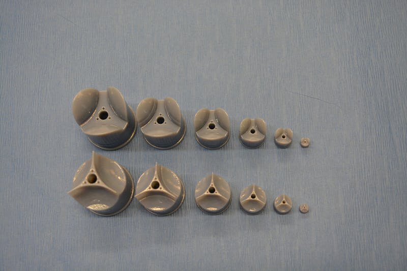
This is the ‘candy floss’ technique at work,

Rotary Jet-Spinning (RJS) works likes a cotton candy machine. A liquid polymer solution is loaded into a reservoir and pushed out through a tiny opening by centrifugal force as the device spins. As the solution leaves the reservoir, the solvent evaporates and the polymers solidify and elongate into small, thin fibers. Courtesy: Harvard University

Rotary Jet-Spinning (RJS) works likes a cotton candy machine. A liquid polymer solution is loaded into a reservoir and pushed out through a tiny opening by centrifugal force as the device spins. As the solution leaves the reservoir, the solvent evaporates and the polymers solidify and elongate into small, thin fibers. Courtesy: Harvard University

Here’s a link to and a citation for the paper,

Production of Synthetic, Para-Aramid and Biopolymer Nanofibers by Immersion Rotary Jet-Spinning by Grant M. Gonzalez, Luke A. MacQueen, Johan U. Lind, Stacey A. Fitzgibbons, Christophe O. Chantre, Isabelle Huggler, Holly M. Golecki, Josue A. Goss, Kevin Kit Parker. Macromolecular Materials and Engineering DOI: 10.1002/mame.201600365 Version of Record online: 7 OCT 2016

© 2016 WILEY-VCH Verlag GmbH & Co. KGaA, Weinheim

This paper is behind a paywall.

Namib beetles, cacti, and pitcher plants teach scientists at Harvard University (US)

In this latest work from Harvard University’s Wyss Institute for Biologically Inspired Engineering, scientists have looked at three desert dwellers for survival strategies in water-poor areas. From a Feb. 25, 2015 news item on Nanowerk,

Organisms such as cacti and desert beetles can survive in arid environments because they’ve evolved mechanisms to collect water from thin air. The Namib desert beetle, for example, collects water droplets on the bumps of its shell while V-shaped cactus spines guide droplets to the plant’s body.

As the planet grows drier, researchers are looking to nature for more effective ways to pull water from air. Now, a team of researchers from the Harvard John A. Paulson School of Engineering and Applied Sciences (SEAS) and the Wyss Institute for Biologically Inspired Engineering at Harvard University have drawn inspiration from these organisms to develop a better way to promote and transport condensed water droplets.

A Feb. 24, 2016 Harvard University press release by Leah Burrows (also on EurekAlert), which originated the news item, expands on the theme,

“Everybody is excited about bioinspired materials research,” said Joanna Aizenberg, the Amy Smith Berylson Professor of Materials Science at SEAS and core faculty member of the Wyss Institute. “However, so far, we tend to mimic one inspirational natural system at a time. Our research shows that a complex bio-inspired approach, in which we marry multiple biological species to come up with non-trivial designs for highly efficient materials with unprecedented properties, is a new, promising direction in biomimetics.”

The new system, described in Nature, is inspired by the bumpy shell of desert beetles, the asymmetric structure of cactus spines and slippery surfaces of pitcher plants. The material harnesses the power of these natural systems, plus Slippery Liquid-Infused Porous Surfaces technology (SLIPS) developed in Aizenberg’s lab, to collect and direct the flow of condensed water droplets.

This approach is promising not only for harvesting water but also for industrial heat exchangers.

“Thermal power plants, for example, rely on condensers to quickly convert steam to liquid water,” said Philseok Kim, co-author of the paper and co-founder and vice president of technology at SEAS spin-off SLIPS Technologies, Inc. “This design could help speed up that process and even allow for operation at a higher temperature, significantly improving the overall energy efficiency.”

The major challenges in harvesting atmospheric water are controlling the size of the droplets, speed in which they form and the direction in which they flow.

For years, researchers focused on the hybrid chemistry of the beetle’s bumps — a hydrophilic top with hydrophobic surroundings — to explain how the beetle attracted water. However, Aizenberg and her team took inspiration from a different possibility – that convex bumps themselves also might be able to harvest water.

“We experimentally found that the geometry of bumps alone could facilitate condensation,” said Kyoo-Chul Park, a postdoctoral researcher and the first author of the paper. “By optimizing that bump shape through detailed theoretical modeling and combining it with the asymmetry of cactus spines and the nearly friction-free coatings of pitcher plants, we were able to design a material that can collect and transport a greater volume of water in a short time compared to other surfaces.”

“Without one of those parameters, the whole system would not work synergistically to promote both the growth and accelerated directional transport of even small, fast condensing droplets,” said Park.

“This research is an exciting first step towards developing a passive system that can efficiently collect water and guide it to a reservoir,” said Kim.

Here’s a link to and a citation for the paper,

Condensation on slippery asymmetric bumps by Kyoo-Chul Park, Philseok Kim, Alison Grinthal, Neil He, David Fox, James C. Weaver, & Joanna Aizenberg. Nature (2016) doi:10.1038/nature16956 Published online 24 February 2016

This paper is behind a paywall.

I have featured the Namib beetle and its water harvesting capabilities most recently in a July 29, 2014 posting and the most recent story I have about SLIPS is in an Oct. 14, 2014 posting.