Tag Archives: John F. Zimmerman

Biohybrid fish made from human cardiac cells could lead to artificial hearts

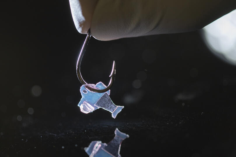
Biohybrid fish on a hook (Photo credit to Michael Rosnach, Keel Yong Lee, Sung-Jin Park, Kevin Kit Parker)

A February 10, 2022 news item on ScienceDaily announces research on a biohybrid fish,

Harvard University researchers, in collaboration with colleagues from Emory University, have developed the first fully autonomous biohybrid fish from human stem-cell derived cardiac muscle cells. The artificial fish swims by recreating the muscle contractions of a pumping heart, bringing researchers one step closer to developing a more complex artificial muscular pump and providing a platform to study heart disease like arrhythmia.

A February 10, 2022 Harvard University John A. Paulson School of Engineering and Applied Sciences news release (also on EurekAlert) by Leah Burrows explains how this research could lead to an artificial heart (Note: Links have been removed),

“Our ultimate goal is to build an artificial heart to replace a malformed heart in a child,” said Kit Parker, the Tarr Family Professor of Bioengineering and Applied Physics at the Harvard John A. Paulson School of Engineering and Applied Sciences (SEAS) and senior author of the paper.  “Most of the work in building heart tissue or hearts, including some work we have done, is focused on replicating the anatomical features or replicating the simple beating of the heart in the engineered tissues. But here, we are drawing design inspiration from the biophysics of the heart, which is harder to do. Now, rather than using heart imaging as a blueprint, we are identifying the key biophysical principles that make the heart work, using them as design criteria, and replicating them in a system, a living, swimming fish, where it is much easier to see if we are successful.”

The research is published in Science

The biohybrid fish developed by the team builds off previous research from Parker’s Disease Biophysics Group. In 2012, the lab used cardiac muscle cells from rats to build a jellyfish-like biohybrid pump and in 2016 the researchers developed a swimming, artificial stingray also from rat heart muscle cells.

In this research, the team built the first autonomous biohybrid device made from human stem-cell derived cardiomyocytes. This device was inspired by the shape and swimming motion of a zebrafish. Unlike previous devices, the biohybrid zebrafish has two layers of muscle cells, one on each side of the tail fin. When one side contracts, the other stretches. That stretch triggers the opening of a mechanosensitive protein channel, which causes a contraction, which triggers a stretch and so on and so forth, leading to a closed loop system that can propel the fish for more than 100 days. 

“By leveraging cardiac mechano-electrical signaling between two layers of muscle, we recreated the cycle where each contraction results automatically as a response to the stretching on the opposite side,” said Keel Yong Lee, a postdoctoral fellow at SEAS and co-first author of the study. “The results highlight the role of feedback mechanisms in muscular pumps such as the heart.”

The researchers also engineered an autonomous pacing node, like a pacemaker, which controls the frequency and rhythm of these spontaneous contractions. Together, the two layers of muscle and the autonomous pacing node enabled the generation of continuous, spontaneous, and coordinated, back-and-forth fin movements.

“Because of the two internal pacing mechanisms, our fish can live longer, move faster and swim more efficiently than previous work,” said Sung-Jin Park, a former postdoctoral fellow in the Disease Biophysics Group at SEAS and co-first author of the study. “This new research provides a model to investigate mechano-electrical signaling as a therapeutic target of heart rhythm management and for understanding pathophysiology in sinoatrial node dysfunctions and cardiac arrhythmia.”

Park is currently an Assistant Professor at the Coulter Department of Biomedical Engineering at Georgia Institute of Technology and Emory University School of Medicine.

Unlike a fish in your refrigerator, this biohybrid fish improves with age. Its muscle contraction amplitude, maximum swimming speed, and muscle coordination all increased for the first month as the cardiomyocyte cells matured.  Eventually, the biohybrid fish reached speeds and swimming efficacy similar to zebrafish in the wild. 

Next, the team aims to build even more complex biohybrid devices from human heart cells. 

“I could build a model heart out of Play-Doh, it doesn’t mean I can build a heart,” said Parker. “You can grow some random tumor cells in a dish until they curdle into a throbbing lump and call it a cardiac organoid. Neither of those efforts is going to, by design, recapitulate the physics of a system that beats over a billion times during your lifetime while simultaneously rebuilding its cells on the fly. That is the challenge. That is where we go to work.”

The research was co-authored by David G. Matthews, Sean L. Kim, Carlos Antonio Marquez, John F. Zimmerman, Herdeline Ann M. Ardona, Andre G. Kleber and George V. Lauder. 

It was supported in part by National Institutes of Health National Center for Advancing Translational Sciences grant UH3TR000522, and National Science Foundation Materials Research Science and Engineering Center grant DMR-142057.

Before giving you a link and a citation for the paper, here’s a little more information about the work from a February 10, 2022 American Association for the Advancement of Science (AAAS) news release on EurekAlert announcing publication of the paper in their journal Science, Note: A link has been removed,

An autonomously swimming biohybrid fish, designed with a focus on two key regulatory features of the human heart, has revealed the importance of feedback mechanisms in muscular pumps (such as the heart). The findings could one day help inform the development of an artificial heart made from living muscle cells. Biohybrid systems – devices containing both biological and artificial components – are an effective way to investigate the physiological control mechanisms in biological organisms and to discover bio-inspired robotic solutions to a host of pressing concerns, including those related to human health. When it comes to natural fluid transport pumps, like those that circulate blood, the performance of biohybrid systems has been lacking, however.  Here, researchers considered whether two functional regulatory features of the heart — mechanoelectrical signaling and automaticity — could be transferred to a synthetic analog of another fluid transport system: a swimming fish. Lee et al. developed an autonomously swimming fish constructed from a bilayer of human cardiac cells; the muscular bilayer was integrated using tissue engineering techniques. Lee and team were able to control muscle contractions in the biohybrid fish using external optogenetic stimulation, allowing the fish analog to swim. In tests, the biohybrid fish outperformed the locomotory speed of previous biohybrid muscular systems, the authors say. It maintained spontaneous activity for 108 days. By contrast, say the authors, biohybrid fish equipped with single-layered muscle showed deteriorating activity within the first month. The data in this study demonstrate the potential of muscular bilayer systems and mechanoelectrical signaling as a means to promote maturation of in vitro muscle tissues, write Lee and colleagues. “Taken together,” the authors conclude, “the technology described here may represent foundational work toward the goal of creating autonomous systems capable of homeostatic regulation and adaptive behavioral control.”

For reporters interested in trends, this work builds upon previous work published in a July 2016 study in Science, in which Sung-jin Park et al. used cardiac cells from rats to develop a self-propelling ray fish analog.

Here’s a link to and a citation for the paper,

An autonomously swimming biohybrid fish designed with human cardiac biophysics by Keel Yong Lee, Sung-Jin Park, David G. Matthews. Sean L. Kim, Carlos Antonio Marquez, John F. Zimmerman, Herdeline Ann M. Ardoña, Andre G. Kleber, George V. Lauder and Kevin Kit Parker. Science • 10 Feb 2022 • Vol 375, Issue 6581 • pp. 639-647 • DOI: 10.1126/science.abh0474

This paper is behind a paywall.

Suit up with nanofiber for protection against explosions and high temperatures

Where explosions are concerned you might expect to see some army research and you would be right. A June 29, 2020 news item on ScienceDaily breaks the news,

Since World War I, the vast majority of American combat casualties has come not from gunshot wounds but from explosions. Today, most soldiers wear a heavy, bullet-proof vest to protect their torso but much of their body remains exposed to the indiscriminate aim of explosive fragments and shrapnel.

Designing equipment to protect extremities against the extreme temperatures and deadly projectiles that accompany an explosion has been difficult because of a fundamental property of materials. Materials that are strong enough to protect against ballistic threats can’t protect against extreme temperatures and vice versa. As a result, much of today’s protective equipment is composed of multiple layers of different materials, leading to bulky, heavy gear that, if worn on the arms and legs, would severely limit a soldier’s mobility.

Now, Harvard University researchers, in collaboration with the U.S. Army Combat Capabilities Development Command Soldier Center (CCDC SC) and West Point, have developed a lightweight, multifunctional nanofiber material that can protect wearers from both extreme temperatures and ballistic threats.

A June 29, 2020 Harvard University news release (also on EurekAlert) by Leah Burrows, which originated the news item, expands on the theme,

“When I was in combat in Afghanistan, I saw firsthand how body armor could save lives,” said senior author Kit Parker, the Tarr Family Professor of Bioengineering and Applied Physics at the Harvard John A. Paulson School of Engineering and Applied Sciences (SEAS) and a lieutenant colonel in the United States Army Reserve. “I also saw how heavy body armor could limit mobility. As soldiers on the battlefield, the three primary tasks are to move, shoot, and communicate. If you limit one of those, you decrease survivability and you endanger mission success.”

“Our goal was to design a multifunctional material that could protect someone working in an extreme environment, such as an astronaut, firefighter or soldier, from the many different threats they face,” said Grant M. Gonzalez, a postdoctoral fellow at SEAS and first author of the paper.

In order to achieve this practical goal, the researchers needed to explore the tradeoff between mechanical protection and thermal insulation, properties rooted in a material’s molecular structure and orientation.

Materials with strong mechanical protection, such as metals and ceramics, have a highly ordered and aligned molecular structure. This structure allows them to withstand and distribute the energy of a direct blow. Insulating materials, on the other hand, have a much less ordered structure, which prevents the transmission of heat through the material.

Kevlar and Twaron are commercial products used extensively in protective equipment and can provide either ballistic or thermal protection, depending on how they are manufactured. Woven Kevlar, for example, has a highly aligned crystalline structure and is used in protective bulletproof vests. Porous Kevlar aerogels, on the other hand, have been shown to have high thermal insulation.

“Our idea was to use this Kevlar polymer to combine the woven, ordered structure of fibers with the porosity of aerogels to make long, continuous fibers with porous spacing in between,” said Gonzalez. “In this system, the long fibers could resist a mechanical impact while the pores would limit heat diffusion.”

The research team used immersion Rotary Jet-Spinning (iRJS), a technique developed by Parker’s Disease Biophysics Group, to manufacture the fibers. In this technique, a liquid polymer solution is loaded into a reservoir and pushed out through a tiny opening by centrifugal force as the device spins. When the polymer solution shoots out of the reservoir, it first passes through an area of open air, where the polymers elongate and the chains align. Then the solution hits a liquid bath that removes the solvent and precipitates the polymers to form solid fibers. Since the bath is also spinning — like water in a salad spinner — the nanofibers follow the stream of the vortex and wrap around a rotating collector at the base of the device.

By tuning the viscosity of the liquid polymer solution, the researchers were able to spin long, aligned nanofibers into porous sheets — providing enough order to protect against projectiles but enough disorder to protect against heat. In about 10 minutes, the team could spin sheets about 10 by 30 centimeters in size.

To test the sheets, the Harvard team turned to their collaborators to perform ballistic tests. Researchers at CCDC SC in Natick, Massachusetts simulated shrapnel impact by shooting large, BB-like projectiles at the sample. The team performed tests by sandwiching the nanofiber sheets between sheets of woven Twaron. They observed little difference in protection between a stack of all woven Twaron sheets and a combined stack of woven Twaron and spun nanofibers.

“The capabilities of the CCDC SC allow us to quantify the successes of our fibers from the perspective of protective equipment for warfighters, specifically,” said Gonzalez.

“Academic collaborations, especially those with distinguished local universities such as Harvard, provide CCDC SC the opportunity to leverage cutting-edge expertise and facilities to augment our own R&D capabilities,” said Kathleen Swana, a researcher at CCDC SC and one of the paper’s authors. “CCDC SC, in return, provides valuable scientific and soldier-centric expertise and testing capabilities to help drive the research forward.”

In testing for thermal protection, the researchers found that the nanofibers provided 20 times the heat insulation capability of commercial Twaron and Kevlar.

“While there are improvements that could be made, we have pushed the boundaries of what’s possible and started moving the field towards this kind of multifunctional material,” said Gonzalez.

“We’ve shown that you can develop highly protective textiles for people that work in harm’s way,” said Parker. “Our challenge now is to evolve the scientific advances to innovative products for my brothers and sisters in arms.”

Harvard’s Office of Technology Development has filed a patent application for the technology and is actively seeking commercialization opportunities.

Here’s a link to and a citation for the paper,

para-Aramid Fiber Sheets for Simultaneous Mechanical and Thermal Protection in Extreme Environments by Grant M. Gonzalez, Janet Ward, John Song, Kathleen Swana, Stephen A. Fossey, Jesse L. Palmer, Felita W. Zhang, Veronica M. Lucian, Luca Cera, John F. Zimmerman, F. John Burpo, Kevin Kit Parker. Matter DOI: https://doi.org/10.1016/j.matt.2020.06.001 Published:June 29, 2020

This paper is behind a paywall.

While this is the first time I’ve featured clothing/armour that’s protective against explosions I have on at least two occasions featured bulletproof clothing in a Canadian context. A November 4, 2013 posting had a story about a Toronto-based tailoring establishment, Garrison Bespoke, that was going to publicly test a bulletproof business suit. Should you be interested, it is possible to order the suit here. There’s also a February 11, 2020 posting announcing research into “Comfortable, bulletproof clothing for Canada’s Department of National Defence.”

Phagocytosis for a bioelectronic future

The process by which a cell engulfs matter is known as phagocytosis. One of the best known examples of failed phagocytosis is that of asbestos fibres in the lungs where lung cells have attempted to engulf a fibre that’s just too big and ends up piercing the cell. When enough of the cells are pierced, the person is diagnosed with mesothelioma.

This particular example of phagocytosis is a happier one according to a Dec. 16, 2016 article by Meghan Rosen for ScienceNews,

Human cells can snack on silicon.

Cells grown in the lab devour nano-sized wires of silicon through an engulfing process known as phagocytosis, scientists report December 16 in Science Advances.

Silicon-infused cells could merge electronics with biology, says John Zimmerman, a biophysicist now at Harvard University. “It’s still very early days,” he adds, but “the idea is to get traditional electronic devices working inside of cells.” Such hybrid devices could one day help control cellular behavior, or even replace electronics used for deep brain stimulation, he says.

Scientists have been trying to load electronic parts inside cells for years. One way is to zap holes in cells with electricity, which lets big stuff, like silicon nanowires linked to bulky materials, slip in. Zimmerman, then at the University of Chicago, and colleagues were looking for a simpler technique, something that would let tiny nanowires in easily and could potentially allow them to travel through a person’s bloodstream — like a drug.

A Dec. 22, 2016 University of Chicago news release by Matt Wood provides more detail,

“You can treat it as a non-genetic, synthetic biology platform,” said Bozhi Tian, PhD, assistant professor of chemistry and senior author of the new study. “Traditionally in biology we use genetic engineering and modify genetic parts. Now we can use silicon parts, and silicon can be internalized. You can target those silicon parts to specific parts of the cell and modulate that behavior with light.”

In the new study, Tian and his team show how cells consume or internalize the nanowires through phagocytosis, the same process they use to engulf and ingest nutrients and other particles in their environment. The nanowires are simply added to cell media, the liquid solution the cells live in, the same way you might administer a drug, and the cells take it from there. Eventually, the goal would be to inject them into the bloodstream or package them into a pill.

Once inside, the nanowires can interact directly with individual parts of the cell, organelles like the mitochondria, nucleus and cytoskeletal filaments. Researchers can then stimulate the nanowires with light to see how individual components of the cell respond, or even change the behavior of the cell. They can last up to two weeks inside the cell before biodegrading.

Seeing how individual parts of a cell respond to stimulation could give researchers insight into how medical treatments that use electrical stimulation work at a more detailed level. For instance, deep brain stimulation helps treat tremors from movement disorders like Parkinson’s disease by sending electrical signals to areas of the brain. Doctors know it works at the level of tissues and brain structures, but seeing how individual components of nerve cells react to these signals could help fine tune and improve the treatment.

The experiments in the study used umbilical vascular endothelial cells, which make up blood vessel linings in the umbilical cord. These cells readily took up the nanowires, but others, like cardiac muscle cells, did not. Knowing that some cells consume the wires and some don’t could also prove useful in experimental settings and give researchers more ways to target specific cell types.

Tian and his team manufactures the nanowires in their lab with a chemical vapor deposition system that grows the silicon structures to different specifications. They can adjust size, shape, and electrical properties as needed, or even add defects on purpose for testing. They can also make wires with porous surfaces that could deliver drugs or genetic material to the cells. The process gives them a variety of ways to manipulate the properties of the nanowires for research.

Here’s a link to and a citation for the paper,

Cellular uptake and dynamics of unlabeled freestanding silicon nanowires by John F. Zimmerman, Ramya Parameswaran, Graeme Murray, Yucai Wang, Michael Burke, and Bozhi Tian. Science Advances  16 Dec 2016: Vol. 2, no. 12, e1601039 DOI: 10.1126/sciadv.1601039

This paper appears to be open access.