Tag Archives: Karlsruhe Institute of Technology (KIT)

Seeing a single nanoparticle catalyst at work

https://www.desy.de/e409/e116959/e119238/media/9833/alloy_kat_np_close-up.jpg
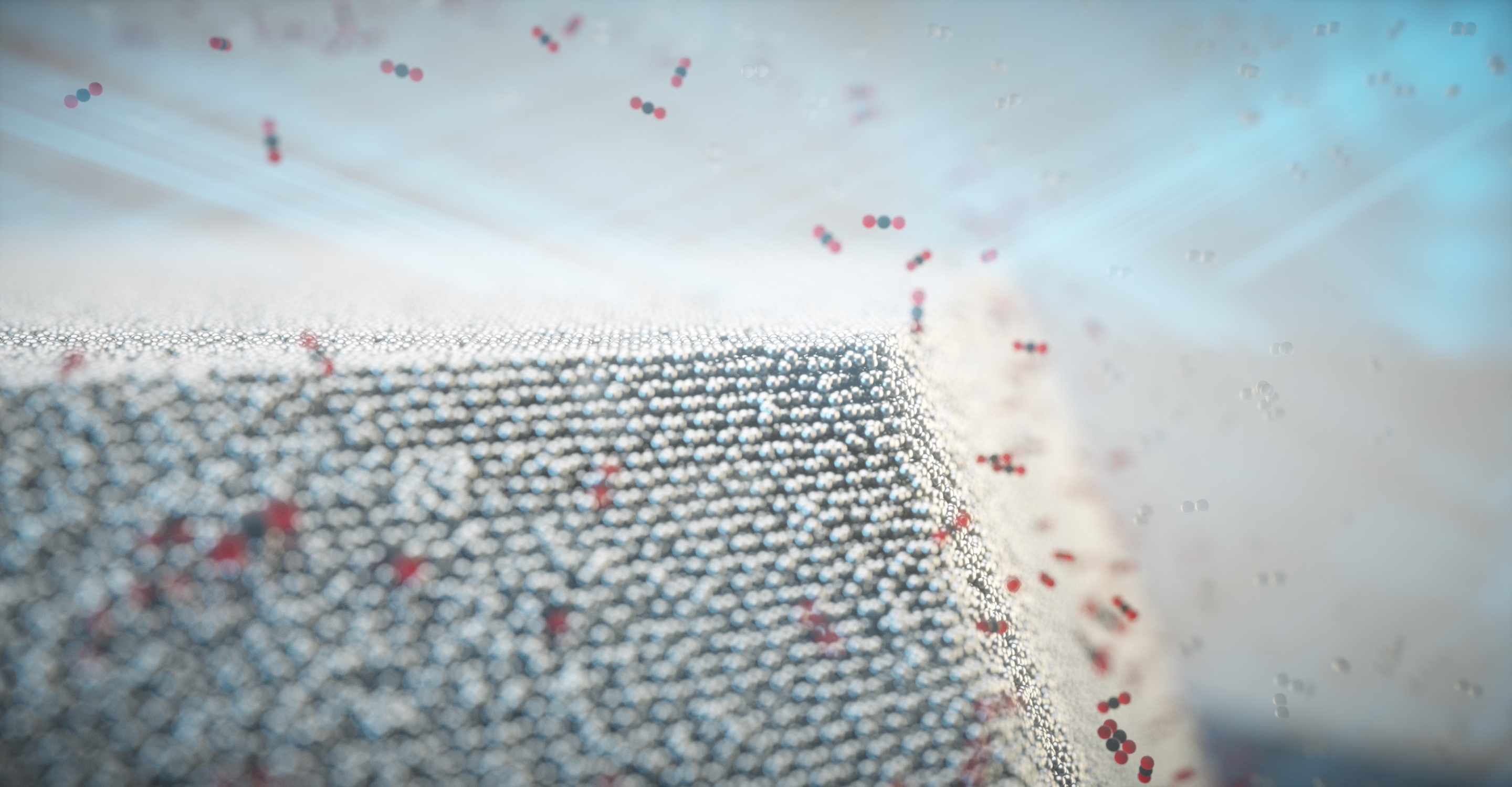
Carbon monoxide oxidises to carbon dioxide on the surface of the nanoparticle. Credit: Science Communication Lab for DESY

An October 1, 2021 news item on ScienceDaily announces research enabling scientists to observe a single nanoparticle at work,

A DESY-led research team has been using high-intensity X-rays to observe a single catalyst nanoparticle at work. The experiment has revealed for the first time how the chemical composition of the surface of an individual nanoparticle changes under reaction conditions, making it more active. The team led by DESY’s Andreas Stierle is presenting its findings in the journal Science Advances. This study marks an important step towards a better understanding of real, industrial catalytic materials.

An October 1, 2021 Deutsches Elektronen-Synchrotron (DESY) press release (also on EurekAlert), which originated the news item, explains why this research is important and provides more technical details,

Catalysts are materials that promote chemical reactions without being consumed themselves. Today, catalysts are used in numerous industrial processes, from fertiliser production to manufacturing plastics. Because of this, catalysts are of huge economic importance. A very well-known example is the catalytic converter installed in the exhaust systems of cars. These contain precious metals such as platinum, rhodium and palladium, which allow highly toxic carbon monoxide (CO) to be converted into carbon dioxide (CO2) and reduce the amount of harmful nitrogen oxides (NOx).

“In spite of their widespread use and great importance, we are still ignorant of many important details of just how the various catalysts work,” explains Stierle, head of the DESY NanoLab. “That’s why we have long wanted to study real catalysts while in operation.” This is not easy, because in order to make the active surface as large as possible, catalysts are typically used in the form of tiny nanoparticles, and the changes that affect their activity occur on their surface.

Surface strain relates to chemical composition

In the framework of the EU project Nanoscience Foundries and Fine Analysis (NFFA), the team from DESY NanoLab has developed a technique for labelling individual nanoparticles and thereby identifying them in a sample. “For the study, we grew nanoparticles of a platinum-rhodium alloy on a substrate in the lab and labelled one specific particle,” says co-author Thomas Keller from DESY NanoLab and in charge of the project at DESY. “The diameter of the labelled particle is around 100 nanometres, and it is similar to the particles used in a car’s catalytic converter.” A nanometre is a millionth of a millimetre.

Using X-rays from the European Synchrotron Radiation Facility ESRF in Grenoble, France, the team was not only able to create a detailed image of the nanoparticle; it also measured the mechanical strain within its surface. “The surface strain is related to the surface composition, in particular the ratio of platinum to rhodium atoms,” explains co-author Philipp Plessow from the Karlsruhe Institute of Technology (KIT), whose group computed strain as a function of surface composition. By comparing the observed and computed facet-dependent strain, conclusions can be drawn concerning the chemical composition at the particle surface. The different surfaces of a nanoparticle are called facets, just like the facets of a cut gemstone.

When the nanoparticle is grown, its surface consists mainly of platinum atoms, as this configuration is energetically favoured. However, the scientists studied the shape of the particle and its surface strain under different conditions, including the operating conditions of an automotive catalytic converter. To do this, they heated the particle to around 430 degrees Celsius and allowed carbon monoxide and oxygen molecules to pass over it. “Under these reaction conditions, the rhodium inside the particle becomes mobile and migrates to the surface because it interacts more strongly with oxygen than the platinum,” explains Plessow. This is also predicted by theory.

“As a result, the surface strain and the shape of the particle change,” reports co-author Ivan Vartaniants, from DESY, whose team converted the X-ray diffraction data into three-dimensional spatial images. “A facet-dependent rhodium enrichment takes place, whereby additional corners and edges are formed.” The chemical composition of the surface, and the shape and size of the particles have a significant effect on their function and efficiency. However, scientists are only just beginning to understand exactly how these are connected and how to control the structure and composition of the nanoparticles. The X-rays allow researchers to detect changes of as little as 0.1 in a thousand in the strain, which in this experiment corresponds to a precision of about 0.0003 nanometres (0.3 picometres).

Crucial step towards analysing industrial catalyst materials

“We can now, for the first time, observe the details of the structural changes in such catalyst nanoparticles while in operation,” says Stierle, Lead Scientist at DESY and professor for nanoscience at the University of Hamburg. “This is a major step forward and is helping us to understand an entire class of reactions that make use of alloy nanoparticles.” Scientists at KIT and DESY now want to explore this systematically at the new Collaborative Research Centre 1441, funded by the German Research Foundation (DFG) and entitled “Tracking the Active Sites in Heterogeneous Catalysis for Emission Control (TrackAct)”.

“Our investigation is an important step towards analysing industrial catalytic materials,” Stierle points out. Until now, scientists have had to grow model systems in the laboratory in order to conduct such investigations. “In this study, we have gone to the limit of what can be done. With DESY’s planned X-ray microscope PETRA IV, we will be able to look at ten times smaller individual particles in real catalysts, and under reaction conditions.”
 
DESY is one of the world’s leading particle accelerator centres and investigates the structure and function of matter – from the interaction of tiny elementary particles and the behaviour of novel nanomaterials and vital biomolecules to the great mysteries of the universe. The particle accelerators and detectors that DESY develops and builds at its locations in Hamburg and Zeuthen are unique research tools. They generate the most intense X-ray radiation in the world, accelerate particles to record energies and open up new windows onto the universe. DESY is a member of the Helmholtz Association, Germany’s largest scientific association, and receives its funding from the German Federal Ministry of Education and Research (BMBF) (90 per cent) and the German federal states of Hamburg and Brandenburg (10 per cent).

Here’s a link to and a citation for the paper,

Single alloy nanoparticle x-ray imaging during a catalytic reaction by Young Yong Kim, Thomas F. Keller, Tiago J. Goncalves, Manuel Abuin, Henning Runge, Luca Gelisio, Jerome Carnis, Vedran Vonk, Philipp N. Plessow, Ivan A. Vartaniants, Andreas Stierle. Science Advances • 1 Oct 2021 • Vol 7, Issue 40 • DOI: 10.1126/sciadv.abh0757

This paper is open access.
Carbon nanotube optics and the quantum

A US-France-Germany collaboration has led to some intriguing work with carbon nanotubes. From a June 18, 2018 news item on ScienceDaily,

Researchers at Los Alamos and partners in France and Germany are exploring the enhanced potential of carbon nanotubes as single-photon emitters for quantum information processing. Their analysis of progress in the field is published in this week’s edition of the journal Nature Materials.

“We are particularly interested in advances in nanotube integration into photonic cavities for manipulating and optimizing light-emission properties,” said Stephen Doorn, one of the authors, and a scientist with the Los Alamos National Laboratory site of the Center for Integrated Nanotechnologies (CINT). “In addition, nanotubes integrated into electroluminescent devices can provide greater control over timing of light emission and they can be feasibly integrated into photonic structures. We are highlighting the development and photophysical probing of carbon nanotube defect states as routes to room-temperature single photon emitters at telecom wavelengths.”

A June 18, 2018 Los Alamos National Laboratory (LANL) news release (also on EurekAlert), which originated the news item, expands on the theme,

The team’s overview was produced in collaboration with colleagues in Paris (Christophe Voisin [Ecole Normale Supérieure de Paris (ENS)]) who are advancing the integration of nanotubes into photonic cavities for modifying their emission rates, and at Karlsruhe (Ralph Krupke [Karlsruhe Institute of Technology (KIT]) where they are integrating nanotube-based electroluminescent devices with photonic waveguide structures. The Los Alamos focus is the analysis of nanotube defects for pushing quantum emission to room temperature and telecom wavelengths, he said.

As the paper notes, “With the advent of high-speed information networks, light has become the main worldwide information carrier. . . . Single-photon sources are a key building block for a variety of technologies, in secure quantum communications metrology or quantum computing schemes.”

The use of single-walled carbon nanotubes in this area has been a focus for the Los Alamos CINT team, where they developed the ability to chemically modify the nanotube structure to create deliberate defects, localizing excitons and controlling their release. Next steps, Doorn notes, involve integration of the nanotubes into photonic resonators, to provide increased source brightness and to generate indistinguishable photons. “We need to create single photons that are indistinguishable from one another, and that relies on our ability to functionalize tubes that are well-suited for device integration and to minimize environmental interactions with the defect sites,” he said.

“In addition to defining the state of the art, we wanted to highlight where the challenges are for future progress and lay out some of what may be the most promising future directions for moving forward in this area. Ultimately, we hope to draw more researchers into this field,” Doorn said.

Here’s a link to and a citation for the paper,

Carbon nanotubes as emerging quantum-light sources by X. He, H. Htoon, S. K. Doorn, W. H. P. Pernice, F. Pyatkov, R. Krupke, A. Jeantet, Y. Chassagneux & C. Voisin. Nature Materials (2018) DOI: https://doi.org/10.1038/s41563-018-0109-2 Published online June 18, 2018

This paper is behind a paywall.

Nanomedicine and an enhanced uptake of nanoparticles

It’s nice to know that a step forward has been taken with regard to improving uptake in  nanoparticle-based drug delivery (see my April 27, 2016 posting titled: How many nanoparticle-based drugs does it take to kill a cancer tumour? More than 1% for insight into the difficulties of f nanoparticle-based drug delivery systems).

Here’s the latest move forward in a March 8, 2017 news item on Nanowerk (Note: A link has been removed),

Nanotechnology has become a growing part of medical research in recent years, with scientists feverishly working to see if tiny particles could revolutionize the world of drug delivery.

But many questions remain about how to effectively transport those particles and associated drugs to cells.
In an article published today in Scientific Reports (“Enhanced cellular uptake of size-separated lipophilic silicon nanoparticles”), FSU Associate Professor of Biological Science Steven Lenhert takes a step forward in the understanding of nanoparticles and how they can best be used to deliver drugs.

After conducting a series of experiments, Lenhert and his colleagues found that it may be possible to boost the efficacy of medicine entering target cells via a nanoparticle.

A March 8, 2017 Florida State University news release by Kathleen Haughney, which originated the news item, provides more detail about the research (an international collaboration involving the University of Toronto [Canada] and the Karlsruhe Institute of Technology [Germany]),

“We can enhance how cells take them up and make more drugs more potent,” Lenhert said.

Initially, Lenhert and his colleagues from the University of Toronto and the Karlsruhe Institute of Technology  wanted to see what happened when they encapsulated silicon nanoparticles in liposomes — or small spherical sacs of molecules — and delivered them to HeLa cells, a standard cancer cell model.

The initial goal was to test the toxicity of silicon-based nanoparticles and get a better understanding of its biological activity.

Silicon is a non-toxic substance and has well-known optical properties that allow their nanostructures to appear fluorescent under an infrared camera, where tissue would be nearly transparent. Scientists believe it has enormous potential as a delivery agent for drugs as well as in medical imaging.

But there are still questions about how silicon behaves at such a small size.

“Nanoparticles change properties as they get smaller, so scientists want to understand the biological activity,” Lenhert said. “For example, how does shape and size affect toxicity?”

Scientists found that 10 out of 18 types of the particles, ranging from 1.5 nanometers to 6 nanometers, were significantly more toxic than crude mixtures of the material.

At first, scientists believed this could be a setback, but they then discovered the reason for the toxicity levels. The more toxic fragments also had enhanced cellular uptake.

That information is more valuable long term, Lenhert said, because it means they could potentially alter nanoparticles to enhance the potency of a given therapeutic.

The work also paves the way for researchers to screen libraries of nanoparticles to see how cells react.

“This is an essential step toward the discovery of novel nanotechnology based therapeutics,” Lenhert said. “There’s big potential here for new therapeutics, but we need to be able to test everything first.”

Here’s a link to and a citation for the paper,

Enhanced cellular uptake of size-separated lipophilic silicon nanoparticles by Aubrey E. Kusi-Appiah, Melanie L. Mastronardi, Chenxi Qian, Kenneth K. Chen, Lida Ghazanfari, Plengchart Prommapan, Christian Kübel, Geoffrey A. Ozin, & Steven Lenhert. Scientific Reports 7, Article number: 43731 (2017) doi:10.1038/srep43731 Published online: 08 March 2017

This paper is open access.

Medieval chain mail inspires physicists

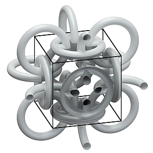
A Feb. 9, 2017 news item on Nanowerk describes new research at the Karlsruhe Institute of Technology (KIT), which takes its inspiration from medieval chain mail,

The Middle Ages certainly were far from being science-friendly: Whoever looked for new findings off the beaten track faced the threat of being burned at the stake. Hence, the contribution of this era to technical progress is deemed to be rather small. Scientists of Karlsruhe Institute of Technology (KIT), however, were inspired by medieval mail armor when producing a new metamaterial with novel properties. They succeeded in reversing the Hall coefficient of a material.

The Hall effect is the occurrence of a transverse electric voltage across an electric conductor passed by current flow, if this conductor is located in a magnetic field. This effect is a basic phenomenon of physics and allows to measure [sic] the strength of magnetic fields. It is the basis of magnetic speed sensors in cars or compasses in smartphones. Apart from measuring magnetic fields, the Hall effect can also be used to characterize metals and semiconductors and in particular to determine charge carrier density of the material. The sign of the measured Hall voltage allows conclusions to be drawn as to whether charge carriers in the semiconductor element carry positive or negative charge.

The ring structure of the metamaterial was inspired by mail armor of medieval knights. (Photo: KIT)

A Feb. ?, 2017 KIT press release (also on EurekAlert), which originated the news item, expands on the theme,

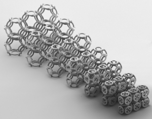
Mathematicians already predicted theoretically that it is possible to reverse the Hall coefficient of a material (such as gold or silicon), i.e. to reverse its sign. This was expected to be achieved by a three-dimensional ring structure resembling medieval mail armor. How-ever, this was considered difficult, as the ring mesh of millionths of a meter in size would have to be composed of three different components.


Die Ringstruktur des Metamaterials wurde von Kettenhemden aus der Ritterzeit inspiriert. (Bild: KIT)
The ring mesh of millionths of a meter in size. (Photo: KIT)

Christian Kern, Muamer Kadic, and Martin Wegener of KIT’s Institute of Applied Physics now found that a single basic material is sufficient, provided that the ring structure chosen follows a certain geometric arrangement. First, they produced polymer scaffolds with a highest-resolution 3D printer. Then, they coated these scaffolds with semiconducting zinc oxide.

The result of the experiment: The scientists can produce meta-materials with a positive coefficient, even though their components have negative coefficients. This sounds a bit like the philosopher’s stone, the formula, by means of which medieval alchemists tried to convert one substance into another. But here, no conversion takes place. “The charge carriers in the metamaterial remain negatively charged electrons,” Christian Kern explains. “Hall measurements only make them appear positively charged, as the structure forces them to take detours.”

Kern admits that this discovery so far is of no practical use. There are sufficient solids with both negative and positive Hall coefficients. But Kern wants to continue research. The next step will be the production of anisotropic structures with a Hall voltage in the direction of the magnetic field. Normally, Hall voltage is directed vertically to current and magnetic fields. Such unconventional materials might be applied in novel sensors for the direct measurement of magnetic field eddies.

The researchers do not seem to have published a paper about this work.

More on the blue tarantula noniridescent photonics

Covered in an Oct. 19, 2016 posting here, some new details have been released about noniridescent photonics and blue tarantulas, this time from the Karlsruhe Institute of Technology (KIT) in a Nov. 17, 2016 (?) press release (also on EurekAlert; h/t Nanowerk Nov. 17, 2016 news item) ,

Colors are produced in a variety of ways. The best known colors are pigments. However, the very bright colors of the blue tarantula or peacock feathers do not result from pigments, but from nanostructures that cause the reflected light waves to overlap. This produces extraordinarily dynamic color effects. Scientists from Karlsruhe Institute of Technology (KIT), in cooperation with international colleagues, have now succeeded in replicating nanostructures that generate the same color irrespective of the viewing angle. DOI: 10.1002/adom.201600599

In contrast to pigments, structural colors are non-toxic, more vibrant and durable. In industrial production, however, they have the drawback of being strongly iridescent, which means that the color perceived depends on the viewing angle. An example is the rear side of a CD. Hence, such colors cannot be used for all applications. Bright colors of animals, by contrast, are often independent of the angle of view. Feathers of the kingfisher always appear blue, no matter from which angle we look. The reason lies in the nanostructures: While regular structures are iridescent, amorphous or irregular structures always produce the same color. Yet, industry can only produce regular nanostructures in an economically efficient way.

Radwanul Hasan Siddique, researcher at KIT in collaboration with scientists from USA and Belgium has now discovered that the blue tarantula does not exhibit iridescence in spite of periodic structures on its hairs. First, their study revealed that the hairs are multi-layered, flower-like structure. Then, the researchers analyzed its reflection behavior with the help of computer simulations. In parallel, they built models of these structures using nano-3D printers and optimized the models with the help of the simulations. In the end, they produced a flower-like structure that generates the same color over a viewing angle of 160 degrees. This is the largest viewing angle of any synthetic structural color reached so far.


Flower-shaped nanostructures generate the color of the blue tarantula. (Graphics: Bill Hsiung, University of Akron)


The 3D print of the optimized flower structure is only 15 µm in dimension. A human hair is about three times as thick. (Photo: Bill Hsiung, Universtiy of Akron)

Apart from the multi-layered structure and rotational symmetry, it is the hierarchical structure from micro to nano that ensures homogeneous reflection intensity and prevents color changes.

Via the size of the “flower,” the resulting color can be adjusted, which makes this coloring method interesting for industry. “This could be a key first step towards a future where structural colorants replace the toxic pigments currently used in textile, packaging, and cosmetic industries,” says Radwanul Hasan Siddique of KIT’s Institute of Microstructure Technology, who now works at the California Institute of Technology. He considers short-term application in textile industry feasible.


The synthetically generated flower structure inspired by the blue tarantula reflects light in the same color over a viewing angle of 160 degrees. (Graphics: Derek Miller)  

Dr. Hendrik Hölscher thinks that the scalability of nano-3D printing is the biggest challenge on the way towards industrial use. Only few companies in the world are able to produce such prints. In his opinion, however, rapid development in this field will certainly solve this problem in the near future.

Once again, here’s a link to and a citation for the paper,

Tarantula-Inspired Noniridescent Photonics with Long-Range Order by Bor-Kai Hsiung, Radwanul Hasan Siddique, Lijia Jiang, Ying Liu, Yongfeng Lu, Matthew D. Shawkey, and Todd A. Blackledge. Advanced Materials DOI: 10.1002/adom.201600599 Version of Record online: 11 OCT 2016

© 2016 WILEY-VCH Verlag GmbH & Co. KGaA, Weinheim

The paper is behind a paywall. You can see the original Oct. 19, 2016 posting for my comments and some excerpts from the paper.

Oil-absorbing hairy fern leaves lead to ‘nanofur’ for oil spill cleanups

German researchers have developed a biomimetic material branded as ‘nanofur’ for oil spill cleanups, according to an Aug. 23, 2016 news item on Nanowerk (Note: A link has been removed),

Some water ferns can absorb large volumes of oil within a short time, because their leaves are strongly water-repellent and, at the same time, highly oil-absorbing. Researchers of Karlsruhe Institute of Technology, together with colleagues of Bonn University, have found that the oil-binding capacity of the water plant results from the hairy microstructure of its leaves (Bioinspiration & Biomimetics, “Microstructures of superhydrophobic plant leaves – inspiration for efficient oil spill cleanup materials”). It is now used as a model to further develop the new Nanofur material for the environmentally friendly cleanup of oil spills.

An Aug.(?) 23 (?), 2016 Karlsruhe Institute of Technology (KIT) press release on EurekAlert, which originated the news item, explains the interest in improving technology for oil spill cleanups and provides insight into this  innovation,

Damaged pipelines, oil tanker disasters, and accidents on oil drilling and production platforms may result in pollutions [sic] of water with crude or mineral oil. Conventional methods to clean up the oil spill are associated with specific drawbacks. Oil combustion or the use of chemical substances to accelerate oil decomposition cause secondary environmental pollution. Many natural materials to take up the oil, such as sawdust or plant fibers, are hardly effective, because they also absorb large amounts of water. On their search for an environmentally friendly alternative to clean up oil spills, the researchers compared various species of aquatic ferns. “We already knew that the leaves of these plants repel water, but for the first time now, we have studied their capacity to absorb oil,” Claudia Zeiger says. She conducted the project at KIT’s Institute of Microstructure Technology.

Damaged pipelines, oil tanker disasters, and accidents on oil drilling and production platforms may result in pollutions of water with crude or mineral oil. Conventional methods to clean up the oil spill are associated with specific drawbacks. Oil combustion or the use of chemical substances to accelerate oil decomposition cause secondary environmental pollution. Many natural materials to take up the oil, such as sawdust or plant fibers, are hardly effective, because they also absorb large amounts of water. On their search for an environmentally friendly alternative to clean up oil spills, the researchers compared various species of aquatic ferns. “We already knew that the leaves of these plants repel water, but for the first time now, we have studied their capacity to absorb oil,” Claudia Zeiger says. She conducted the project at KIT’s Institute of Microstructure Technology.

Aquatic ferns originally growing in tropical and subtropical regions can now also be found in parts of Europe. As they reproduce strongly, they are often considered weed. However, they have a considerable potential as low-cost, rapid, and environmentally friendly oil absorbers, which is obvious from a short video (see below). ”The plants might be used in lakes to absorb accidental oil spills,” Zeiger says. After less than 30 seconds, the leaves reach maximum absorption and can be skimmed off together with the absorbed oil. The water plant named salvinia has trichomes on the leaf surface – hairy extensions of 0.3 to 2.5 mm in length. Comparison of different salvinia species revealed that leaves with the longest hairs did not absorb the largest amounts of oil. “Oil-absorbing capacity is determined by the shape of the hair ends,” Zeiger emphasizes. The largest quantity of oil was absorbed by leaves of the water fern salvinia molesta, whose hair ends are shaped like an eggbeater.

Based on this new knowledge on the relationship between surface structure of leaves and their oil-absorbing capacity, the researchers improved the ‘Nanofur’ material developed at their institute. This plastic nanofur mimics the water-repellent and oil-absorbing effect of salvinia to separate oil and water. “We study nanostructures and microstructures in nature for potential technical developments,” says Hendrik Hölscher, Head of the Biomimetic Surfaces Group of the Institute of Microstructure Technology of KIT. He points out that different properties of plants made of the same material frequently result from differences of their finest structures.

Here’s a link to and a citation for the paper,

Microstructures of superhydrophobic plant leaves – inspiration for efficient oil spill cleanup materials by Claudia Zeiger, Isabelle C Rodrigues da Silva, Matthias Mail, Maryna N Kavalenka, Wilhelm Barthlott, and Hendrik Hölscher. Bioinspiration & Biomimetics, Volume 11, Number 5 DOI: http://dx.doi.org/10.1088/1748-3190/11/5/056003

Published 16 August 2016, © 2016 IOP Publishing Ltd

This article appears to be open access.

There is also a video demonstration of the material,

Enjoy!

Photovoltaics as rose petals

Where solar cells (photovoltaics) are concerned, mimimicking plants is a longstanding pursuit. The latest  plant material to be mimicked is the rose petal’s surface. From a June 24, 2016 news item on ScienceDaily,

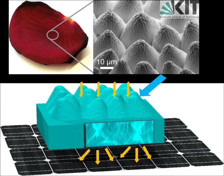
With a surface resembling that of plants, solar cells improve light-harvesting and thus generate more power. Scientists of KIT (Karlsruhe Institute of Technology) reproduced the epidermal cells of rose petals that have particularly good antireflection properties and integrated the transparent replicas into an organic solar cell. This resulted in a relative efficiency gain of twelve percent. …

Caption: Biomimetics: the epidermis of a rose petal is replicated in a transparent layer which is then integrated into the front of a solar cell. Credit Illustration: Guillaume Gomard, KIT

Caption: Biomimetics: the epidermis of a rose petal is replicated in a transparent layer which is then integrated into the front of a solar cell.
Credit Illustration: Guillaume Gomard, KIT

A June 24, 2016 KIT press release on EurekAlert, which originated the news item, expands on the theme,

Photovoltaics works in a similar way as the photosynthesis of plants. Light energy is absorbed and converted into a different form of energy. In this process, it is important to use a possibly large portion of the sun’s light spectrum and to trap the light from various incidence angles as the angle changes with the sun’s position. Plants have this capability as a result of a long evolution process – reason enough for photovoltaics researchers to look closely at nature when developing solar cells with a broad absorption spectrum and a high incidence angle tolerance.

Scientists at the KIT and the ZSW (Center for Solar Energy and Hydrogen Research Baden-Württemberg) now suggest in their article published in the Advanced Optical Materials journal to replicate the outermost tissue of the petals of higher plants, the so-called epidermis, in a transparent layer and integrate that layer into the front of solar cells in order to increase their efficiency.

First, the researchers at the Light Technology Institute (LTI), the Institute of Microstructure Technology (IMT), the Institute of Applied Physics (APH), and the Zoological Institute (ZOO) of KIT as well as their colleagues from the ZSW investigated the optical properties, and above all, the antireflection effect of the epidermal cells of different plant species. These properties are particularly pronounced in rose petals where they provide stronger color contrasts and thus increase the chance of pollination. As the scientists found out under the electron microscope, the epidermis of rose petals consists of a disorganized arrangement of densely packed microstructures, with additional ribs formed by randomly positioned nanostructures.

In order to exactly replicate the structure of these epidermal cells over a larger area, the scientists transferred it to a mold made of polydimethylsiloxane, a silicon-based polymer, pressed the resulting negative structure into optical glue which was finally left to cure under UV light. “This easy and cost-effective method creates microstructures of a depth and density that are hardly achievable with artificial techniques,” says Dr. Guillaume Gomard, Group Leader “Nanopothonics” at KIT’s LTI.

The scientists then integrated the transparent replica of the rose petal epidermis into an organic solar cell. This resulted in power conversion efficiency gains of twelve percent for vertically incident light. At very shallow incidence angles, the efficiency gain was even higher. The scientists attribute this gain primarily to the excellent omnidirectional antireflection properties of the replicated epidermis that is able to reduce surface reflection to a value below five percent, even for a light incidence angle of nearly 80 degrees. In addition, as examinations using a confocal laser microscope showed, every single replicated epidermal cell works as a microlense. The microlense effect extends the optical path within the solar cell, enhances the light-matter-interaction, and increases the probability that the photons will be absorbed.

“Our method is applicable to both other plant species and other PV technologies,” Guillaume Gomard explains. “Since the surfaces of plants have multifunctional properties, it might be possible in the future to apply multiple of these properties in a single step.” The results of this research lead to another basic question: What is the role of disorganization in complex photonic structures? Further studies are now examining this issue with the perspective that the next generation of solar cells might benefit from their results.

Here’s a link to and a citation for the paper,

Flower Power: Exploiting Plants’ Epidermal Structures for Enhanced Light Harvesting in Thin-Film Solar Cells by Ruben Hünig, Adrian Mertens, Moritz Stephan, Alexander Schulz, Benjamin Richter, Michael Hetterich, Michael Powalla, Uli Lemmer, Alexander Colsmann, and Guillaume Gomard. Advanced Optical Materials  Version of Record online: 30 MAY 2016 DOI: 10.1002/adom.201600046

© 2016 WILEY-VCH Verlag GmbH & Co. KGaA, Weinheim

This paper is behind a paywall.

Invisibility cloak effective in fog and milk

It’s an intriguing notion, an invisibility cloak that’s effective in milk and I suspect that I’ve never entirely understood the implications of the research featured in a June 6, 2014 news item on Nanowerk,

Real invisibility cloaks are rather complex and work in certain situations only. The laws of physics prevent an optical invisibility cloak from making objects in air invisible for any directions, colors, and polarizations. If the medium is changed, however, it becomes much easier to hide objects. KIT physicists have now succeeded in manufacturing with relatively simple means and testing an ideal invisibility cloak for diffusive light-scattering media, such as fog or milk. …

A June 6, 2014 Karlsruhe Institute of Technology (KIT) press release, which originated the news item, provides more details,

In diffusive media, light does no longer propagate linearly, but is scattered permanently by the particles in the medium. Examples are fog, clouds, or frosted glass panes that let the light in, but hide the light source. “This property of light-scattering media can be used to hide objects inside,” says Robert Schittny, first author of the study. “The new invisibility cloaks have a rather simple structure.”

In the experiment, Schittny used an extended light source to illuminate a Plexiglas tank of a few centimeters in width from the back. The tank was filled with a white, turbid liquid. Objects inside cast a visible shadow onto the tank wall. Simple metal cylinders or spheres of a few centimeters in diameter were used as test objects. To hide them, they were first coated with a white dispersion paint, such that the light was reflected in a diffusive manner. To pass the light around the object, the researchers applied a thin shell made of the transparent silicon material PDMS, to which a certain concentration of light-scattering melamine microparticles was added. The silicon/melamine shell caused a quicker diffusion than in the environment and, thus, passed the light around the objects. Hence, they did no longer cast a shadow. “Disappearance of the shadow indicates successful cloaking.”

“Ideal optical invisibility cloaks in air have a drawback,” Martin Wegener points out. He conducts research at the KIT Institute of Applied Physics and the KIT Institute of Nanotechnology. “They violate Albert Einstein’s theory of relativity that prescribes an upper limit for the speed of light. “In diffuse media, in which light is scattered several times, however, the effective speed of light is reduced. Here, ideal invisibility cloaks can be realized.”

The researchers have provided this image to illustrate their work,

In a diffusive light-scattering medium, light moves on random paths (see magnifying glass). A normal object (left) casts a shadow, an object with an invisibility cloak (right) does not. (Image: R. Schittny / KIT)

In a diffusive light-scattering medium, light moves on random paths (see magnifying glass). A normal object (left) casts a shadow, an object with an invisibility cloak (right) does not. (Image: R. Schittny / KIT)

I’m not convinced that there’s actually a cloaked object in that image but I appreciate the call to use my imagination.

Here’s a link to and a citation for the paper,

Invisibility cloaking in a diffusive light scattering medium by Robert Schittny, Muamer Kadic, Tiemo Bückmann, and Martin Wegener. Science DOI: 10.1126/science.1254524 Published Online June 5 2014

This article is behind a paywall.

Chemistry of Cyborgs: review of the state of the art by German researchers

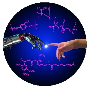
Communication between man and machine – a fascinating area at the interface of chemistry, biomedicine, and engineering. (Figure: KIT/S. Giselbrecht, R. Meyer, B. Rapp)

Communication between man and machine – a fascinating area at the interface of chemistry, biomedicine, and engineering. (Figure: KIT/S. Giselbrecht, R. Meyer, B. Rapp)

German researchers from the Karlsruhe Institute of Technology (KIT), Professor Christof M. Niemeyer and Dr. Stefan Giselbrecht of the Institute for Biological Interfaces 1 (IBG 1) and Dr. Bastian E. Rapp, Institute of Microstructure Technology (IMT) have written a good overview of the current state of cyborgs while pointing out some of the ethical issues associated with this field. From the Jan. 10, 2014 news item on ScienceDaily,

Medical implants, complex interfaces between brain and machine or remotely controlled insects: Recent developments combining machines and organisms have great potentials, but also give rise to major ethical concerns. In a new review, KIT scientists discuss the state of the art of research, opportunities, and risks.

The Jan. ?, 2014 KIT press release (also on EurekAlert with a release date of Jan. 10, 2014), which originated the news item, describes the innovations and the work at KIT in more detail,

They are known from science fiction novels and films – technically modified organisms with extraordinary skills, so-called cyborgs. This name originates from the English term “cybernetic organism”. In fact, cyborgs that combine technical systems with living organisms are already reality. The KIT researchers Professor Christof M. Niemeyer and Dr. Stefan Giselbrecht of the Institute for Biological Interfaces 1 (IBG 1) and Dr. Bastian E. Rapp, Institute of Microstructure Technology (IMT), point out that this especially applies to medical implants.

In recent years, medical implants based on smart materials that automatically react to changing conditions, computer-supported design and fabrication based on magnetic resonance tomography datasets or surface modifications for improved tissue integration allowed major progress to be achieved. For successful tissue integration and the prevention of inflammation reactions, special surface coatings were developed also by the KIT under e.g. the multidisciplinary Helmholtz program “BioInterfaces”.

Progress in microelectronics and semiconductor technology has been the basis of electronic implants controlling, restoring or improving the functions of the human body, such as cardiac pacemakers, retina implants, hearing implants, or implants for deep brain stimulation in pain or Parkinson therapies. Currently, bioelectronic developments are being combined with robotics systems to design highly complex neuroprostheses. Scientists are working on brain-machine interfaces (BMI) for the direct physical contacting of the brain. BMI are used among others to control prostheses and complex movements, such as gripping. Moreover, they are important tools in neurosciences, as they provide insight into the functioning of the brain. Apart from electric signals, substances released by implanted micro- and nanofluidic systems in a spatially or temporarily controlled manner can be used for communication between technical devices and organisms.

BMI are often considered data suppliers. However, they can also be used to feed signals into the brain, which is a highly controversial issue from the ethical point of view. “Implanted BMI that feed signals into nerves, muscles or directly into the brain are already used on a routine basis, e.g. in cardiac pacemakers or implants for deep brain stimulation,” Professor Christof M. Niemeyer, KIT, explains. “But these signals are neither planned to be used nor suited to control the entire organism – brains of most living organisms are far too complex.”

Brains of lower organisms, such as insects, are less complex. As soon as a signal is coupled in, a certain movement program, such as running or flying, is started. So-called biobots, i.e. large insects with implanted electronic and microfluidic control units, are used in a new generation of tools, such as small flying objects for monitoring and rescue missions. In addition, they are applied as model systems in neurosciences in order to understand basic relationships.

Electrically active medical implants that are used for longer terms depend on reliable power supply. Presently, scientists are working on methods to use the patient body’s own thermal, kinetic, electric or chemical energy.

In their review the KIT researchers sum up that developments combining technical devices with organisms have a fascinating potential. They may considerably improve the quality of life of many people in the medical sector in particular. However, ethical and social aspects always have to be taken into account.

After briefly reading the paper, I can say the researchers are most interested in the science and technology aspects but they do have this to say about ethical and social issues in the paper’s conclusion (Note: Links have been removed),

The research and development activities summarized here clearly raise significant social and ethical concerns, in particular, when it comes to the use of BMIs for signal injection into humans, which may lead to modulation or even control of behavior. The ethical issues of this new technology have been discussed in the excellent commentary of Jens Clausen,33 which we highly recommend for further reading. The recently described engineering of a synthetic polymer construct, which is capable of propulsion in water through a collection of adhered rat cardiomyocytes,77 a “medusoid” also described as a “cyborg jellyfish with a rat heart”, brings up an additional ethical aspect. The motivation of the work was to reverse-engineer muscular pumps, and it thus represents fundamental research in tissue engineering for biomedical applications. However, it is also an impressive, early demonstration that autonomous control of technical devices can be achieved through small populations of cells or microtissues. It seems reasonable that future developments along this line will strive, for example, to control complex robots through the use of brain tissue. Given the fact that the robots of today are already capable of autonomously performing complex missions, even in unknown territories,78 this approach might indeed pave the way for yet another entirely new generation of cybernetic organisms.

Here’s a link to and a citation for the English language version of the paper, which is open access (as of Jan. 10, 2014),

The Chemistry of Cyborgs—Interfacing Technical Devices with Organisms by Dr. Stefan Giselbrecht, Dr. Bastian E. Rapp, & Prof.Dr. Christof M. Niemeyer. Angewandte Chemie International Edition Volume 52, Issue 52, pages 13942–13957, December 23, 2013 Article first published online: 29 NOV 2013 DOI: 10.1002/anie.201307495

Copyright © 2013 WILEY-VCH Verlag GmbH & Co. KGaA, Weinheim

For those with German language skills,

Chemie der Cyborgs – zur Verknüpfung technischer Systeme mit Lebewesen.  by Stefan Giselbrecht, Bastian E. Rapp, and Christof M. Niemeyer. Angewandte Chemie. Volume 125, issue 52, pages 14190, December 23, 2013. DOI: 10.1002/ange.201307495

I have written many times about cyborgs and neuroprosthetics including this Aug. 30, 2011 posting titled:  Eye, arm, & leg prostheses, cyborgs, eyeborgs, Deus Ex, and ableism, where I mention Gregor Wolbring, a Canadian academic (University of Calgary) who has written extensively on the social and ethical issues of human enhancement technologies. You can find out more on his blog, Nano and Nano- Bio, Info, Cogno, Neuro, Synbio, Geo, Chem…

For anyone wanting to search this blog for these pieces, try using the term machine/flesh as a tag, as well as, human enhancement, neuroprostheses, cyborgs …