Tag Archives: Lawrence Berkeley National Laboratory

Training your bacterium to perform photosynthesis

A Jan. 4, 2016 news item on Nanotechnology Now announces a rather distinctive approach to artificial photosynthesis,

Trainers of dogs, horses, and other animal performers take note: a bacterium named Moorella thermoacetica has been induced to perform only a single trick, but it’s a doozy. Berkeley Lab researchers are using M. thermoacetica to perform photosynthesis – despite being non-photosynthetic – and also to synthesize semiconductor nanoparticles in a hybrid artificial photosynthesis system for converting sunlight into valuable chemical products.

“We’ve demonstrated the first self-photosensitization of a non-photosynthetic bacterium, M. thermoacetica, with cadmium sulfide nanoparticles to produce acetic acid from carbon dioxide at efficiencies and yield that are comparable to or may even exceed the capabilities of natural photosynthesis,” says Peidong Yang, a chemist with Berkeley Lab’s Materials Sciences Division, who led this work.

“The bacteria/inorganic-semiconductor hybrid artificial photosynthesis system we’ve created is self-replicating through the bio-precipitation of cadmium sulfide nanoparticles, which serve as the light harvester to sustain cellular metabolism,” Yang says. “Demonstrating this cyborgian ability to self-augment the functionality of biological systems through inorganic chemistry opens up the integration of biotic and abiotic components for the next generation of advanced solar-to-chemical conversion technologies.”

A Jan. 1, 2016 Berkeley Lab news release, which originated the news item, provides a little more detail,

Photosynthesis is the process by which nature harvests sunlight and uses the solar energy to synthesize carbohydrates from carbon dioxide and water. Artificial versions of photosynthesis are being explored for the clean, green and sustainable production of chemical products now made from petroleum, primarily fuels and plastics. Yang and his research group have been at the forefront of developing artificial photosynthetic technologies that can realize the full potential of solar-to-chemical synthesis.

“In our latest study, we combined the highly efficient light harvesting of an inorganic semiconductor with the high specificity, low cost, and self-replication and self-repair of a biocatalyst,” Yang says. “By inducing the self-photosensitization of M. thermoacetica with cadmium sulfide nanoparticles, we enabled the photosynthesis of acetic acid from carbon dioxide over several days of light-dark cycles at relatively high quantum yields, demonstrating a self-replicating route toward solar-to-chemical carbon dioxide reduction.”

Cadmium sulfide is a well-studied semiconductor with a band structure and that is well-suited for photosynthesis. As both an “electrograph” (meaning it can undergo direct electron transfers from an electrode), and an “acetogen” (meaning it can direct nearly 90-percent of its photosynthetic products towards acetic acid), M. thermoacetica serves as the ideal model organism for demonstrating the capabilities of this hybrid artificial photosynthesis system.

“Our hybrid system combines the best of both worlds: the light-harvesting capabilities of semiconductors with the catalytic power of biology,” Yang says. “In this study, we’ve demonstrated not only that biomaterials can be of sufficient quality to carry out useful photochemistry, but that in some ways they may be even more advantageous in biological applications.”

Here’s a link to and a citation for the paper,

Self-photosensitization of nonphotosynthetic bacteria for solar-to-chemical production by Kelsey K. Sakimoto, Andrew Barnabas Wong, Peidong Yang. Science 1 January 2016: Vol. 351 no. 6268 pp. 74-77 DOI: 10.1126/science.aad3317

This paper is behind a paywall.

A fatigue-free stretchable conductor for foldable electronics

There’s been a lot of talk about foldable, stretchable, and/or bendable electronics, which is exciting in itself but I find this work on developing a fatigue-free conductor particularly intriguing. After all, who hasn’t purchased something that stretches, folds, etc. only to find that it becomes ‘fatigued’ and is now ‘stretched out’.

A Sept. 23, 2015 news item on Azonano describes the new conductors,

Researchers have discovered a new stretchable, transparent conductor that can be folded or stretched and released, resulting in a large curvature or a significant strain, at least 10,000 times without showing signs of fatigue.

This is a crucial step in creating a new generation of foldable electronics – think a flat-screen television that can be rolled up for easy portability – and implantable medical devices. The work, published Monday [Sept. 21, 2015] in the Proceedings of the National Academy of Sciences, pairs gold nanomesh with a stretchable substrate made with polydimethylsiloxane, or PDMS.

The research is the result of an international collaboration including the University of Houston (US), Harvard University (US), Methodist Research Institute (US), Zhengzhou University (China), Lawrence Berkeley National Laboratory (LBNL; US).

A Sept. 22, 2015 University of Houston news release by Jeannie Kever, which originated the news item, describes this -fatigue-free material in more detail,

The substrate is stretched before the gold nanomesh is placed on it – a process known as “prestretching” – and the material showed no sign of fatigue when cyclically stretched to a strain of more than 50 percent.

The gold nanomesh also proved conducive to cell growth, indicating it is a good material for implantable medical devices.

Fatigue is a common problem for researchers trying to develop a flexible, transparent conductor, making many materials that have good electrical conductivity, flexibility and transparency – all three are needed for foldable electronics – wear out too quickly to be practical, said Zhifeng Ren, a physicist at the University of Houston and principal investigator at the Texas Center for Superconductivity, who was the lead author for the paper.

The new material, produced by grain boundary lithography, solves that problem, he said.

In addition to Ren, other researchers on the project included Chuan Fei Guo and Ching-Wu “Paul” Chu, both from UH; Zhigang Suo, Qihan Liu and Yecheng Wang, all from Harvard University, and Guohui Wang and Zhengzheng Shi, both from the Houston Methodist Research Institute.

In materials science, “fatigue” is used to describe the structural damage to a material caused by repeated movement or pressure, known as “strain cycling.” Bend a material enough times, and it becomes damaged or breaks.    That means the materials aren’t durable enough for consumer electronics or biomedical devices.

“Metallic materials often exhibit high cycle fatigue, and fatigue has been a deadly disease for metals,” the researchers wrote.

“We weaken the constraint of the substrate by making the interface between the Au (gold) nanomesh and PDMS slippery, and expect the Au nanomesh to achieve superstretchability and high fatigue resistance,” they wrote in the paper. “Free of fatigue here means that both the structure and the resistance do not change or have little change after many strain cycles.”

As a result, they reported, “the Au nanomesh does not exhibit strain fatigue when it is stretched to 50 percent for 10,000 cycles.”

Many applications require a less dramatic stretch – and many materials break with far less stretching – so the combination of a sufficiently large range for stretching and the ability to avoid fatigue over thousands of cycles indicates a material that would remain productive over a long period of time, Ren said.

The grain boundary lithography involved a bilayer lift-off metallization process, which included an indium oxide mask layer and a silicon oxide sacrificial layer and offers good control over the dimensions of the mesh structure.

The researchers used mouse embryonic fibroblast cells to determine biocompatibility; that, along with the fact that the stretchability of gold nanomesh on a slippery substrate resembles the bioenvironment of tissue or organ surfaces, suggest the nanomesh “might be implanted in the body as a pacemaker electrode, a connection to nerve endings or the central nervous system, a beating heart, and so on,” they wrote.

Here’s a link to and citation for the paper,

Fatigue-free, superstretchable, transparent, and biocompatible metal electrodes by Chuan Fei Guo, Qihan Liu, Guohui Wang, Yecheng Wang, Zhengzheng Shi, Zhigang Suo, Ching-Wu Chu, and Zhifeng Ren. PNAS (Proceedings of the National Academy of Sciences)  doi: 10.1073/pnas.1516873112 Published online Sept. 21, 2015

This paper appears to be open access.

SINGLE (3D Structure Identification of Nanoparticles by Graphene Liquid Cell Electron Microscopy) and the 3D structures of two individual platinum nanoparticles in solution

It seems to me there’s been an explosion of new imaging techniques lately. This one from the Lawrence Berkelely National Laboratory is all about imaging colloidal nanoparticles (nanoparticles in solution), from a July 20, 2015 news item on Azonano,

Just as proteins are one of the basic building blocks of biology, nanoparticles can serve as the basic building blocks for next generation materials. In keeping with this parallel between biology and nanotechnology, a proven technique for determining the three dimensional structures of individual proteins has been adapted to determine the 3D structures of individual nanoparticles in solution.

A multi-institutional team of researchers led by the U.S. Department of Energy (DOE)’s Lawrence Berkeley National Laboratory (Berkeley Lab), has developed a new technique called “SINGLE” that provides the first atomic-scale images of colloidal nanoparticles. SINGLE, which stands for 3D Structure Identification of Nanoparticles by Graphene Liquid Cell Electron Microscopy, has been used to separately reconstruct the 3D structures of two individual platinum nanoparticles in solution.

A July 16, 2015 Berkeley Lab news release, which originated the news item, reveals more details about the reason for the research and the research itself,

“Understanding structural details of colloidal nanoparticles is required to bridge our knowledge about their synthesis, growth mechanisms, and physical properties to facilitate their application to renewable energy, catalysis and a great many other fields,” says Berkeley Lab director and renowned nanoscience authority Paul Alivisatos, who led this research. “Whereas most structural studies of colloidal nanoparticles are performed in a vacuum after crystal growth is complete, our SINGLE method allows us to determine their 3D structure in a solution, an important step to improving the design of nanoparticles for catalysis and energy research applications.”

Alivisatos, who also holds the Samsung Distinguished Chair in Nanoscience and Nanotechnology at the University of California Berkeley, and directs the Kavli Energy NanoScience Institute at Berkeley (Kavli ENSI), is the corresponding author of a paper detailing this research in the journal Science. The paper is titled “3D Structure of Individual Nanocrystals in Solution by Electron Microscopy.” The lead co-authors are Jungwon Park of Harvard University, Hans Elmlund of Australia’s Monash University, and Peter Ercius of Berkeley Lab. Other co-authors are Jong Min Yuk, David Limmer, Qian Chen, Kwanpyo Kim, Sang Hoon Han, David Weitz and Alex Zettl.

Colloidal nanoparticles are clusters of hundreds to thousands of atoms suspended in a solution whose collective chemical and physical properties are determined by the size and shape of the individual nanoparticles. Imaging techniques that are routinely used to analyze the 3D structure of individual crystals in a material can’t be applied to suspended nanomaterials because individual particles in a solution are not static. The functionality of proteins are also determined by their size and shape, and scientists who wanted to image 3D protein structures faced a similar problem. The protein imaging problem was solved by a technique called “single-particle cryo-electron microscopy,” in which tens of thousands of 2D transmission electron microscope (TEM) images of identical copies of an individual protein or protein complex frozen in random orientations are recorded then computationally combined into high-resolution 3D reconstructions. Alivisatos and his colleagues utilized this concept to create their SINGLE technique.

“In materials science, we cannot assume the nanoparticles in a solution are all identical so we needed to develop a hybrid approach for reconstructing the 3D structures of individual nanoparticles,” says co-lead author of the Science paper Peter Ercius, a staff scientist with the National Center for Electron Microscopy (NCEM) at the Molecular Foundry, a DOE Office of Science User Facility.

“SINGLE represents a combination of three technological advancements from TEM imaging in biological and materials science,” Ercius says. “These three advancements are the development of a graphene liquid cell that allows TEM imaging of nanoparticles rotating freely in solution, direct electron detectors that can produce movies with millisecond frame-to-frame time resolution of the rotating nanocrystals, and a theory for ab initio single particle 3D reconstruction.”

The graphene liquid cell (GLC) that helped make this study possible was also developed at Berkeley Lab under the leadership of Alivisatos and co-author Zettl, a physicist who also holds joint appointments with Berkeley Lab, UC Berkeley and Kavli ENSI. TEM imaging uses a beam of electrons rather than light for illumination and magnification but can only be used in a high vacuum because molecules in the air disrupt the electron beam. Since liquids evaporate in high vacuum, samples in solutions must be hermetically sealed in special solid containers – called cells – with a very thin viewing window before being imaged with TEM. In the past, liquid cells featured silicon-based viewing windows whose thickness limited resolution and perturbed the natural state of the sample materials. The GLC developed at Berkeley lab features a viewing window made from a graphene sheet that is only a single atom thick.

“The GLC provides us with an ultra-thin covering of our nanoparticles while maintaining liquid conditions in the TEM vacuum,” Ercius says. “Since the graphene surface of the GLC is inert, it does not adsorb or otherwise perturb the natural state of our nanoparticles.”

Working at NCEM’s TEAM I, the world’s most powerful electron microscope, Ercius, Alivisatos and their colleagues were able to image in situ the translational and rotational motions of individual nanoparticles of platinum that were less than two nanometers in diameter. Platinum nanoparticles were chosen because of their high electron scattering strength and because their detailed atomic structure is important for catalysis.

“Our earlier GLC studies of platinum nanocrystals showed that they grow by aggregation, resulting in complex structures that are not possible to determine by any previously developed method,” Ercius says. “Since SINGLE derives its 3D structures from images of individual nanoparticles rotating freely in solution, it enables the analysis of heterogeneous populations of potentially unordered nanoparticles that are synthesized in solution, thereby providing a means to understand the structure and stability of defects at the nanoscale.”

The next step for SINGLE is to recover a full 3D atomic resolution density map of colloidal nanoparticles using a more advanced camera installed on TEAM I that can provide 400 frames-per-second and better image quality.

“We plan to image defects in nanoparticles made from different materials, core shell particles, and also alloys made of two different atomic species,” Ercius says. [emphasis mine]

“Two different atomic species?”, they really are pushing that biology analogy.

Here’s a link to and a citation for the paper,

3D structure of individual nanocrystals in solution by electron microscopy by Jungwon Park, Hans Elmlund, Peter Ercius, Jong Min Yuk, David T. Limme, Qian Chen, Kwanpyo Kim, Sang Hoon Han, David A. Weitz, A. Zettl, A. Paul Alivisatos. Science 17 July 2015: Vol. 349 no. 6245 pp. 290-295 DOI: 10.1126/science.aab1343

This paper is behind a paywall.

Nanoscale imaging gets rough

Smooth is easier than rough when imaging at the nanoscale according to a June 17, 2015 Northwestern University news release by Megan Fellman (also on EurekAlert),

A multi-institutional team of scientists has taken an important step in understanding where atoms are located on the surfaces of rough materials, information that could be very useful in diverse commercial applications, such as developing green energy and understanding how materials rust.

Researchers from Northwestern University, Brookhaven National Laboratory, Lawrence Berkeley National Laboratory and the University of Melbourne, Australia, have developed a new imaging technique that uses atomic resolution secondary electron images in a quantitative way to determine the arrangement of atoms on the surface.

Many important processes take place at surfaces, ranging from the catalysis used to generate energy-dense fuels from sunlight and carbon dioxide to how bridges and airplanes corrode, or rust. Every material interacts with the world through its surface, which is often different in both structure and chemistry from the bulk of the material.

The real focus of the work is on corrosion, according to the news release,

“We are excited by the possibilities of applying our imaging technique to corrosion and catalysis problems,” said Laurence Marks, a co-author of the paper and a professor of materials science and engineering at Northwestern’s McCormick School of Engineering and Applied Science. “The cost of corrosion to industry and the military is enormous, and we do not understand everything that is taking place. We must learn more, so we can produce materials that will last longer.”

To understand these processes and improve material performance, it is vital to know how the atoms are arranged on surfaces. While there are many good methods for obtaining this information for rather flat surfaces, most currently available tools are limited in what they can reveal when the surfaces are rough.

Scanning electron microscopes are widely used to produce images of many different materials, and roughness of the surface is not that important. Until very recently, instruments could not obtain clear atomic images of surfaces until a group at Brookhaven managed in 2011 to get the first images that seemed to show the surfaces very clearly. However, it was not clear to what extent they really were able to image the surface, as there was no theory for the imaging and many uncertainties.

The new work has answered all these questions, Marks said, providing a definitive way of understanding the surfaces in detail. What was needed was to use a carefully controlled sample of strontium titanate and perform a large range of different types of imaging to unravel the precise details of how secondary electron images are produced.

“We started this work by investigating a well-studied material,” said Jim Ciston, a staff scientist at Lawrence Berkeley National Laboratory and the lead author of the paper, who obtained the experimental images. “This new technique is so powerful that we had to revise much of what was already thought to be well-known. This is an exciting prospect because the surface of every material can act as its own nanomaterial coating, which can greatly change the chemistry and behavior.”

“The beauty of the technique is that we can image surface atoms and bulk atoms simultaneously,” said Yimei Zhu, a scientist at Brookhaven National Laboratory. “Currently, no existing methods can achieve that.”

Les Allen, who led the theoretical and modeling aspects of the new imaging technique in Melbourne, said, “We now have a sophisticated understanding of what the images mean. It now will be full steam ahead to apply them to many different types of problems.”

Here’s a link to and citation for the paper,

Surface determination through atomically resolved secondary-electron imaging by J. Ciston, H. G. Brown, A. J. D’Alfonso, P. Koirala, C. Ophus, Y. Lin, Y. Suzuki, H. Inada, Y. Zhu, L. J. Allen, & L. D. Marks. Nature Communications 6, Article number: 7358 doi:10.1038/ncomms8358 Published 17 June 2015

This paper is open access.

Kavli nanoscience and microbiomes

It’s been a while since I’ve mentioned the Kavli Foundation, which is dedicated to “advancing basic science for humanity.” On this occasion,  there’s a Feb. 12, 2015 news item on Nanowerk featuring a Kavli Foundation discussion about nanoscience and microbiomes,

Microbiomes, communities of one-celled organisms, are everywhere in nature. They play important roles in health and agriculture, yet we know surprisingly little about them. Nanoscience might help.

In a far-ranging discussion, two top researchers spoke with the Kavli Foundation about how nanoscience can help us understand and manipulate natural microbiomes.

Microbiomes are communities of bacteria, fungi, protozoa, algae, other one-celled microbes, and viruses that interact with one another in complex ways. These ecosystems are enormously complex. A few grams of soil or marine sediment might contain as many as several hundred thousand different species of microbes.

“There are all these amazing chemistries that microbes perform that can do really wonderful things for humanity, like providing new antibiotics and nutrients for crops. It’s pretty much an unlimited resource of novelty and chemistry—if we can develop improved tools to tap into it,” said Eoin Brodie, a staff scientist in Lawrence Berkeley National Laboratory’s Ecology Department.

In the past, researchers have sought to understand these communities by growing different microbes in cultures and observing their behaviors. Yet only a small fraction of these microorganisms grow in pure cultures.

Nanoscience could provide new ways to unravel these complex ecosystems, according to Jack Gilbert, a principle investigator at Argonne National Laboratory’s Biosciences Division.

You can continue reading either on Nanowerk or here on the Kavli website where you’ll find the Kavli Foundation is having a series of conversations about microbiomes, which you may want to check out. This conversation with Brodie and Gilbert seems to be in aid of an upcoming Google Hangout,

Spotlight Live: Thinking Smaller – How Nanoscience Can Help Us Understand Nature’s Many Microbiomes
Wednesday, March 4 – 11:00 am PST

Join us here on March 4 for a live Google Hangout with Eoin Brodie and Jack A. Gilbert. Questions can be submitted by email or via Twitter with the hashtag: #KavliLive. For updates, follow The Kavli Foundation on Twitter and Facebook.

Not ageing gracefully; the lithium-ion battery story

There’s an alphabet soup’s worth of agencies involved in research on lithium-ion battery ageing which has resulted in two papers as noted in a May 30, 2014 news item Azonano,

Batteries do not age gracefully. The lithium ions that power portable electronics cause lingering structural damage with each cycle of charge and discharge, making devices from smartphones to tablets tick toward zero faster and faster over time. To stop or slow this steady degradation, scientists must track and tweak the imperfect chemistry of lithium-ion batteries with nanoscale precision.

In two recent Nature Communications papers, scientists from several U.S. Department of Energy national laboratories—Lawrence Berkeley, Brookhaven, SLAC, and the National Renewable Energy Laboratory—collaborated to map these crucial billionths-of-a-meter dynamics and lay the foundation for better batteries.

A May 29, 2014 Brookhaven National Laboratory news release by Justin Eure, which originated the news item, describes the research techniques in more detail,

“We discovered surprising and never-before-seen evolution and degradation patterns in two key battery materials,” said Huolin Xin, a materials scientist at Brookhaven Lab’s Center for Functional Nanomaterials (CFN) and coauthor on both studies. “Contrary to large-scale observation, the lithium-ion reactions actually erode the materials non-uniformly, seizing upon intrinsic vulnerabilities in atomic structure in the same way that rust creeps unevenly across stainless steel.”

Xin used world-leading electron microscopy techniques in both studies to directly visualize the nanoscale chemical transformations of battery components during each step of the charge-discharge process. In an elegant and ingenious setup, the collaborations separately explored a nickel-oxide anode and a lithium-nickel-manganese-cobalt-oxide cathode—both notable for high capacity and cyclability—by placing samples inside common coin-cell batteries running under different voltages.

“Armed with a precise map of the materials’ erosion, we can plan new ways to break the patterns and improve performance,” Xin said.

In these experiments, lithium ions traveled through an electrolyte solution, moving into an anode when charging and a cathode when discharging. The processes were regulated by electrons in the electrical circuit, but the ions’ journeys—and the battery structures—subtly changed each time.

The news release first describes the research involving the nickel-oxide anode, one of the two areas of interest,

For the nickel-oxide anode, researchers submerged the batteries in a liquid organic electrolyte and closely controlled the charging rates. They stopped at predetermined intervals to extract and analyze the anode. Xin and his collaborators rotated 20-nanometer-thick sheets of the post-reaction material inside a carefully calibrated transmission electron microscope (TEM) grid at CFN to catch the contours from every angle—a process called electron tomography.

To see the way the lithium-ions reacted with the nickel oxide, the scientists used a suite of custom-written software to digitally reconstruct the three-dimensional nanostructures with single-nanometer resolution. Surprisingly, the reactions sprang up at isolated spatial points rather than sweeping evenly across the surface.

“Consider the way snowflakes only form around tiny particles or bits of dirt in the air,” Xin said. “Without an irregularity to glom onto, the crystals cannot take shape. Our nickel oxide anode only transforms into metallic nickel through nanoscale inhomogeneities or defects in the surface structure, a bit like chinks in the anode’s armor.”

The electron microscopy provided a crucial piece of the larger puzzle assembled in concert with Berkeley Lab materials scientists and soft x-ray spectroscopy experiments conducted at SLAC’s Stanford Synchrotron Radiation Lightsource (SSRL). The combined data covered the reactions on the nano-, meso-, and microscales.

Next, there’s this about the second area of interest, a lithium-nickel-manganese-cobalt-oxide (NMC) cathode (from the news release),

In the other study, scientists sought the voltage sweet-spot for the high-performing lithium-nickel-manganese-cobalt-oxide (NMC) cathode: How much power can be stored, at what intensity, and across how many cycles?

The answers hinged on intrinsic material qualities and the structural degradation caused by cycles at 4.7 volts and 4.3 volts, as measured against a lithium metal standard.

As revealed through another series of coin-cell battery tests, 4.7 volts caused rapid decomposition of the electrolytes and poor cycling—the higher power comes at a price. A 4.3-volt battery, however, offered a much longer cycling lifetime at the cost of lower storage and more frequent recharges.

In both cases, the chemical evolution exhibited sprawling surface asymmetries, though not without profound patterns.

“As the lithium ions race through the reaction layers, they cause clumping crystallization—a kind of rock-salt matrix builds up over time and begins limiting performance,” Xin said. “We found that these structures tended to form along the lithium-ion reaction channels, which we directly visualized under the TEM. The effect was even more pronounced at higher voltages, explaining the more rapid deterioration.”

Identifying this crystal-laden reaction pathways hints at a way forward in battery design.

“It may be possible to use atomic deposition to coat the NMC cathodes with elements that resist crystallization, creating nanoscale boundaries within the micron-sized powders needed at the cutting-edge of industry,” Xin said. “In fact, Berkeley Lab battery experts Marca Doeff and Feng Lin are working on that now.”

Shirley Meng, a professor at UC San Diego’s Department of NanoEngineering, added, “This beautiful study combines several complementary tools that probe both the bulk and surface of the NMC layered oxide—one of the most promising cathode materials for high-voltage operation that enables higher energy density in lithium-ion batteries. The meaningful insights provided by this study will significantly impact the optimization strategies for this type of cathode material.”

The TEM measurements revealed the atomic structures while electron energy loss spectroscopy helped pinpoint the chemical evolution—both carried out at the CFN….

The scientists next want to observe these changes in real-time which will necessitate the custom design of some new equipment (“electrochemical contacts and liquid flow holders”).

Here are links to and citations for the papers,

Phase evolution for conversion reaction electrodes in lithium-ion batteries by Feng Lin, Dennis Nordlund, Tsu-Chien Weng, Ye Zhu, Chunmei Ban, Ryan M. Richards, & Huolin L. Xin. Nature Communications 5, Article number: 3358 doi:10.1038/ncomms4358 Published 24 February 2014

Surface reconstruction and chemical evolution of stoichiometric layered cathode materials for lithium-ion batteries by Feng Lin, Isaac M. Markus, Dennis Nordlund, Tsu-Chien Weng, Mark D. Asta, Huolin L. Xin & Marca M. Doeff. Nature Communications 5, Article number: 3529 doi:10.1038/ncomms4529 Published 27 March 2014

Both of these articles are behind a paywall and they both offer previews via ReadCube Access.

Interplanetary invaders (dust particles) may be delivery system for water and organics to earth

Researchers at the University of Hawaii and their colleagues in other institutions have determined that interplanetary dust particles (IDP) can deliver solar wind-generated water in addition to the organics which it is known they carry according to a Jan. 24, 2014 news item on ScienceDaily,

Researchers from the University of Hawaii — Manoa (UHM) School of Ocean and Earth Science and Technology (SOEST), Lawrence Livermore National Laboratory, Lawrence Berkeley National Laboratory, and University of California — Berkeley discovered that interplanetary dust particles (IDPs) could deliver water and organics to Earth and other terrestrial planets.

Interplanetary dust, dust that has come from comets, asteroids, and leftover debris from the birth of the solar system, continually rains down on Earth and other Solar System bodies. These particles are bombarded by solar wind, predominately hydrogen ions. This ion bombardment knocks the atoms out of order in the silicate mineral crystal and leaves behind oxygen that is more available to react with hydrogen, for example, to create water molecules.

“It is a thrilling possibility that this influx of dust has acted as a continuous rainfall of little reaction vessels containing both the water and organics needed for the eventual origin of life on Earth and possibly Mars,” said Hope Ishii, new Associate Researcher in the Hawaii Institute of Geophysics and Planetology (HIGP) at UHM SOEST and co-author of the study. This mechanism of delivering both water and organics simultaneously would also work for exoplanets, worlds that orbit other stars. These raw ingredients of dust and hydrogen ions from their parent star would allow the process to happen in almost any planetary system.

The Jan. 24, 2013 University of Hawaii news release (also on EurekAlert), which originated the news item, describes the implications of the research,

Implications of this work are potentially huge: Airless bodies in space such as asteroids and the Moon, with ubiquitous silicate minerals, are constantly being exposed to solar wind irradiation that can generate water. In fact, this mechanism of water formation would help explain remotely sensed data of the Moon, which discovered OH and preliminary water, and possibly explains the source of water ice in permanently shadowed regions of the Moon.

“Perhaps more exciting,” said Hope Ishii, Associate Researcher in HIGP and co-author of the study, “interplanetary dust, especially dust from primitive asteroids and comets, has long been known to carry organic carbon species that survive entering the Earth’s atmosphere, and we have now demonstrated that it also carries solar-wind-generated water. So we have shown for the first time that water and organics can be delivered together.”

The news release provides some background information and a few details about how the research was conducted,

It has been known since the Apollo-era, when astronauts brought back rocks and soil from the Moon, that solar wind causes the chemical makeup of the dust’s surface layer to change. Hence, the idea that solar wind irradiation might produce water-species has been around since then, but whether it actually does produce water has been debated.  The reasons for the uncertainty are that the amount of water produced is small and it is localized in very thin rims on the surfaces of silicate minerals so that older analytical techniques were unable to confirm the presence of water.

Using a state-of-the-art transmission electron microscope, the scientists have now actually detected water produced by solar-wind irradiation in the space-weathered rims on silicate minerals in interplanetary dust particles.  Futher, on the bases of laboratory-irradiated minerals that have similar amorphous rims, they were able to conclude that the water forms from the interaction of solar wind hydrogen ions (H+) with oxygen in the silicate mineral grains.

This recent work does not suggest how much water may have been delivered to Earth in this manner from IDPs.

“In no way do we suggest that it was sufficient to form oceans, for example,” said Ishii. “However, the relevance of our work is not the origin of the Earth’s oceans but that we have shown continuous, co-delivery of water and organics intimately intermixed.”

Here’s a citation for the paper and a link to the abstract,

Detection of solar wind-produced water in irradiated rims on silicate minerals by John Bradley, Hope Ishii, Jeffrey Gillis-Davis, James Ciston, Michael Nielsen, Hans Bechtel, and Michael Martin. Proceedings of the National Academy of Sciences, doi: 10.1073/pnas.1320115111

I believe this paper is behind a paywall.

Cooling it—an application using carbon nanotubes and a theory that hotter leads to cooler

The only thing these two news items have in common is their focus on cooling down electronic devices. Well, there’s also the fact that the work is being done at the nanoscale.

First, there’s a Jan. 23, 2014 news item on Azonano about a technique using carbon nanotubes to cool down microprocessors,

“Cool it!” That’s a prime directive for microprocessor chips and a promising new solution to meeting this imperative is in the offing. Researchers with the U.S. Department of Energy (DOE)’s Lawrence Berkeley National Laboratory (Berkeley Lab) have developed a “process friendly” technique that would enable the cooling of microprocessor chips through carbon nanotubes.

Frank Ogletree, a physicist with Berkeley Lab’s Materials Sciences Division, led a study in which organic molecules were used to form strong covalent bonds between carbon nanotubes and metal surfaces. This improved by six-fold the flow of heat from the metal to the carbon nanotubes, paving the way for faster, more efficient cooling of computer chips. The technique is done through gas vapor or liquid chemistry at low temperatures, making it suitable for the manufacturing of computer chips.

The Jan. 22, 2014 Berkeley Lab news release (also on EurekAlert), which originated the news item, describes the nature  of the problem in more detail,

Overheating is the bane of microprocessors. As transistors heat up, their performance can deteriorate to the point where they no longer function as transistors. With microprocessor chips becoming more densely packed and processing speeds continuing to increase, the overheating problem looms ever larger. The first challenge is to conduct heat out of the chip and onto the circuit board where fans and other techniques can be used for cooling. Carbon nanotubes have demonstrated exceptionally high thermal conductivity but their use for cooling microprocessor chips and other devices has been hampered by high thermal interface resistances in nanostructured systems.

“The thermal conductivity of carbon nanotubes exceeds that of diamond or any other natural material but because carbon nanotubes are so chemically stable, their chemical interactions with most other materials are relatively weak, which makes for  high thermal interface resistance,” Ogletree says. “Intel came to the Molecular Foundry wanting to improve the performance of carbon nanotubes in devices. Working with Nachiket Raravikar and Ravi Prasher, who were both Intel engineers when the project was initiated, we were able to increase and strengthen the contact between carbon nanotubes and the surfaces of other materials. This reduces thermal resistance and substantially improves heat transport efficiency.”

The news release then describes the proposed solution,

Sumanjeet Kaur, lead author of the Nature Communications paper and an expert on carbon nanotubes, with assistance from co-author and Molecular Foundry chemist Brett Helms, used reactive molecules to bridge the carbon nanotube/metal interface – aminopropyl-trialkoxy-silane (APS) for oxide-forming metals, and cysteamine for noble metals. First vertically aligned carbon nanotube arrays were grown on silicon wafers, and thin films of aluminum or gold were evaporated on glass microscope cover slips. The metal films were then “functionalized” and allowed to bond with the carbon nanotube arrays. Enhanced heat flow was confirmed using a characterization technique developed by Ogletree that allows for interface-specific measurements of heat transport.

“You can think of interface resistance in steady-state heat flow as being an extra amount of distance the heat has to flow through the material,” Kaur says. “With carbon nanotubes, thermal interface resistance adds something like 40 microns of distance on each side of the actual carbon nanotube layer. With our technique, we’re able to decrease the interface resistance so that the extra distance is around seven microns at each interface.”

Although the approach used by Ogletree, Kaur and their colleagues substantially strengthened the contact between a metal and individual carbon nanotubes within an array, a majority of the nanotubes within the array may still fail to connect with the metal. The Berkeley team is now developing a way to improve the density of carbon nanotube/metal contacts. Their technique should also be applicable to single and multi-layer graphene devices, which face the same cooling issues.

For anyone who’s never heard of the Molecular Foundry before (from the news release),

The Molecular Foundry is one of five DOE [Department of Energy] Nanoscale Science Research Centers (NSRCs), national user facilities for interdisciplinary research at the nanoscale, supported by the DOE Office of Science. Together the NSRCs comprise a suite of complementary facilities that provide researchers with state-of-the-art capabilities to fabricate, process, characterize, and model nanoscale materials, and constitute the largest infrastructure investment of the National Nanotechnology Initiative. The NSRCs are located at DOE’s Argonne, Brookhaven, Lawrence Berkeley, Oak Ridge and Sandia and Los Alamos national laboratories.

My second item comes from the University of Buffalo (UB), located in the US. From a Jan. 21, 2014 University of Buffalo news release by Cory Nealon (also on EurekAlert),

Heat in electronic devices is generated by the movement of electrons through transistors, resistors and other elements of an electrical network. Depending on the network, there are a variety of ways, such as cooling fans and heat sinks, to prevent the circuits from overheating.

But as more integrated circuits and transistors are added to devices to boost their computing power, it’s becoming more difficult to keep those elements cool. Most nanoelectrics research centers are working to develop advanced materials that are capable of withstanding the extreme environment inside smartphones, laptops and other devices.

While advanced materials show tremendous potential, the UB research suggests there may still be room within the existing paradigm of electronic devices to continue developing more powerful computers.

To support their findings, the researchers fabricated nanoscale semiconductor devices in a state-of-the-art gallium arsenide crystal provided to UB by Sandia’s Reno [John L. Reno, Center for Integrated Nanotechnologies at Sandia National Laboratories]. The researchers then subjected the chip to a large voltage, squeezing an electrical current through the nanoconductors. This, in turn, increased the amount of heat circulating through the chip’s nanotransistor.

But instead of degrading the device, the nanotransistor spontaneously transformed itself into a quantum state that was protected from the effect of heating and provided a robust channel of electric current. To help explain, Bird [Jonathan Bird, UB professor of electrical engineering] offered an analogy to Niagara Falls.

“The water, or energy, comes from a source; in this case, the Great Lakes. It’s channeled into a narrow point (the Niagara River) and ultimately flows over Niagara Falls. At the bottom of waterfall is dissipated energy. But unlike the waterfall, this dissipated energy recirculates throughout the chip and changes how heat affects, or in this case doesn’t affect, the network’s operation.”

While this behavior may seem unusual, especially conceptualizing it in terms of water flowing over a waterfall, it is the direct result of the quantum mechanical nature of electronics when viewed on the nanoscale. The current is made up of electrons which spontaneously organize to form a narrow conducting filament through the nanoconductor. It is this filament that is so robust against the effects of heating.

“We’re not actually eliminating the heat, but we’ve managed to stop it from affecting the electrical network. In a way, this is an optimization of the current paradigm,” said Han [J. E. Han, UB Dept. of Physics], who developed the theoretical models which explain the findings.

What an interesting and counter-intuitive approach to managing the heat in our devices.

For those who want more, here’s a link to and citation for the carbon nanotube paper,

Enhanced thermal transport at covalently functionalized carbon nanotube array interfaces by Sumanjeet Kaur, Nachiket Raravikar, Brett A. Helms, Ravi Prasher, & D. Frank Ogletree. Nature Communications 5, Article number: 3082 doi:10.1038/ncomms4082 Published 22 January 2014

This paper is behind a paywall.

Now here’s a link to and a citation for the ‘making it hotter to make it cooler’ paper,

Formation of a protected sub-band for conduction in quantum point contacts under extreme biasing by J. Lee, J. E. Han, S. Xiao, J. Song, J. L. Reno, & J. P. Bird. Nature Nanotechnology (2014) doi:10.1038/nnano.2013.297 Published online 19 January 2014

This paper is behind a paywall although there is an option to preview it for free via ReadCube Access.

Get yourself some e-whiskers for improved tactile sensing

E-whiskers are highly responsive tactile sensor networks made from carbon nanotubes and silver nanoparticles that resemble the whiskers of cats and other mammals. Courtesy: Berkeley Labs [downloaded from http://newscenter.lbl.gov/science-shorts/2014/01/20/e-whiskers/]

E-whiskers are highly responsive tactile sensor networks made from carbon nanotubes and silver nanoparticles that resemble the whiskers of cats and other mammals. Courtesy: Berkeley Labs [downloaded from http://newscenter.lbl.gov/science-shorts/2014/01/20/e-whiskers/]

A Jan. 21, 2014 news item on Azonano features work from researchers who have simulated the sensitivity of cat’s and rat’s whiskers by creating e-whiskers,

Researchers with Berkeley Lab and the University of California (UC) Berkeley have created tactile sensors from composite films of carbon nanotubes and silver nanoparticles similar to the highly sensitive whiskers of cats and rats. These new e-whiskers respond to pressure as slight as a single Pascal, about the pressure exerted on a table surface by a dollar bill. Among their many potential applications is giving robots new abilities to “see” and “feel” their surrounding environment.

The Jan. 20, 2014 Lawrence Berkeley National Laboratory (Berkeley Lab) ‘science short’ by Lynn Yarris, which originated the news item,  provides more information about the research,

“Whiskers are hair-like tactile sensors used by certain mammals and insects to monitor wind and navigate around obstacles in tight spaces,” says the leader of this research Ali Javey, a faculty scientist in Berkeley Lab’s Materials Sciences Division and a UC Berkeley professor of electrical engineering and computer science.  “Our electronic whiskers consist of high-aspect-ratio elastic fibers coated with conductive composite films of nanotubes and nanoparticles. In tests, these whiskers were 10 times more sensitive to pressure than all previously reported capacitive or resistive pressure sensors.”

Javey and his research group have been leaders in the development of e-skin and other flexible electronic devices that can interface with the environment. In this latest effort, they used a carbon nanotube paste to form an electrically conductive network matrix with excellent bendability. To this carbon nanotube matrix they loaded a thin film of silver nanoparticles that endowed the matrix with high sensitivity to mechanical strain.

“The strain sensitivity and electrical resistivity of our composite film is readily tuned by changing the composition ratio of the carbon nanotubes and the silver nanoparticles,” Javey says. “The composite can then be painted or printed onto high-aspect-ratio elastic fibers to form e-whiskers that can be integrated with different user-interactive systems.”

Javey notes that the use of elastic fibers with a small spring constant as the structural component of the whiskers provides large deflection and therefore high strain in response to the smallest applied pressures. As proof-of-concept, he and his research group successfully used their e-whiskers to demonstrate highly accurate 2D and 3D mapping of wind flow. In the future, e-whiskers could be used to mediate tactile sensing for the spatial mapping of nearby objects, and could also lead to wearable sensors for measuring heartbeat and pulse rate.

“Our e-whiskers represent a new type of highly responsive tactile sensor networks for real time monitoring of environmental effects,” Javey says. “The ease of fabrication, light weight and excellent performance of our e-whiskers should have a wide range of applications for advanced robotics, human-machine user interfaces, and biological applications.”

The researchers’ paper has been published in the Proceedings of the National Academy of Sciences and is titled: “Highly sensitive electronic whiskers based on patterned carbon nanotube and silver nanoparticle composite films.”

Here’s what the e-whiskers look like,

An array of seven vertically placed e-whiskers was used for 3D mapping of the wind by Ali Javey and his group [ Kuniharu Takei, Zhibin Yu, Maxwell Zheng, Hiroki Ota and Toshitake Takahashi].  Courtesy: Berkeley Lab

An array of seven vertically placed e-whiskers was used for 3D mapping of the wind by Ali Javey and his group [ Kuniharu Takei, Zhibin Yu, Maxwell Zheng, Hiroki Ota and Toshitake Takahashi]. Courtesy: Berkeley Lab

Finding a successor to graphene

The folks at the Lawrence Berkeley National Laboratory (Berkeley Lab) have announced a ‘natural’ 3D counterpart of graphene in a Jan. 16, 2014 Berkeley Lab news release (also on EurekAlert and on Azonano dated Jan. 17, 2014),

The discovery of what is essentially a 3D version of graphene – the 2D sheets of carbon through which electrons race at many times the speed at which they move through silicon – promises exciting new things to come for the high-tech industry, including much faster transistors and far more compact hard drives. A collaboration of researchers at the U.S Department of Energy (DOE)’s Lawrence Berkeley National Laboratory (Berkeley Lab) has discovered that sodium bismuthate can exist as a form of quantum matter called a three-dimensional topological Dirac semi-metal (3DTDS). This is the first experimental confirmation of 3D Dirac fermions in the interior or bulk of a material, a novel state that was only recently proposed by theorists.

The news release provides a description of graphene and the search for alternatives (counterparts),

Two of the most exciting new materials in the world of high technology today are graphene and topological insulators, crystalline materials that are electrically insulating in the bulk but conducting on the surface. Both feature 2D Dirac fermions (fermions that aren’t their own antiparticle), which give rise to extraordinary and highly coveted physical properties. Topological insulators also possess a unique electronic structure, in which bulk electrons behave like those in an insulator while surface electrons behave like those in graphene.

“The swift development of graphene and topological insulators has raised questions as to whether there are 3D counterparts and other materials with unusual topology in their electronic structure,” says Chen [Yulin Chen, a physicist from the University of Oxford who led this study working with Berkeley Lab’s Advanced Light Source (ALS)]. “Our discovery answers both questions. In the sodium bismuthate we studied, the bulk conduction and valence bands touch only at discrete points and disperse linearly along all three momentum directions to form bulk 3D Dirac fermions. Furthermore, the topology of a 3DTSD electronic structure is also as unique as those of topological insulators.”

I’m a bit puzzled as to how this new material can be described as “essentially a 3D version of graphene” as my understanding is that graphene must be composed of carbon and have a 2-dimensiional honeycomb structure to merit the name. In any event, this new material, sodium bismuthate, has some disadvantages but the discovery is an encouraging development (from the news release),

Sodium bismuthate is too unstable to be used in devices without proper packaging, but it triggers the exploration for the development of other 3DTDS materials more suitable for everyday devices, a search that is already underway. Sodium bismuthate can also be used to demonstrate potential applications of 3DTDS systems, which offer some distinct advantages over graphene.

“A 3DTDS system could provide a significant improvement in efficiency in many applications over graphene because of its 3D volume,” Chen says. “Also, preparing large-size atomically thin single domain graphene films is still a challenge. It could be easier to fabricate graphene-type devices for a wider range of applications from 3DTDS systems.”

In addition, Chen says, a 3DTDS system also opens the door to other novel physical properties, such as giant diamagnetism that diverges when energy approaches the 3D Dirac point, quantum magnetoresistance in the bulk, unique Landau level structures under strong magnetic fields, and oscillating quantum spin Hall effects. All of these novel properties can be a boon for future electronic technologies. Future 3DTDS systems can also serve as an ideal platform for applications in spintronics.

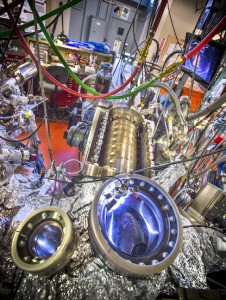
While I don’t understand (again) the image the researchers have included as an illustration of their work, I do find the ‘blue jewels in a pile of junk’ very appealing,

Beamline 10.0.1 at Berkeley Lab’s Advanced Light Source is optimized for the study of for electron structures and correlated electron systems. (Photo by Roy Kaltschmidt) Courtesy: Berkeley Lab

Beamline 10.0.1 at Berkeley Lab’s Advanced Light Source is optimized for the study of for electron structures and correlated electron systems. (Photo by Roy Kaltschmidt) Courtesy: Berkeley Lab

Here’s a link to and a citation for the paper,

Discovery of a Three-dimensional Topological Dirac Semimetal, Na3Bi by Zhongkai Liu, Bo Zhou, Yi Zhang, Zhijun Wang, Hongming Weng, Dharmalingam Prabhakaran, Sung-Kwan Mo, Zhi-Xun Shen, Zhong Fang, Xi Dai, and Zahid Hussain. Published Online January 16 2014 Science DOI: 10.1126/science.1245085

This paper is behind a paywall.