Tag Archives: Leah Burrows

Biohybrid fish made from human cardiac cells could lead to artificial hearts

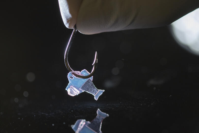
Biohybrid fish on a hook (Photo credit to Michael Rosnach, Keel Yong Lee, Sung-Jin Park, Kevin Kit Parker)

A February 10, 2022 news item on ScienceDaily announces research on a biohybrid fish,

Harvard University researchers, in collaboration with colleagues from Emory University, have developed the first fully autonomous biohybrid fish from human stem-cell derived cardiac muscle cells. The artificial fish swims by recreating the muscle contractions of a pumping heart, bringing researchers one step closer to developing a more complex artificial muscular pump and providing a platform to study heart disease like arrhythmia.

A February 10, 2022 Harvard University John A. Paulson School of Engineering and Applied Sciences news release (also on EurekAlert) by Leah Burrows explains how this research could lead to an artificial heart (Note: Links have been removed),

“Our ultimate goal is to build an artificial heart to replace a malformed heart in a child,” said Kit Parker, the Tarr Family Professor of Bioengineering and Applied Physics at the Harvard John A. Paulson School of Engineering and Applied Sciences (SEAS) and senior author of the paper.  “Most of the work in building heart tissue or hearts, including some work we have done, is focused on replicating the anatomical features or replicating the simple beating of the heart in the engineered tissues. But here, we are drawing design inspiration from the biophysics of the heart, which is harder to do. Now, rather than using heart imaging as a blueprint, we are identifying the key biophysical principles that make the heart work, using them as design criteria, and replicating them in a system, a living, swimming fish, where it is much easier to see if we are successful.”

The research is published in Science

The biohybrid fish developed by the team builds off previous research from Parker’s Disease Biophysics Group. In 2012, the lab used cardiac muscle cells from rats to build a jellyfish-like biohybrid pump and in 2016 the researchers developed a swimming, artificial stingray also from rat heart muscle cells.

In this research, the team built the first autonomous biohybrid device made from human stem-cell derived cardiomyocytes. This device was inspired by the shape and swimming motion of a zebrafish. Unlike previous devices, the biohybrid zebrafish has two layers of muscle cells, one on each side of the tail fin. When one side contracts, the other stretches. That stretch triggers the opening of a mechanosensitive protein channel, which causes a contraction, which triggers a stretch and so on and so forth, leading to a closed loop system that can propel the fish for more than 100 days. 

“By leveraging cardiac mechano-electrical signaling between two layers of muscle, we recreated the cycle where each contraction results automatically as a response to the stretching on the opposite side,” said Keel Yong Lee, a postdoctoral fellow at SEAS and co-first author of the study. “The results highlight the role of feedback mechanisms in muscular pumps such as the heart.”

The researchers also engineered an autonomous pacing node, like a pacemaker, which controls the frequency and rhythm of these spontaneous contractions. Together, the two layers of muscle and the autonomous pacing node enabled the generation of continuous, spontaneous, and coordinated, back-and-forth fin movements.

“Because of the two internal pacing mechanisms, our fish can live longer, move faster and swim more efficiently than previous work,” said Sung-Jin Park, a former postdoctoral fellow in the Disease Biophysics Group at SEAS and co-first author of the study. “This new research provides a model to investigate mechano-electrical signaling as a therapeutic target of heart rhythm management and for understanding pathophysiology in sinoatrial node dysfunctions and cardiac arrhythmia.”

Park is currently an Assistant Professor at the Coulter Department of Biomedical Engineering at Georgia Institute of Technology and Emory University School of Medicine.

Unlike a fish in your refrigerator, this biohybrid fish improves with age. Its muscle contraction amplitude, maximum swimming speed, and muscle coordination all increased for the first month as the cardiomyocyte cells matured.  Eventually, the biohybrid fish reached speeds and swimming efficacy similar to zebrafish in the wild. 

Next, the team aims to build even more complex biohybrid devices from human heart cells. 

“I could build a model heart out of Play-Doh, it doesn’t mean I can build a heart,” said Parker. “You can grow some random tumor cells in a dish until they curdle into a throbbing lump and call it a cardiac organoid. Neither of those efforts is going to, by design, recapitulate the physics of a system that beats over a billion times during your lifetime while simultaneously rebuilding its cells on the fly. That is the challenge. That is where we go to work.”

The research was co-authored by David G. Matthews, Sean L. Kim, Carlos Antonio Marquez, John F. Zimmerman, Herdeline Ann M. Ardona, Andre G. Kleber and George V. Lauder. 

It was supported in part by National Institutes of Health National Center for Advancing Translational Sciences grant UH3TR000522, and National Science Foundation Materials Research Science and Engineering Center grant DMR-142057.

Before giving you a link and a citation for the paper, here’s a little more information about the work from a February 10, 2022 American Association for the Advancement of Science (AAAS) news release on EurekAlert announcing publication of the paper in their journal Science, Note: A link has been removed,

An autonomously swimming biohybrid fish, designed with a focus on two key regulatory features of the human heart, has revealed the importance of feedback mechanisms in muscular pumps (such as the heart). The findings could one day help inform the development of an artificial heart made from living muscle cells. Biohybrid systems – devices containing both biological and artificial components – are an effective way to investigate the physiological control mechanisms in biological organisms and to discover bio-inspired robotic solutions to a host of pressing concerns, including those related to human health. When it comes to natural fluid transport pumps, like those that circulate blood, the performance of biohybrid systems has been lacking, however.  Here, researchers considered whether two functional regulatory features of the heart — mechanoelectrical signaling and automaticity — could be transferred to a synthetic analog of another fluid transport system: a swimming fish. Lee et al. developed an autonomously swimming fish constructed from a bilayer of human cardiac cells; the muscular bilayer was integrated using tissue engineering techniques. Lee and team were able to control muscle contractions in the biohybrid fish using external optogenetic stimulation, allowing the fish analog to swim. In tests, the biohybrid fish outperformed the locomotory speed of previous biohybrid muscular systems, the authors say. It maintained spontaneous activity for 108 days. By contrast, say the authors, biohybrid fish equipped with single-layered muscle showed deteriorating activity within the first month. The data in this study demonstrate the potential of muscular bilayer systems and mechanoelectrical signaling as a means to promote maturation of in vitro muscle tissues, write Lee and colleagues. “Taken together,” the authors conclude, “the technology described here may represent foundational work toward the goal of creating autonomous systems capable of homeostatic regulation and adaptive behavioral control.”

For reporters interested in trends, this work builds upon previous work published in a July 2016 study in Science, in which Sung-jin Park et al. used cardiac cells from rats to develop a self-propelling ray fish analog.

Here’s a link to and a citation for the paper,

An autonomously swimming biohybrid fish designed with human cardiac biophysics by Keel Yong Lee, Sung-Jin Park, David G. Matthews. Sean L. Kim, Carlos Antonio Marquez, John F. Zimmerman, Herdeline Ann M. Ardoña, Andre G. Kleber, George V. Lauder and Kevin Kit Parker. Science • 10 Feb 2022 • Vol 375, Issue 6581 • pp. 639-647 • DOI: 10.1126/science.abh0474

This paper is behind a paywall.
Suit up with nanofiber for protection against explosions and high temperatures

Where explosions are concerned you might expect to see some army research and you would be right. A June 29, 2020 news item on ScienceDaily breaks the news,

Since World War I, the vast majority of American combat casualties has come not from gunshot wounds but from explosions. Today, most soldiers wear a heavy, bullet-proof vest to protect their torso but much of their body remains exposed to the indiscriminate aim of explosive fragments and shrapnel.

Designing equipment to protect extremities against the extreme temperatures and deadly projectiles that accompany an explosion has been difficult because of a fundamental property of materials. Materials that are strong enough to protect against ballistic threats can’t protect against extreme temperatures and vice versa. As a result, much of today’s protective equipment is composed of multiple layers of different materials, leading to bulky, heavy gear that, if worn on the arms and legs, would severely limit a soldier’s mobility.

Now, Harvard University researchers, in collaboration with the U.S. Army Combat Capabilities Development Command Soldier Center (CCDC SC) and West Point, have developed a lightweight, multifunctional nanofiber material that can protect wearers from both extreme temperatures and ballistic threats.

A June 29, 2020 Harvard University news release (also on EurekAlert) by Leah Burrows, which originated the news item, expands on the theme,

“When I was in combat in Afghanistan, I saw firsthand how body armor could save lives,” said senior author Kit Parker, the Tarr Family Professor of Bioengineering and Applied Physics at the Harvard John A. Paulson School of Engineering and Applied Sciences (SEAS) and a lieutenant colonel in the United States Army Reserve. “I also saw how heavy body armor could limit mobility. As soldiers on the battlefield, the three primary tasks are to move, shoot, and communicate. If you limit one of those, you decrease survivability and you endanger mission success.”

“Our goal was to design a multifunctional material that could protect someone working in an extreme environment, such as an astronaut, firefighter or soldier, from the many different threats they face,” said Grant M. Gonzalez, a postdoctoral fellow at SEAS and first author of the paper.

In order to achieve this practical goal, the researchers needed to explore the tradeoff between mechanical protection and thermal insulation, properties rooted in a material’s molecular structure and orientation.

Materials with strong mechanical protection, such as metals and ceramics, have a highly ordered and aligned molecular structure. This structure allows them to withstand and distribute the energy of a direct blow. Insulating materials, on the other hand, have a much less ordered structure, which prevents the transmission of heat through the material.

Kevlar and Twaron are commercial products used extensively in protective equipment and can provide either ballistic or thermal protection, depending on how they are manufactured. Woven Kevlar, for example, has a highly aligned crystalline structure and is used in protective bulletproof vests. Porous Kevlar aerogels, on the other hand, have been shown to have high thermal insulation.

“Our idea was to use this Kevlar polymer to combine the woven, ordered structure of fibers with the porosity of aerogels to make long, continuous fibers with porous spacing in between,” said Gonzalez. “In this system, the long fibers could resist a mechanical impact while the pores would limit heat diffusion.”

The research team used immersion Rotary Jet-Spinning (iRJS), a technique developed by Parker’s Disease Biophysics Group, to manufacture the fibers. In this technique, a liquid polymer solution is loaded into a reservoir and pushed out through a tiny opening by centrifugal force as the device spins. When the polymer solution shoots out of the reservoir, it first passes through an area of open air, where the polymers elongate and the chains align. Then the solution hits a liquid bath that removes the solvent and precipitates the polymers to form solid fibers. Since the bath is also spinning — like water in a salad spinner — the nanofibers follow the stream of the vortex and wrap around a rotating collector at the base of the device.

By tuning the viscosity of the liquid polymer solution, the researchers were able to spin long, aligned nanofibers into porous sheets — providing enough order to protect against projectiles but enough disorder to protect against heat. In about 10 minutes, the team could spin sheets about 10 by 30 centimeters in size.

To test the sheets, the Harvard team turned to their collaborators to perform ballistic tests. Researchers at CCDC SC in Natick, Massachusetts simulated shrapnel impact by shooting large, BB-like projectiles at the sample. The team performed tests by sandwiching the nanofiber sheets between sheets of woven Twaron. They observed little difference in protection between a stack of all woven Twaron sheets and a combined stack of woven Twaron and spun nanofibers.

“The capabilities of the CCDC SC allow us to quantify the successes of our fibers from the perspective of protective equipment for warfighters, specifically,” said Gonzalez.

“Academic collaborations, especially those with distinguished local universities such as Harvard, provide CCDC SC the opportunity to leverage cutting-edge expertise and facilities to augment our own R&D capabilities,” said Kathleen Swana, a researcher at CCDC SC and one of the paper’s authors. “CCDC SC, in return, provides valuable scientific and soldier-centric expertise and testing capabilities to help drive the research forward.”

In testing for thermal protection, the researchers found that the nanofibers provided 20 times the heat insulation capability of commercial Twaron and Kevlar.

“While there are improvements that could be made, we have pushed the boundaries of what’s possible and started moving the field towards this kind of multifunctional material,” said Gonzalez.

“We’ve shown that you can develop highly protective textiles for people that work in harm’s way,” said Parker. “Our challenge now is to evolve the scientific advances to innovative products for my brothers and sisters in arms.”

Harvard’s Office of Technology Development has filed a patent application for the technology and is actively seeking commercialization opportunities.

Here’s a link to and a citation for the paper,

para-Aramid Fiber Sheets for Simultaneous Mechanical and Thermal Protection in Extreme Environments by Grant M. Gonzalez, Janet Ward, John Song, Kathleen Swana, Stephen A. Fossey, Jesse L. Palmer, Felita W. Zhang, Veronica M. Lucian, Luca Cera, John F. Zimmerman, F. John Burpo, Kevin Kit Parker. Matter DOI: https://doi.org/10.1016/j.matt.2020.06.001 Published:June 29, 2020

This paper is behind a paywall.

While this is the first time I’ve featured clothing/armour that’s protective against explosions I have on at least two occasions featured bulletproof clothing in a Canadian context. A November 4, 2013 posting had a story about a Toronto-based tailoring establishment, Garrison Bespoke, that was going to publicly test a bulletproof business suit. Should you be interested, it is possible to order the suit here. There’s also a February 11, 2020 posting announcing research into “Comfortable, bulletproof clothing for Canada’s Department of National Defence.”

Ouchies no more! Not from bandages, anyway.

An adhesive that US and Chinese scientists have developed shows great promise not just for bandages but wearable robotics too. From a December 14, 2018 news item on Nanowerk,

Researchers from the Harvard John A. Paulson School of Engineering and Applied Sciences (SEAS) and Xi’an Jiaotong University in China have developed a new type of adhesive that can strongly adhere wet materials — such as hydrogel and living tissue — and be easily detached with a specific frequency of light.

The adhesives could be used to attach and painlessly detach wound dressings, transdermal drug delivery devices, and wearable robotics.

A December 18, 2018 SEAS news release by Leah Burrows (also on EurekAlert but published Dec. 14, 2018), which originated the news item, delves further,

“Strong adhesion usually requires covalent bonds, physical interactions, or a combination of both,” said Yang Gao, first author of the paper and researcher at Xi’an Jiaotong University. “Adhesion through covalent bonds is hard to remove and adhesion through physical interactions usually requires solvents, which can be time-consuming and environmentally harmful. Our method of using light to trigger detachment is non-invasive and painless.”

The adhesive uses an aqueous solution of polymer chains spread between two, non-sticky materials — like jam between two slices of bread. On their own, the two materials adhere poorly together but the polymer chains act as a molecular suture, stitching the two materials together by forming a network with the two preexisting polymer networks. This process is known as topological entanglement.

When exposed to ultra-violet light, the network of stitches dissolves, separating the two materials.

The researchers, led by Zhigang Suo, the Allen E. and Marilyn M. Puckett Professor of Mechanics and Materials at SEAS, tested adhesion and detachment on a range of materials, sticking together hydrogels; hydrogels and organic tissue; elastomers; hydrogels and elastomers; and hydrogels and inorganic solids.

“Our strategy works across a range of materials and may enable broad applications,” said Kangling Wu, co-lead author and researcher at Xi’an Jiaotong University in China.
While the researchers focused on using UV light to trigger detachment, their work suggests the possibility that the stitching polymer could detach with near-infrared light, a feature which could be applied to a range of new medical procedures.

“In nature, wet materials don’t like to adhere together,” said Suo. “We have discovered a general approach to overcome this challenge. Our molecular sutures can strongly adhere wet materials together. Furthermore, the strong adhesion can be made permanent, transient, or detachable on demand, in response to a cue. So, as we see it, nature is full of loopholes, waiting to be stitched.”

Here’s a link to and  a citation for the paper,

Photodetachable Adhesion by Yang Gao, Kangling Wu, Zhigang Suo. https://doi.org/10.1002/adma.201806948 First published: 14 December 2018

This paper is behind a paywall.

Portable nanofibre fabrication device (point-of-use manufacturing)

A portable nanofiber fabrication device is quite an achievement although it seems it’s not quite ready for prime time yet. From a March 1, 2017 news item on Nanowerk (Note: A link has been removed),

Harvard researchers have developed a lightweight, portable nanofiber fabrication device that could one day be used to dress wounds on a battlefield or dress shoppers in customizable fabrics. The research was published recently in Macromolecular Materials and Engineering (“Design and Fabrication of Fibrous Nanomaterials Using Pull Spinning”)

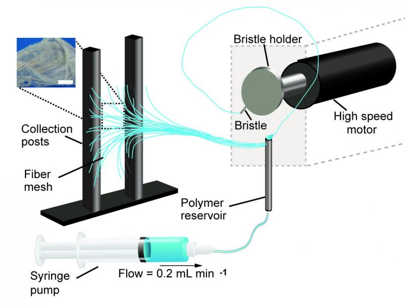
A schematic of the pull spinning apparatus with a side view illustration of a fiber being pulled from the polymer reservoir. The pull spinning system consists of a rotating bristle that dips and pulls a polymer jet in a spiral trajectory (Leila Deravi/Harvard University)

A March 1, 2017 Harvard University news release (also on EurekAlert) by Leah Burrow,, which originated the news item, describes the current process for nanofiber fabrication and explains how this technique is an improvement,

There are many ways to make nanofibers. These versatile materials — whose target applications include everything from tissue engineering to bullet proof vests — have been made using centrifugal force, capillary force, electric field, stretching, blowing, melting, and evaporation.

Each of these fabrication methods has pros and cons. For example, Rotary Jet-Spinning (RJS) and Immersion Rotary Jet-Spinning (iRJS) are novel manufacturing techniques developed in the Disease Biophysics Group at the Harvard John A. Paulson School of Engineering and Applied Sciences (SEAS) and the Wyss Institute for Biologically Inspired Engineering. Both RJS and iRJS dissolve polymers and proteins in a liquid solution and use centrifugal force or precipitation to elongate and solidify polymer jets into nanoscale fibers. These methods are great for producing large amounts of a range of materials – including DNA, nylon, and even Kevlar – but until now they haven’t been particularly portable.

The Disease Biophysics Group recently announced the development of a hand-held device that can quickly produce nanofibers with precise control over fiber orientation. Regulating fiber alignment and deposition is crucial when building nanofiber scaffolds that mimic highly aligned tissue in the body or designing point-of-use garments that fit a specific shape.

“Our main goal for this research was to make a portable machine that you could use to achieve controllable deposition of nanofibers,” said Nina Sinatra, a graduate student in the Disease Biophysics Group and co-first author of the paper. “In order to develop this kind of point-and-shoot device, we needed a technique that could produce highly aligned fibers with a reasonably high throughput.”

The new fabrication method, called pull spinning, uses a high-speed rotating bristle that dips into a polymer or protein reservoir and pulls a droplet from solution into a jet. The fiber travels in a spiral trajectory and solidifies before detaching from the bristle and moving toward a collector. Unlike other processes, which involve multiple manufacturing variables, pull spinning requires only one processing parameter — solution viscosity — to regulate nanofiber diameter. Minimal process parameters translate to ease of use and flexibility at the bench and, one day, in the field.

Pull spinning works with a range of different polymers and proteins. The researchers demonstrated proof-of-concept applications using polycaprolactone and gelatin fibers to direct muscle tissue growth and function on bioscaffolds, and nylon and polyurethane fibers for point-of-wear apparel.

“This simple, proof-of-concept study demonstrates the utility of this system for point-of-use manufacturing,” said Kit Parker, the Tarr Family Professor of Bioengineering and Applied Physics and director of the Disease Biophysics Group. “Future applications for directed production of customizable nanotextiles could extend to spray-on sportswear that gradually heats or cools an athlete’s body, sterile bandages deposited directly onto a wound, and fabrics with locally varying mechanical properties.”

Here’s a link to and a citation for the paper,

Design and Fabrication of Fibrous Nanomaterials Using Pull Spinning by Leila F. Deravi, Nina R. Sinatra, Christophe O. Chantre, Alexander P. Nesmith, Hongyan Yuan, Sahm K. Deravi, Josue A. Goss, Luke A. MacQueen, Mohammad R. Badrossamy, Grant M. Gonzalez, Michael D. Phillips, and Kevin Kit Parker. Macromolecular Materials and Engineering DOI: 10.1002/mame.201600404 Version of Record online: 17 JAN 2017

© 2017 WILEY-VCH Verlag GmbH & Co. KGaA, Weinheim

This paper is behind a paywall.

What is a multiregional brain-on-a-chip?

In response to having created a multiregional brain-on-a-chip, there’s an explanation from the team at Harvard University (which answers my question) in a Jan. 13, 2017 Harvard John A. Paulson School of Engineering and Applied Sciences news release (also on EurekAlert) by Leah Burrows,

Harvard University researchers have developed a multiregional brain-on-a-chip that models the connectivity between three distinct regions of the brain. The in vitro model was used to extensively characterize the differences between neurons from different regions of the brain and to mimic the system’s connectivity.

“The brain is so much more than individual neurons,” said Ben Maoz, co-first author of the paper and postdoctoral fellow in the Disease Biophysics Group in the Harvard John A. Paulson School of Engineering and Applied Sciences (SEAS). “It’s about the different types of cells and the connectivity between different regions of the brain. When modeling the brain, you need to be able to recapitulate that connectivity because there are many different diseases that attack those connections.”

“Roughly twenty-six percent of the US healthcare budget is spent on neurological and psychiatric disorders,” said Kit Parker, the Tarr Family Professor of Bioengineering and Applied Physics Building at SEAS and Core Faculty Member of the Wyss Institute for Biologically Inspired Engineering at Harvard University. “Tools to support the development of therapeutics to alleviate the suffering of these patients is not only the human thing to do, it is the best means of reducing this cost.”

Researchers from the Disease Biophysics Group at SEAS and the Wyss Institute modeled three regions of the brain most affected by schizophrenia — the amygdala, hippocampus and prefrontal cortex.

They began by characterizing the cell composition, protein expression, metabolism, and electrical activity of neurons from each region in vitro.

“It’s no surprise that neurons in distinct regions of the brain are different but it is surprising just how different they are,” said Stephanie Dauth, co-first author of the paper and former postdoctoral fellow in the Disease Biophysics Group. “We found that the cell-type ratio, the metabolism, the protein expression and the electrical activity all differ between regions in vitro. This shows that it does make a difference which brain region’s neurons you’re working with.”

Next, the team looked at how these neurons change when they’re communicating with one another. To do that, they cultured cells from each region independently and then let the cells establish connections via guided pathways embedded in the chip.

The researchers then measured cell composition and electrical activity again and found that the cells dramatically changed when they were in contact with neurons from different regions.

“When the cells are communicating with other regions, the cellular composition of the culture changes, the electrophysiology changes, all these inherent properties of the neurons change,” said Maoz. “This shows how important it is to implement different brain regions into in vitro models, especially when studying how neurological diseases impact connected regions of the brain.”

To demonstrate the chip’s efficacy in modeling disease, the team doped different regions of the brain with the drug Phencyclidine hydrochloride — commonly known as PCP — which simulates schizophrenia. The brain-on-a-chip allowed the researchers for the first time to look at both the drug’s impact on the individual regions as well as its downstream effect on the interconnected regions in vitro.

The brain-on-a-chip could be useful for studying any number of neurological and psychiatric diseases, including drug addiction, post traumatic stress disorder, and traumatic brain injury.

“To date, the Connectome project has not recognized all of the networks in the brain,” said Parker. “In our studies, we are showing that the extracellular matrix network is an important part of distinguishing different brain regions and that, subsequently, physiological and pathophysiological processes in these brain regions are unique. This advance will not only enable the development of therapeutics, but fundamental insights as to how we think, feel, and survive.”

Here’s an image from the researchers,

Caption: Image of the in vitro model showing three distinct regions of the brain connected by axons. Credit: Disease Biophysics Group/Harvard University

Here’s a link to and a citation for the paper,

Neurons derived from different brain regions are inherently different in vitro: A novel multiregional brain-on-a-chip by Stephanie Dauth, Ben M Maoz, Sean P Sheehy, Matthew A Hemphill, Tara Murty, Mary Kate Macedonia, Angie M Greer, Bogdan Budnik, Kevin Kit Parker. Journal of Neurophysiology Published 28 December 2016 Vol. no. [?] , DOI: 10.1152/jn.00575.2016

This paper is behind a paywall and they haven’t included the vol. no. in the citation I’ve found.

Superconductivity with spin

Vivid lines of light tracing a pattern reminiscent of a spinning top toy Courtesy: Harvard University

Vivid lines of light tracing a pattern reminiscent of a spinning top toy Courtesy: Harvard University

An Oct. 14, 2016 Harvard University John A. Paulson School of Engineering and Applied Sciences (SEAS) press release (also on EurekAlert) by Leah Burrows describes how scientists have discovered a way to transmit spin information through supercapacitors,

Researchers from the Harvard John A. Paulson School of Engineering and Applied Sciences (SEAS) have made a discovery that could lay the foundation for quantum superconducting devices. Their breakthrough solves one the main challenges to quantum computing: how to transmit spin information through superconducting materials.

Every electronic device — from a supercomputer to a dishwasher — works by controlling the flow of charged electrons. But electrons can carry so much more information than just charge; electrons also spin, like a gyroscope on axis.

Harnessing electron spin is really exciting for quantum information processing because not only can an electron spin up or down — one or zero — but it can also spin any direction between the two poles. Because it follows the rules of quantum mechanics, an electron can occupy all of those positions at once. Imagine the power of a computer that could calculate all of those positions simultaneously.

A whole field of applied physics, called spintronics, focuses on how to harness and measure electron spin and build spin equivalents of electronic gates and circuits.

By using superconducting materials through which electrons can move without any loss of energy, physicists hope to build quantum devices that would require significantly less power.

But there’s a problem.

According to a fundamental property of superconductivity, superconductors can’t transmit spin. Any electron pairs that pass through a superconductor will have the combined spin of zero.

In work published recently in Nature Physics, the Harvard researchers found a way to transmit spin information through superconducting materials.

“We now have a way to control the spin of the transmitted electrons in simple superconducting devices,” said Amir Yacoby, Professor of Physics and of Applied Physics at SEAS and senior author of the paper.

It’s easy to think of superconductors as particle super highways but a better analogy would be a super carpool lane as only paired electrons can move through a superconductor without resistance.

These pairs are called Cooper Pairs and they interact in a very particular way. If the way they move in relation to each other (physicists call this momentum) is symmetric, then the pair’s spin has to be asymmetric — for example, one negative and one positive for a combined spin of zero. When they travel through a conventional superconductor, Cooper Pairs’ momentum has to be zero and their orbit perfectly symmetrical.

But if you can change the momentum to asymmetric — leaning toward one direction — then the spin can be symmetric. To do that, you need the help of some exotic (aka weird) physics.

Superconducting materials can imbue non-superconducting materials with their conductive powers simply by being in close proximity. Using this principle, the researchers built a superconducting sandwich, with superconductors on the outside and mercury telluride in the middle. The atoms in mercury telluride are so heavy and the electrons move so quickly, that the rules of relativity start to apply.

“Because the atoms are so heavy, you have electrons that occupy high-speed orbits,” said Hechen Ren, coauthor of the study and graduate student at SEAS. “When an electron is moving this fast, its electric field turns into a magnetic field which then couples with the spin of the electron. This magnetic field acts on the spin and gives one spin a higher energy than another.”

So, when the Cooper Pairs hit this material, their spin begins to rotate.

“The Cooper Pairs jump into the mercury telluride and they see this strong spin orbit effect and start to couple differently,” said Ren. “The homogenous breed of zero momentum and zero combined spin is still there but now there is also a breed of pairs that gains momentum, breaking the symmetry of the orbit. The most important part of that is that the spin is now free to be something other than zero.”

The team could measure the spin at various points as the electron waves moved through the material. By using an external magnet, the researchers could tune the total spin of the pairs.

“This discovery opens up new possibilities for storing quantum information. Using the underlying physics behind this discovery provides also new possibilities for exploring the underlying nature of superconductivity in novel quantum materials,” said Yacoby.

Here’s a link to and a citation for the paper,

Controlled finite momentum pairing and spatially varying order parameter in proximitized HgTe quantum wells by Sean Hart, Hechen Ren, Michael Kosowsky, Gilad Ben-Shach, Philipp Leubner, Christoph Brüne, Hartmut Buhmann, Laurens W. Molenkamp, Bertrand I. Halperin, & Amir Yacoby. Nature Physics (2016) doi:10.1038/nphys3877 Published online 19 September 2016

This paper is behind a paywall.

Stronger more robust nanofibers for everything from bulletproof vests to cellular scaffolds (tissue engineering)

This work on a new technique for producing nanofibers comes from Harvard University’s School of Engineering and Applied Sciences and the Wyss Institute for Biologically Inspired Engineering (also at Harvard University). From an Oct. 10, 2016 news item on phys.org,

Fibrous materials—known for their toughness, durability and pliability—are used in everything from bulletproof vests to tires, filtration systems and cellular scaffolds for tissue engineering and regenerative medicine.

The properties of these materials are such that the smaller the fibers are, the stronger and tougher they become. But making certain fibers very small has been an engineering challenge.

Now, researchers from the Harvard John A. Paulson School of Engineering and Applied Sciences (SEAS) and the Wyss Institute for Biologically Inspired Engineering at Harvard have developed a new method to make and collect nanofibers and control their size and morphology. This could lead to stronger, more durable bulletproof vests and armor and more robust cellular scaffolding for tissue repair.

An Oct. 7, 2016 Harvard University press release by Leah Burrows, which originated the news item, describes the research in more detail (Note: A link has been removed),

Nanofibers are smaller than one micrometer in diameter.  Most nanofiber production platforms rely on dissolving polymers in a solution, which then evaporates as the fiber forms.

Rotary Jet-Spinning (RJS), the technique developed by Kit Parker’s Disease Biophysics Group, works likes a cotton candy machine. Parker is Tarr Family Professor of Bioengineering and Applied Physics at SEAS and a Core Member of the Wyss Institute. A liquid polymer solution is loaded into a reservoir and pushed out through a tiny opening by centrifugal force as the device spins. As the solution leaves the reservoir, the solvent evaporates and the polymers solidify and elongate into small, thin fibers.

“This advance is important because it allows us to manufacture ballistic protection that is much lighter, more flexible and more functional than what is available today,” said Parker, who in addition to his Harvard role is a lieutenant colonel in the United States Army Reserve and was motivated by his own combat experiences in Afghanistan. “Not only could it save lives but for the warfighter, it also could help reduce the repetitive injury motions that soldiers, sailors, marines and airmen have suffered over the last 15 years of the war on terror.”

“Rotary Jet-Spinning is great for most polymer fibers you want to make,” said Grant Gonzalez, a graduate student at SEAS and first author of the paper.  “However, some fibers require a solvent that doesn’t evaporate easily. Para-aramid, the polymer used in Kevlar® for example, is dissolved in sulfuric acid, which doesn’t evaporate off. The solution just splashes against the walls of the device without forming fibers.”

Nanofibers are smaller than one micrometer in diameter.  Most nanofiber production platforms rely on dissolving polymers in a solution, which then evaporates as the fiber forms.

Rotary Jet-Spinning (RJS), the technique developed by Kit Parker’s Disease Biophysics Group, works likes a cotton candy machine. Parker is Tarr Family Professor of Bioengineering and Applied Physics at SEAS and a Core Member of the Wyss Institute. A liquid polymer solution is loaded into a reservoir and pushed out through a tiny opening by centrifugal force as the device spins. As the solution leaves the reservoir, the solvent evaporates and the polymers solidify and elongate into small, thin fibers.

“This advance is important because it allows us to manufacture ballistic protection that is much lighter, more flexible and more functional than what is available today,” said Parker, who in addition to his Harvard role is a lieutenant colonel in the United States Army Reserve and was motivated by his own combat experiences in Afghanistan. “Not only could it save lives but for the warfighter, it also could help reduce the repetitive injury motions that soldiers, sailors, marines and airmen have suffered over the last 15 years of the war on terror.”

“Rotary Jet-Spinning is great for most polymer fibers you want to make,” said Grant Gonzalez, a graduate student at SEAS and first author of the paper.  “However, some fibers require a solvent that doesn’t evaporate easily. Para-aramid, the polymer used in Kevlar® for example, is dissolved in sulfuric acid, which doesn’t evaporate off. The solution just splashes against the walls of the device without forming fibers.”

Other methods, such as electrospinning, which uses an electric field to pull the polymer into a thin fiber, also have poor results with Kevlar and other polymers such as alginate used for tissue scaffolding and DNA.

The Harvard team overcame these challenges by developing a wet-spinning platform, which uses the same principles as the RJS system but relies on precipitation rather than evaporation to separate the solvent from the polymer.

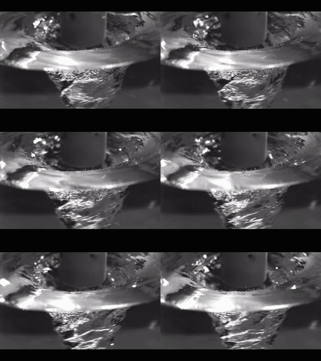
In this system, called immersion Rotary Jet-Spinning (iRJS), when the polymer solution shoots out of the reservoir, it first passes through an area of open air, where the polymers elongate and the chains align. Then the solution hits a liquid bath that removes the solvent and precipitates the polymers to form solid fibers. Since the bath is also spinning — like water in a salad spinner — the nanofibers follow the stream of the vortex and wrap around a rotating collector at the base of the device.

Using this system, the team produced Nylon, DNA, alginate and ballistic resistant para-aramid nanofibers. The team could tune the fiber’s diameter by changing the solution concentration, the rotational speed and the distance the polymer traveled from the reservoir to the bath.

“By being able to modulate fiber strength, we can create a cellular scaffold that can mimic skeleton muscle and native tissues,” said Gonzalez.  “This platform could enable us to create a wound dressing out of alginate material or seed and mature cells on scaffolding for tissue engineering.”

Because the fibers were collected by a spinning vortex, the system also produced well-aligned sheets of nanofibers, which is important for scaffolding and ballistic resistant materials.

This is the ‘candy floss’ technique at work,

Rotary Jet-Spinning (RJS) works likes a cotton candy machine. A liquid polymer solution is loaded into a reservoir and pushed out through a tiny opening by centrifugal force as the device spins. As the solution leaves the reservoir, the solvent evaporates and the polymers solidify and elongate into small, thin fibers. Courtesy: Harvard University

Rotary Jet-Spinning (RJS) works likes a cotton candy machine. A liquid polymer solution is loaded into a reservoir and pushed out through a tiny opening by centrifugal force as the device spins. As the solution leaves the reservoir, the solvent evaporates and the polymers solidify and elongate into small, thin fibers. Courtesy: Harvard University

Here’s a link to and a citation for the paper,

Production of Synthetic, Para-Aramid and Biopolymer Nanofibers by Immersion Rotary Jet-Spinning by Grant M. Gonzalez, Luke A. MacQueen, Johan U. Lind, Stacey A. Fitzgibbons, Christophe O. Chantre, Isabelle Huggler, Holly M. Golecki, Josue A. Goss, Kevin Kit Parker. Macromolecular Materials and Engineering DOI: 10.1002/mame.201600365 Version of Record online: 7 OCT 2016

© 2016 WILEY-VCH Verlag GmbH & Co. KGaA, Weinheim

This paper is behind a paywall.

Tune your windows for privacy

Caption: With an applied voltage, the nanowires on either side of the glass become attracted to each other and move toward each other, squeezing and deforming the soft elastomer. Because the nanowires are scattered unevenly across the surface, the elastomer deforms unevenly. That uneven roughness causes light to scatter, turning the glass opaque. Credit: David Clarke/Harvard SEAS [School of Engineering and Applied Sciences]

Right now, this is my favourite science illustration. A March 14, 2016 news item on Nanowerk announces Harvard’s new technology that can turn a clear window into an opaque one at the touch of a switch,

Say goodbye to blinds.

Researchers at the Harvard John A. Paulson School of Engineering and Applied Sciences have developed a technique that can quickly change the opacity of a window, turning it cloudy, clear or somewhere in between with the flick of a switch.

Tunable windows aren’t new but most previous technologies have relied on electrochemical reactions achieved through expensive manufacturing. This technology, developed by David Clarke, the Extended Tarr Family Professor of Materials, and postdoctoral fellow Samuel Shian, uses geometry [to] adjust the transparency of a window.

A March 14, 2016 Harvard University news release (also on EurekAlert) by Leah Burrows, which originated the news item, describes the technology in more detail,

The tunable window is comprised of a sheet of glass or plastic, sandwiched between transparent, soft elastomers sprayed with a coating of silver nanowires, too small to scatter light on their own.

But apply an electric voltage and things change quickly.

With an applied voltage, the nanowires on either side of the glass are energized to move toward each other, squeezing and deforming the soft elastomer. Because the nanowires are distributed unevenly across the surface, the elastomer deforms unevenly. The resulting uneven roughness causes light to scatter, turning the glass opaque.

The change happens in less than a second.

It’s like a frozen pond, said Shian.

“If the frozen pond is smooth, you can see through the ice. But if the ice is heavily scratched, you can’t see through,” said Shian.

Clarke and Shian found that the roughness of the elastomer surface depended on the voltage, so if you wanted a window that is only light clouded, you would apply less voltage than if you wanted a totally opaque window.

“Because this is a physical phenomenon rather than based on a chemical reaction, it is a simpler and potentially cheaper way to achieve commercial tunable windows,” said Clarke.

Current chemical-based controllable windows use vacuum deposition to coat the glass, a process that deposits layers of a material molecule by molecule. It’s expensive and painstaking. In Clarke and Shian’s method, the nanowire layer can be sprayed or peeled onto the elastomer, making the technology scalable for larger architectural projects.

Next the team is working on incorporating thinner elastomers, which would require lower voltages, more suited for standard electronical supplies.

Here’s a link to and a citation for the paper,

Electrically tunable window device by Samuel Shian and David R. Clarke. Optics Letters Vol. 41, Issue 6, pp. 1289-1292 (2016) •doi: 10.1364/OL.41.001289

This is an open access paper.

Origami and our pop-up future

They should have declared Jan. 25, 2016 ‘L. Mahadevan Day’ at Harvard University. The researcher was listed as an author on two major papers. I covered the first piece of research, 4D printed hydrogels, in this Jan. 26, 2016 posting. Now for Mahadevan’s other work, from a Jan. 27, 2016 news item on Nanotechnology Now,

What if you could make any object out of a flat sheet of paper?

That future is on the horizon thanks to new research by L. Mahadevan, the Lola England de Valpine Professor of Applied Mathematics, Organismic and Evolutionary Biology, and Physics at the Harvard John A. Paulson School of Engineering and Applied Sciences (SEAS). He is also a core faculty member of the Wyss Institute for Biologically Inspired Engineering, and member of the Kavli Institute for Bionano Science and Technology, at Harvard University.

Mahadevan and his team have characterized a fundamental origami fold, or tessellation, that could be used as a building block to create almost any three-dimensional shape, from nanostructures to buildings. …

A Jan. 26, 2016 Harvard University news release by Leah Burrows, which originated the news item, provides more detail about the specific fold the team has been investigating,

The folding pattern, known as the Miura-ori, is a periodic way to tile the plane using the simplest mountain-valley fold in origami. It was used as a decorative item in clothing at least as long ago as the 15th century. A folded Miura can be packed into a flat, compact shape and unfolded in one continuous motion, making it ideal for packing rigid structures like solar panels.  It also occurs in nature in a variety of situations, such as in insect wings and certain leaves.

“Could this simple folding pattern serve as a template for more complicated shapes, such as saddles, spheres, cylinders, and helices?” asked Mahadevan.

“We found an incredible amount of flexibility hidden inside the geometry of the Miura-ori,” said Levi Dudte, graduate student in the Mahadevan lab and first author of the paper. “As it turns out, this fold is capable of creating many more shapes than we imagined.”

Think surgical stents that can be packed flat and pop-up into three-dimensional structures once inside the body or dining room tables that can lean flat against the wall until they are ready to be used.

“The collapsibility, transportability and deployability of Miura-ori folded objects makes it a potentially attractive design for everything from space-bound payloads to small-space living to laparoscopic surgery and soft robotics,” said Dudte.

Here’s a .gif demonstrating the fold,

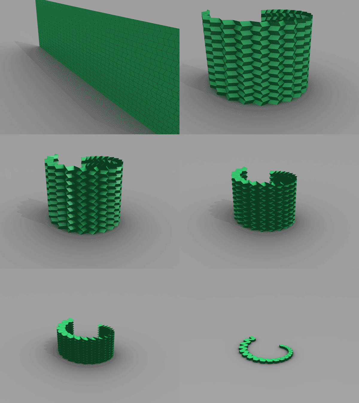
This spiral folds rigidly from flat pattern through the target surface and onto the flat-folded plane (Image courtesy of Mahadevan Lab) Harvard University

This spiral folds rigidly from flat pattern through the target surface and onto the flat-folded plane (Image courtesy of Mahadevan Lab) Harvard University

The news release offers some details about the research,

To explore the potential of the tessellation, the team developed an algorithm that can create certain shapes using the Miura-ori fold, repeated with small variations. Given the specifications of the target shape, the program lays out the folds needed to create the design, which can then be laser printed for folding.

The program takes into account several factors, including the stiffness of the folded material and the trade-off between the accuracy of the pattern and the effort associated with creating finer folds – an important characterization because, as of now, these shapes are all folded by hand.

“Essentially, we would like to be able to tailor any shape by using an appropriate folding pattern,” said Mahadevan. “Starting with the basic mountain-valley fold, our algorithm determines how to vary it by gently tweaking it from one location to the other to make a vase, a hat, a saddle, or to stitch them together to make more and more complex structures.”

“This is a step in the direction of being able to solve the inverse problem – given a functional shape, how can we design the folds on a sheet to achieve it,” Dudte said.

“The really exciting thing about this fold is it is completely scalable,” said Mahadevan. “You can do this with graphene, which is one atom thick, or you can do it on the architectural scale.”

Co-authors on the study include Etienne Vouga, currently at the University of Texas at Austin, and Tomohiro Tachi from the University of Tokyo. …

Here’s a link to and a citation for the paper,

Programming curvature using origami tessellations by Levi H. Dudte, Etienne Vouga, Tomohiro Tachi, & L. Mahadevan. Nature Materials (2016) doi:10.1038/nmat4540 Published online 25 January 2016

This paper is behind a paywall.