Tag Archives: ligands

Organic-inorganic nanohybrids: organic ligands attached to colloidal inorganic nanocrystals

A May 24, 2023 news item on phys.org introduces organic-inorganic nanohybrids for optoelectronic devices,

When designing optoelectronic devices, such as solar cells, photocatalysts, and photodetectors, scientists usually prioritize materials that are stable and possess tunable properties. This allows them precise control over optical characteristics of the materials and ensures retention of their properties over time, despite varying environmental conditions.

Organic-inorganic nanohybrids, which are made up of organic ligands attached to the surface of colloidal inorganic nanocrystals via coordinate bonds, are promising in this regard. They are known to exhibit enhanced stability owing to the formation of a protective layer by organic ligands around the reactive inorganic nanocrystal. However, the incorporation of organic ligands has been found to lower the conductivity and photon absorption efficiency of inorganic nanocrystals.

In a breakthrough study on ligand-nanocrystal interactions, researchers from Japan now demonstrate a quasi-reversible displacement of organic ligands on the surface of nanocrystals. Their findings, published in ACS Nano, provide a new perspective to the common belief that the organic ligands are anchored to the surface of the nanocrystals.

A May 22, 2023 Ritsumeikan University (Japan) press release (also on EurekAlert but published May 24, 2023), which originated the news item, provides more detail,

… The research team, led by Professor Yoichi Kobayashi from Ritsumeikan University, Japan, found that the coordination bond between perylene bisimide with a carboxyl group (PBI) and inorganic zinc sulfide (ZnS) nanocrystals can be reversibly displaced by exposing the material to visible light.

Shedding light on this novel behavior of organic-inorganic nanohybrids, Prof. Kobayashi says, “We explored the ligand properties of organic-inorganic nanohybrid systems by using perylene bisimide with a carboxyl group (PBI)-coordinated zinc sulfide (ZnS) NCs (PBI–ZnS) as a model system. Our findings provide the first example of photoinduced displacement of aromatic ligands with semiconductor nanocrystals.”

In their study, the researchers carried out both theoretical analysis and experimental investigations to understand the material’s unique photoinducible characteristics. They first conducted density functional theory calculations to study the structure and orbitals of PBI–ZnS ([PBI-Zn25S31]) in both its ground and first excited states. Next, they performed time-resolved impulsive stimulated Raman spectroscopy to excite the sample with an ultrafast laser. This helped them analyze the corresponding Raman spectrum that revealed the nature of the excited state of PBI–ZnS.

The experimental observations and calculations showed that, upon photoexcitation, an electron is excited from the PBI molecule, and the corresponding “hole”(the vacancy formed due to the absence of the electron) rapidly moves from the aromatic ligand (PBI) to ZnS. This results in a long-lived, negatively-charged PBI ion that is displaced from the surface of the ZnS nanocrystal. Over time, however, the displaced ligands recombine with the surface defects of the ZnS nanocrystal, leading to a quasi-reversible photoinduced displacement of coordinated PBI. Notably, the dynamic behavior of coordinated ligand molecules observed in this study is different from that observed for typical photoinduced charge transfer processes in which the hole typically remains on the donor molecule, enabling it to recombine with the electron quickly.

Explaining the significance of these findings, Prof. Kobayashi says, “The precise understanding of ligand-nanocrystal interaction is important not only for fundamental nanoscience but also for developing advanced photofunctional materials using nanomaterials. These include photocatalysts for the decomposition of persistent chemicals using visible light and photoconductive microcircuit patterning for wearable devices.”

Indeed, the results of this study present a promising avenue for enhancing the tunability and functionality of inorganic materials with aromatic molecules. This, in turn, could significantly impact the field of fundamental nanoscience and photochemistry in the times to come.

Here’s a link to and a citation for the paper,

Quasi-Reversible Photoinduced Displacement of Aromatic Ligands from Semiconductor Nanocrystals by Daisuke Yoshioka, Yusuke Yoneda, I-Ya Chang, Hikaru Kuramochi, Kim Hyeon-Deuk, and Yoichi Kobayashi. ACS Nano 2023, 17, 12, 11309–11317 DOI: https://doi.org/10.1021/acsnano.2c12578 Publication Date:May 9, 2023 Copyright © 2023 American Chemical Society

This paper is behind a paywall.
Using natural proteins to grow gold nanoclusters for hybrid bionanomaterials

While there’s a January 10, 2022 news item on Nanowerk, the research being announced was made available online in the Fall of 2021 and is now available in print,

Gold nanoclusters are groups of a few gold atoms with interesting photoluminescent properties. The features of gold nanoclusters depend not only on their structure, but their size and also by the ligands coordinated to them. These inorganic nanomaterials have been used in sensing, biomedicine and optics and their coordination with biomolecules can endow multiple capabilities in biological media.

A research collaboration between the groups of Dr. Juan Cabanillas, Research Professor at IMDEA Nanociencia and Dr. Aitziber L. Cortajarena, Ikerbasque Professor and Principal Investigator at CIC biomaGUNE have explored the use of natural proteins to grow gold nanoclusters, resulting in hybrid bionanomaterials with tunable photoluminescent properties and with a plethora of potential applications.

A January 10, 2022 IMDEA Nanociencia press release, which originated the news item, provides more technical detail about the research,

The nanoclusters –with less than 2 nm in size- differentiate from larger nanoparticles (plasmonic) since they present discrete energy levels coupled optically. The groups of amino acids within the proteins coordinate the gold atoms and allow the groups to be arranged around the gold nanocluster, facilitating the stabilization and adding an extra level of tailoring. These nanoclusters have interesting energy harvesting features. Since the discrete energy levels are optically coupled, the absorption of a photon leads to promotion of an electron to higher levels, which can trigger a photophysical process or a photochemical reaction.  

The results by Cabanillas and Cortajarena groups, published in Advanced Optical Materials and Nano Letters, explore the origin of the photoluminescence in protein-designed gold nanoclusters and shed light into the strong influence of environmental conditions on the nature of luminescence. Nanocluster capping by two types of amino acids (histidine and cysteine) allow for changing the emission spectral range from blue to red, paving the way to tune the optical properties by an appropriate ligand choice. The nature of emission is also changed with capping, from fluorescence to phosphorescence, respectively. The synergistic protein-nanocluster effects on emission are still not clear, and the groups at IMDEA Nanociencia and CIC biomaGUNE are working to elucidate the mechanisms behind. There are potential applications for the aforementioned nanoclusters, in solid state as active medium in laser cavities. Optical gain properties from these nanoclusters are yet to be demonstrated, which could pave the way to a new generation of potentially interesting laser devices. As the combination of gold plus proteins is potentially biocompatible, many potential applications in biomedicine can also be envisaged.

A related publication of the groups in Nano Letters demonstrates that the insertion of tryptophans, amino acids with high electron density, in the vicinity of the nanocluster boosts its photoluminescence quantum efficiency up to 40% in some cases, values relevant for solid state light emission applications. Researchers also observed an antenna effect: the tryptophans can absorb light in a discrete manner and transfer the energy to the cluster. This effect has interest for energy harvesting and for sensing purposes as well.

The proteins through the biocapping enable the synthesis of the nanoclusters and largely improve their quantum efficiency. “The photoluminescence quantum efficiency is largely improved when using the biocapping” Dr. Cabanillas says. He believes this research work means “a new field opening for the tuning of optical properties of nanoclusters through protein engineering, and much work is ahead for the understanding of the amplification mechanism”. Dr. Cortajarena emphasizes “we have already demonstrated the great potential of engineered photoluminescent protein-nanocluster in biomedical and technological fields, and understanding the fundamental emission mechanisms is pivotal for future applications“. A variety of further applications include biosensors, as the protein admits functionalization with recognition molecules, energy harvesting, imaging and photodynamic therapies. Further work is ahead this opening avenue for photophysics research.

This research is a collaboration led by Dr. Juan Cabanillas and Dr. Aitziber L. Cortajarena research groups at IMDEA Nanociencia and CIC biomaGUNE, with contributions from researchers at the Diamond Light Source Ltd. [synchrotron] and DIPC. It has been cofounded by the projects AMAPOLA, NMAT2D, FULMATEN, Atracción de Talento from Comunidad de Madrid and the Severo Ochoa Centre of Excellence award to IMDEA Nanociencia. CIC biomaGUNE acknowledges support by the projects ERC-ProNANO, ERC-NIMM, ProTOOLs and the Maria de Maeztu Units of Excellence Programme.

Here are links to and citations for the papers,

Tuning the Optical Properties of Au Nanoclusters by Designed Proteins by Elena Lopez-Martinez, Diego Gianolio, Saül Garcia-Orrit, Victor Vega-Mayoral, Juan Cabanillas-Gonzalez, Carlos Sanchez-Cano, Aitziber L. Cortajarena. Advanced Optical Materials Volume 10, Issue 1 January 4, 2022 2101332 DOI: https://doi.org/10.1002/adom.202101332 First published: 31 October 2021

This paper is open access.

Boosting the Photoluminescent Properties of Protein-Stabilized Gold Nanoclusters through Protein Engineering by Antonio Aires, Ahmad Sousaraei, Marco Möller, Juan Cabanillas-Gonzalez, and Aitziber L. Cortajarena. Nano Lett. 2021, 21, 21, 9347–9353 DOI: https://doi.org/10.1021/acs.nanolett.1c03768 Publication Date: November 1, 2021 Copyright © 2021 American Chemical Society

This paper is behind a paywall.

Not being familiar with either of the two research institutions mentioned in the press release, I did a little digging.

Here’s a little information about IMDEA Nanociencia (IMDEA Nanoscience Institute), from its Wikipedia entry, Note: All links have been removed,

IMDEA Nanoscience Institute is a private non-profit foundation within the IMDEA Institutes network, created in 2006-2007 as a result of collaboration agreement between the Community of Madrid and Spanish Ministry of Education and Science. The foundation manages IMDEA-Nanoscience Institute,[1] a scientific centre dedicated to front-line research in nanoscience, nanotechnology and molecular design and aiming at transferable innovations and close contact with industries. IMDEA Nanoscience is a member of the Campus of International excellence, a consortium of research institutes promoted by the Autonomous University of Madrid and Spanish National Research Council (UAM/CSIC).[2]

As for CIC biomaGUNE, here’s more from its institutional profile on the science.eus website,

The Centre for Cooperative Research in Biomaterials-CIC biomaGUNE, located in San Sebastian (Spain), was officially opened in December 2006. CIC biomaGUNE is a non-profit research organization created to promote scientific research and technological innovation at the highest levels in the Basque Country following the BioBasque policy in order to create a new business sector based on biosciences. Established by the Department of Industry, Technology & Innovation of the Government of the Autonomous Community of the Basque Country, CIC biomaGUNE constitutes one of the Centres of the CIC network, the largest Basque Country research network on specific strategic areas, having the mission to contribute to the economical and social development of the country through the generation of knowledge and speeding up the process that leads to technological innovation.

Brain-inspired electronics with organic memristors for wearable computing

I went down a rabbit hole while trying to figure out the difference between ‘organic’ memristors and standard memristors. I have put the results of my investigation at the end of this post. First, there’s the news.

An April 21, 2020 news item on ScienceDaily explains why researchers are so focused on memristors and brainlike computing,

The advent of artificial intelligence, machine learning and the internet of things is expected to change modern electronics and bring forth the fourth Industrial Revolution. The pressing question for many researchers is how to handle this technological revolution.

“It is important for us to understand that the computing platforms of today will not be able to sustain at-scale implementations of AI algorithms on massive datasets,” said Thirumalai Venkatesan, one of the authors of a paper published in Applied Physics Reviews, from AIP Publishing.

“Today’s computing is way too energy-intensive to handle big data. We need to rethink our approaches to computation on all levels: materials, devices and architecture that can enable ultralow energy computing.”

An April 21, 2020 American Institute of Physics (AIP) news release (also on EurekAlert), which originated the news item, describes the authors’ approach to the problems with organic memristors,

Brain-inspired electronics with organic memristors could offer a functionally promising and cost- effective platform, according to Venkatesan. Memristive devices are electronic devices with an inherent memory that are capable of both storing data and performing computation. Since memristors are functionally analogous to the operation of neurons, the computing units in the brain, they are optimal candidates for brain-inspired computing platforms.

Until now, oxides have been the leading candidate as the optimum material for memristors. Different material systems have been proposed but none have been successful so far.

“Over the last 20 years, there have been several attempts to come up with organic memristors, but none of those have shown any promise,” said Sreetosh Goswami, lead author on the paper. “The primary reason behind this failure is their lack of stability, reproducibility and ambiguity in mechanistic understanding. At a device level, we are now able to solve most of these problems,”

This new generation of organic memristors is developed based on metal azo complex devices, which are the brainchild of Sreebata Goswami, a professor at the Indian Association for the Cultivation of Science in Kolkata and another author on the paper.

“In thin films, the molecules are so robust and stable that these devices can eventually be the right choice for many wearable and implantable technologies or a body net, because these could be bendable and stretchable,” said Sreebata Goswami. A body net is a series of wireless sensors that stick to the skin and track health.

The next challenge will be to produce these organic memristors at scale, said Venkatesan.

“Now we are making individual devices in the laboratory. We need to make circuits for large-scale functional implementation of these devices.”

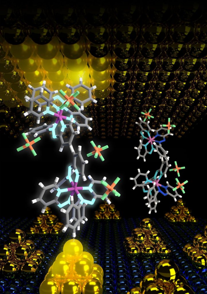
Caption: The device structure at a molecular level. The gold nanoparticles on the bottom electrode enhance the field enabling an ultra-low energy operation of the molecular device. Credit Sreetosh Goswami, Sreebrata Goswami and Thirumalai Venky Venkatesan

Here’s a link to and a citation for the paper,

An organic approach to low energy memory and brain inspired electronics by Sreetosh Goswami, Sreebrata Goswami, and T. Venkatesan. Applied Physics Reviews 7, 021303 (2020) DOI: https://doi.org/10.1063/1.5124155

This paper is open access.

Basics about memristors and organic memristors

This undated article on Nanowerk provides a relatively complete and technical description of memristors in general (Note: A link has been removed),

A memristor (named as a portmanteau of memory and resistor) is a non-volatile electronic memory device that was first theorized by Leon Ong Chua in 1971 as the fourth fundamental two-terminal circuit element following the resistor, the capacitor, and the inductor (IEEE Transactions on Circuit Theory, “Memristor-The missing circuit element”).

Its special property is that its resistance can be programmed (resistor function) and subsequently remains stored (memory function). Unlike other memories that exist today in modern electronics, memristors are stable and remember their state even if the device loses power.

However, it was only almost 40 years later that the first practical device was fabricated. This was in 2008, when a group led by Stanley Williams at HP Research Labs realized that switching of the resistance between a conducting and less conducting state in metal-oxide thin-film devices was showing Leon Chua’s memristor behavior. …

The article on Nanowerk includes an embedded video presentation on memristors given by Stanley Williams (also known as R. Stanley Williams).

Mention of an ‘organic’memristor can be found in an October 31, 2017 article by Ryan Whitwam,

The memristor is composed of the transition metal ruthenium complexed with “azo-aromatic ligands.” [emphasis mine] The theoretical work enabling this material was performed at Yale, and the organic molecules were synthesized at the Indian Association for the Cultivation of Sciences. …

I highlighted ‘ligands’ because that appears to be the difference. However, there is more than one type of ligand on Wikipedia.

First, there’s the Ligand (biochemistry) entry (Note: Links have been removed),

In biochemistry and pharmacology, a ligand is a substance that forms a complex with a biomolecule to serve a biological purpose. …

Then, there’s the Ligand entry,

In coordination chemistry, a ligand[help 1] is an ion or molecule (functional group) that binds to a central metal atom to form a coordination complex …

Finally, there’s the Ligand (disambiguation) entry (Note: Links have been removed),

  • Ligand, an atom, ion, or functional group that donates one or more of its electrons through a coordinate covalent bond to one or more central atoms or ions
  • Ligand (biochemistry), a substance that binds to a protein
  • a ‘guest’ in host–guest chemistry

I did take a look at the paper and did not see any references to proteins or other biomolecules that I could recognize as such. I’m not sure why the researchers are describing their device as an ‘organic’ memristor but this may reflect a shortcoming in the definitions I have found or shortcomings in my reading of the paper rather than an error on their parts.

Hopefully, more research will be forthcoming and it will be possible to better understand the terminology.

Liquid circuitry, shape-shifting fluids and more

I’d have to see it to believe it but researchers at the US Dept. of Energy (DOE) Lawrence Berkeley National Laboratory (LBNL) have developed a new kind of ‘bijel’ which would allow for some pretty nifty robotics. From a Sept. 25, 2017 news item on ScienceDaily,

A new two-dimensional film, made of polymers and nanoparticles and developed by researchers at the Department of Energy’s Lawrence Berkeley National Laboratory (Berkeley Lab), can direct two different non-mixing liquids into a variety of exotic architectures. This finding could lead to soft robotics, liquid circuitry, shape-shifting fluids, and a host of new materials that use soft, rather than solid, substances.

The study, reported today in the journal Nature Nanotechnology, presents the newest entry in a class of substances known as bicontinuous jammed emulsion gels, or bijels, which hold promise as a malleable liquid that can support catalytic reactions, electrical conductivity, and energy conversion.

A Sept. 25, 2017 LBNL news release (also on EurekAlert), which originated the news item, expands on the theme,

Bijels are typically made of immiscible, or non-mixing, liquids. People who shake their bottle of vinaigrette before pouring the dressing on their salad are familiar with such liquids. As soon as the shaking stops, the liquids start to separate again, with the lower density liquid – often oil – rising to the top.

Trapping, or jamming, particles where these immiscible liquids meet can prevent the liquids from completely separating, stabilizing the substance into a bijel. What makes bijels remarkable is that, rather than just making the spherical droplets that we normally see when we try to mix oil and water, the particles at the interface shape the liquids into complex networks of interconnected fluid channels.

Bijels are notoriously difficult to make, however, involving exact temperatures at precisely timed stages. In addition, the liquid channels are normally more than 5 micrometers across, making them too large to be useful in energy conversion and catalysis.

“Bijels have long been of interest as next-generation materials for energy applications and chemical synthesis,” said study lead author Caili Huang. “The problem has been making enough of them, and with features of the right size. In this work, we crack that problem.”

Huang started the work as a graduate student with Thomas Russell, the study’s principal investigator, at Berkeley Lab’s Materials Sciences Division, and he continued the project as a postdoctoral researcher at DOE’s Oak Ridge National Laboratory.

Creating a new bijel recipe

The method described in this new study simplifies the bijel process by first using specially coated particles about 10-20 nanometers in diameter. The smaller-sized particles line the liquid interfaces much more quickly than the ones used in traditional bijels, making the smaller channels that are highly valued for applications.

Illustration shows key stages of bijel formation. Clockwise from top left, two non-mixing liquids are shown. Ligands (shown in yellow) with amine groups are dispersed throughout the oil or solvent, and nanoparticles coated with carboxylic acids (shown as blue dots) are scattered in the water. With vigorous shaking, the nanoparticles and ligands form a “supersoap” that gets trapped at the interface of the two liquids. The bottom panel is a magnified view of the jammed nanoparticle supersoap. (Credit: Caili Huang/ORNL)

“We’ve basically taken liquids like oil and water and given them a structure, and it’s a structure that can be changed,” said Russell, a visiting faculty scientist at Berkeley Lab. “If the nanoparticles are responsive to electrical, magnetic, or mechanical stimuli, the bijels can become reconfigurable and re-shaped on demand by an external field.”

The researchers were able to prepare new bijels from a variety of common organic, water-insoluble solvents, such as toluene, that had ligands dissolved in it, and deionized water, which contained the nanoparticles. To ensure thorough mixing of the liquids, they subjected the emulsion to a vortex spinning at 3,200 revolutions per minute.

“This extreme shaking creates a whole bunch of new places where these particles and polymers can meet each other,” said study co-author Joe Forth, a postdoctoral fellow at Berkeley Lab’s Materials Sciences Division. “You’re synthesizing a lot of this material, which is in effect a thin, 2-D coating of the liquid surfaces in the system.”

The liquids remained a bijel even after one week, a sign of the system’s stability.

Russell, who is also a professor of polymer science and engineering at the University of Massachusetts-Amherst, added that these shape-shifting characteristics would be valuable in microreactors, microfluidic devices, and soft actuators.

Nanoparticle supersoap

Nanoparticles had not been seriously considered in bijels before because their small size made them hard to trap in the liquid interface. To resolve that problem, the researchers coated nano-sized particles with carboxylic acids and put them in water. They then took polymers with an added amine group – a derivative of ammonia – and dissolved them in the toluene.

At left is a vial of bijel stabilized with nanoparticle surfactants. On the right is the same vial after a week of inversion, showing that the nanoparticle kept the liquids from moving. (Credit: Caili Huang/ORNL)

This configuration took advantage of the amine group’s affinity to water, a characteristic that is comparable to surfactants, like soap. Their nanoparticle “supersoap” was designed so that the nanoparticles join ligands, forming an octopus-like shape with a polar head and nonpolar legs that get jammed at the interface, the researchers said.

“Bijels are really a new material, and also excitingly weird in that they are kinetically arrested in these unusual configurations,” said study co-author Brett Helms, a staff scientist at Berkeley Lab’s Molecular Foundry. “The discovery that you can make these bijels with simple ingredients is a surprise. We all have access to oils and water and nanocrystals, allowing broad tunability in bijel properties. This platform also allows us to experiment with new ways to control their shape and function since they are both responsive and reconfigurable.”

The nanoparticles were made of silica, but the researchers noted that in previous studies they used graphene and carbon nanotubes to form nanoparticle surfactants.

“The key is that the nanoparticles can be made of many materials,” said Russell.  “The most important thing is what’s on the surface.”

This is an animation of the bijel

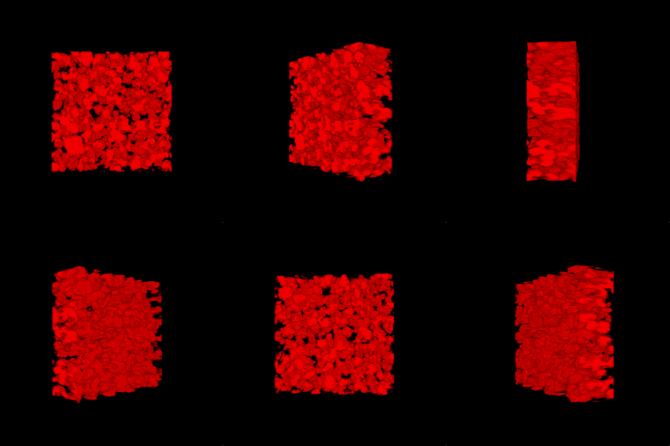
3-D rendering of the nanoparticle bijel taken by confocal microscope. (Credit: Caili Huang/ORNL [Oak Ridge National Laboratory] and Joe Forth/Berkeley Lab)

Here’s a link to and a citation for the paper,

Bicontinuous structured liquids with sub-micrometre domains using nanoparticle surfactants by Caili Huang, Joe Forth, Weiyu Wang, Kunlun Hong, Gregory S. Smith, Brett A. Helms & Thomas P. Russell. Nature Nanotechnology (2017) doi:10.1038/nnano.2017.182 25 September 2017

This paper is behind a paywall.

Nano-decoy for human influenza A virus

While the implications for this research are exciting, keep in mind that so far they’ve been testing immune-compromised mice. An Oct. 24, 2016 news item on Nanowerk announces the research,

To infect its victims, influenza A heads for the lungs, where it latches onto sialic acid on the surface of cells. So researchers created the perfect decoy: A carefully constructed spherical nanoparticle coated in sialic acid lures the influenza A virus to its doom. When misted into the lungs, the nanoparticle traps influenza A, holding it until the virus self-destructs.

An Oct. 24, 2015 Rensselaer Polytechnic Institute press release by Mary L. Martialay, which originated the news item, describes the research (Note: Links have been removed),

In a study on immune-compromised mice, the treatment reduced influenza A mortality from 100 percent to 25 percent over 14 days. The novel approach, which is radically different from existing influenza A vaccines, and treatments based on neuraminidase inhibitors, could be extended to a host of viruses that use a similar approach to infecting humans, such as Zika, HIV, and malaria. …

“Instead of blocking the virus, we mimicked its target – it’s a completely novel approach,” said Robert Linhardt, a glycoprotein expert and Rensselaer Polytechnic Institute professor who led the research. “It is effective with influenza and we have reason to believe it will function with many other viruses. This could be a therapeutic in cases where vaccine is not an option, such as exposure to an unanticipated strain, or with immune-compromised patients.”

The project is a collaboration between researchers within the Center for Biotechnology and Interdisciplinary Studies (CBIS) at Rensselaer and several institutions in South Korea including Kyungpook National University. Lead author Seok-Joon Kwon, a CBIS research scientist, coordinated the project across borders, enabling the South Korean institutions to test a drug designed and characterized at Rensselaer. …

To access the interior of a cell and replicate itself, influenza A must first bind to the cell surface, and then cut itself free. It binds with the protein hemagglutinin, and severs that tie with the enzyme neuraminidase. Influenza A produces numerous variations each of hemagglutinin and neuraminidase, all of which are antigens within the pathogen that provoke an immune system response. Strains of influenza A are characterized according to the variation of hemagglutinin and neuraminidase they carry, thus the origin of the familiar H1N1 or H3N2 designations.

Medications to counter the virus do exist, but all are vulnerable to the continual antigenic evolution of the virus. A yearly vaccine is effective only if it matches the strain of virus that infects the body. And the virus has shown an ability to develop resistance to a class of therapeutics based on neuraminidase inhibitors, which bind to and block neuraminidase.

The new solution targets an aspect of infection that does not change: all hemagglutinin varieties of influenza A must bind to human sialic acid. To trap the virus, the team designed a dendrimer, a spherical nanoparticle with treelike branches emanating from its core. On the outermost branches, they attached molecules, or “ligands,” of sialic acid.

The research found that the size of the dendrimer and the spacing between the ligands is integral to the function of the nanoparticle. Hemagglutinin occurs in clusters of three, or “trimers,” on the surface of the virus, and researchers found that a spacing of 3 nanometers between ligands resulted in the strongest binding to the trimers. Once bound to the densely packed dendrimer, viral neuraminidase is unable to sever the link. The coat of the virus contains millions of trimers, but the research revealed that only a few links provokes the virus to discharge its genetic cargo and ultimately self-destruct.

A different approach, using a less structured nanoparticle, had been previously tested in unrelated research, but the nanoparticle selected proved both toxic, and could be inactivated by neuraminidase. The new approach is far more promising.

“The major accomplishment was in designing an architecture that is optimized to bind so tightly to the hemagglutinin, the neuraminidase can’t squeeze in and free the virus,” said Linhardt. “It’s trapped.”

Here’s a link to and a citation for the paper,

Nanostructured glycan architecture is important in the inhibition of influenza A virus infection by Seok-Joon Kwon, Dong Hee Na, Jong Hwan Kwak, Marc Douaisi, Fuming Zhang, Eun Ji Park, Jong-Hwan Park, Hana Youn, Chang-Seon Song, Ravi S. Kane, Jonathan S. Dordick, Kyung Bok Lee, & Robert J. Linhardt. Nature Nanotechnology (2016)  doi:10.1038/nnano.2016.181 Published online 24 October 2016

This paper is behind a paywall.

Gold unzips your DNA but not in a sexy way

The animation that the scientists from North Carolina have provided makes the gold nanoparticles look downright mean as that DNA definitely does not want to be unzipped but perhaps your mileage varies,

The June 20, 2012 news item on Nanowerk provides more detail,

New research from North Carolina State University finds that gold nanoparticles with a slight positive charge work collectively to unravel DNA’s double helix. This finding has ramifications for gene therapy research and the emerging field of DNA-based electronics.

The research team introduced gold nanoparticles, approximately 1.5 nanometers in diameter, into a solution containing double-stranded DNA. The nanoparticles were coated with organic molecules called ligands. Some of the ligands held a positive charge, while others were hydrophobic – meaning they were repelled by water.

Because the gold nanoparticles had a slight positive charge from the ligands, and DNA is always negatively charged, the DNA and nanoparticles were pulled together into complex packages.

“However, we found that the DNA was actually being unzipped by the gold nanoparticles,” Melechko [Dr. Anatoli Melechko, an associate professor of materials science and engineering at NC State and co-author of the paper] says. The positively-charged ligands on the nanoparticles attached to the DNA as predicted, but the hydrophobic ligands of the nanoparticles became tangled with each other. As this tangling pulled the nanoparticles into clusters, the nanoparticles pulled the DNA apart.

The implications for this ‘unzipping’ are,

“We think gold nanoparticles still hold promise for gene therapy,” says Dr. Yaroslava Yingling, an assistant professor of materials science and engineering at NC State and co-author of the paper. “But it’s clear that we need to tailor the ligands, charge and chemistry of these materials to ensure the DNA’s structural integrity is not compromised.”

The finding is also relevant to research on DNA-based electronics, which hopes to use DNA as a template for creating nanoelectronic circuits. Because some work in that field involves placing metal nanoparticles on DNA, this finding indicates that researchers will have to pay close attention to the characteristics of those nanoparticles – or risk undermining the structural integrity of the DNA.