Tag Archives: liver

Precision targeting of the liver for gene editing

Apparently the magic is in the lipid nanoparticles. A March 1, 2021 news item on Nanowerk announced research into lipid nanoparticles as a means to deliver CRISPR (clustered regularly interspaced short palindromic repeats) to specific organs (Note: A link has been removed),

The genome editing technology CRISPR has emerged as a powerful new tool that can change the way we treat disease. The challenge when altering the genetics of our cells, however, is how to do it safely, effectively, and specifically targeted to the gene, tissue and organ that needs treatment.

Scientists at Tufts University and the Broad Institute of Harvard [University] and MIT [Massachusetts Institute of Technology] have developed unique nanoparticles comprised of lipids — fat molecules — that can package and deliver gene editing machinery specifically to the liver.

In a study published in the Proceedings of the National Academy of Sciences [PNAS] (“Lipid nanoparticle-mediated codelivery of Cas9 mRNA and single-guide RNA achieves liver-specific in vivo genome editing of Angptl3”), they have shown that they can use the lipid nanoparticles (LNPs) to efficiently deliver the CRISPR machinery into the liver of mice, resulting in specific genome editing and the reduction of blood cholesterol levels by as much as 57% — a reduction that can last for at least several months with just one shot.

A March 2, 2021 Tufts University news release (also on EurekAlert but published March 1, 2021), which originated the news item, provides greater insight into and technical detail about the research,

The problem of high cholesterol plagues more than 29 million Americans, according to the Centers for Disease Control and Prevention. The condition is complex and can originate from multiple genes as well as nutritional and lifestyle choices, so it is not easy to treat. The Tufts and Broad researchers, however, have modified one gene that could provide a protective effect against elevated cholesterol if it can be shut down by gene editing.

The gene that the researchers focused on codes for the angiopoietin-like 3 enzyme (Angptl3). That enzyme tamps down the activity of other enzymes – lipases – that help break down cholesterol. If researchers can knock out the Angptl3 gene, they can let the lipases do their work and reduce levels of cholesterol in the blood. It turns out that some lucky people have a natural mutation in their Angptl3 gene, leading to consistently low levels of triglycerides and low-density lipoprotein (LDL) cholesterol, commonly called “bad” cholesterol, in their bloodstream without any known clinical downsides.

“If we can replicate that condition by knocking out the angptl3 gene in others, we have a good chance of having a safe and long term solution to high cholesterol,” said Qiaobing Xu, associate professor of biomedical engineering at Tufts’ School of Engineering and corresponding author of the study. “We just have to make sure we deliver the gene editing package specifically to the liver so as not to create unwanted side effects.”

Xu’s team was able to do precisely that in mouse models. After a single injection of lipid nanoparticles packed with mRNA coding for CRISPR-Cas9 and a single-guide RNA targeting Angptl3, they observed a profound reduction in LDL cholesterol by as much as 57% and triglyceride levels by about 29 %, both of which remained at those lowered levels for at least 100 days. The researchers speculate that the effect may last much longer than that, perhaps limited only by the slow turnover of cells in the liver, which can occur over a period of about a year. The reduction of cholesterol and triglycerides is dose dependent, so their levels could be adjusted by injecting fewer or more LNPs in the single shot, the researchers said.

By comparison, an existing, FDA [US Food and Drug Administration]-approved version of CRISPR mRNA-loaded LNPs could only reduce LDL cholesterol by at most 15.7% and triglycerides by 16.3% when it was tested in mice, according to the researchers.

The trick to making a better LNP was in customizing the components – the molecules that come together to form bubbles around the mRNA. The LNPs are made up of long chain lipids that have a charged or polar head that is attracted to water, a carbon chain tail that points toward the middle of the bubble containing the payload, and a chemical linker between them. Also present are polyethylene glycol, and yes, even some cholesterol – which has a normal role in lipid membranes to make them less leaky – to hold their contents better.

The researchers found that the nature and relative ratio of these components appeared to have profound effects on the delivery of mRNA into the liver, so they tested LNPs with many combinations of heads, tails, linkers and ratios among all components for their ability to target liver cells. Because the in vitro potency of an LNP formulation rarely reflects its in vivo performance, they directly evaluated the delivery specificity and efficacy in mice that have a reporter gene in their cells that lights up red when genome editing occurs. Ultimately, they found a CRISPR mRNA-loaded LNP that lit up just the liver in mice, showing that it could specifically and efficiently deliver gene-editing tools into the liver to do their work.

The LNPs were built upon earlier work at Tufts, where Xu and his team developed LNPs with as much as 90% efficiency in delivering mRNA into cells. A unique feature of those nanoparticles was the presence of disulfide bonds between the long lipid chains. Outside the cells, the LNPs form a stable spherical structure that locks in their contents. When they are inside a cell, the environment within breaks the disulfide bonds to disassemble the nanoparticles. The contents are then quickly and efficiently released into the cell. By preventing loss outside the cell, the LNPs can have a much higher yield in delivering their contents.

“CRISPR is one of the most powerful therapeutic tools for the treatment of diseases with a genetic etiology. We have recently seen the first human clinical trail for CRISPR therapy enabled by LNP delivery to be administered systemically to edit genes inside the human body. Our LNP platform developed here holds great potential for clinical translation,” said Min Qiu, post-doctoral researcher in Xu’s lab at Tufts.  “We envision that with this LNP platform in hand, we could now make CRISPR a practical and safe approach to treat a broad spectrum of liver diseases or disorders,” said Zachary Glass, graduate student in the Xu lab. Qiu and Glass are co-first authors of the study.

Here’s a link to and a citation for the paper,

Lipid nanoparticle-mediated codelivery of Cas9 mRNA and single-guide RNA achieves liver-specific in vivo genome editing of Angptl3 by Min Qiu, Zachary Glass, Jinjin Chen, Mary Haas, Xin Jin, Xuewei Zhao, Xuehui Rui, Zhongfeng Ye, Yamin Li, Feng Zhang, and Qiaobing Xu. PNAS March 9, 2021 118 (10) e2020401118 DOI: https://doi.org/10.1073/pnas.2020401118

This paper appears to be behind a paywall.
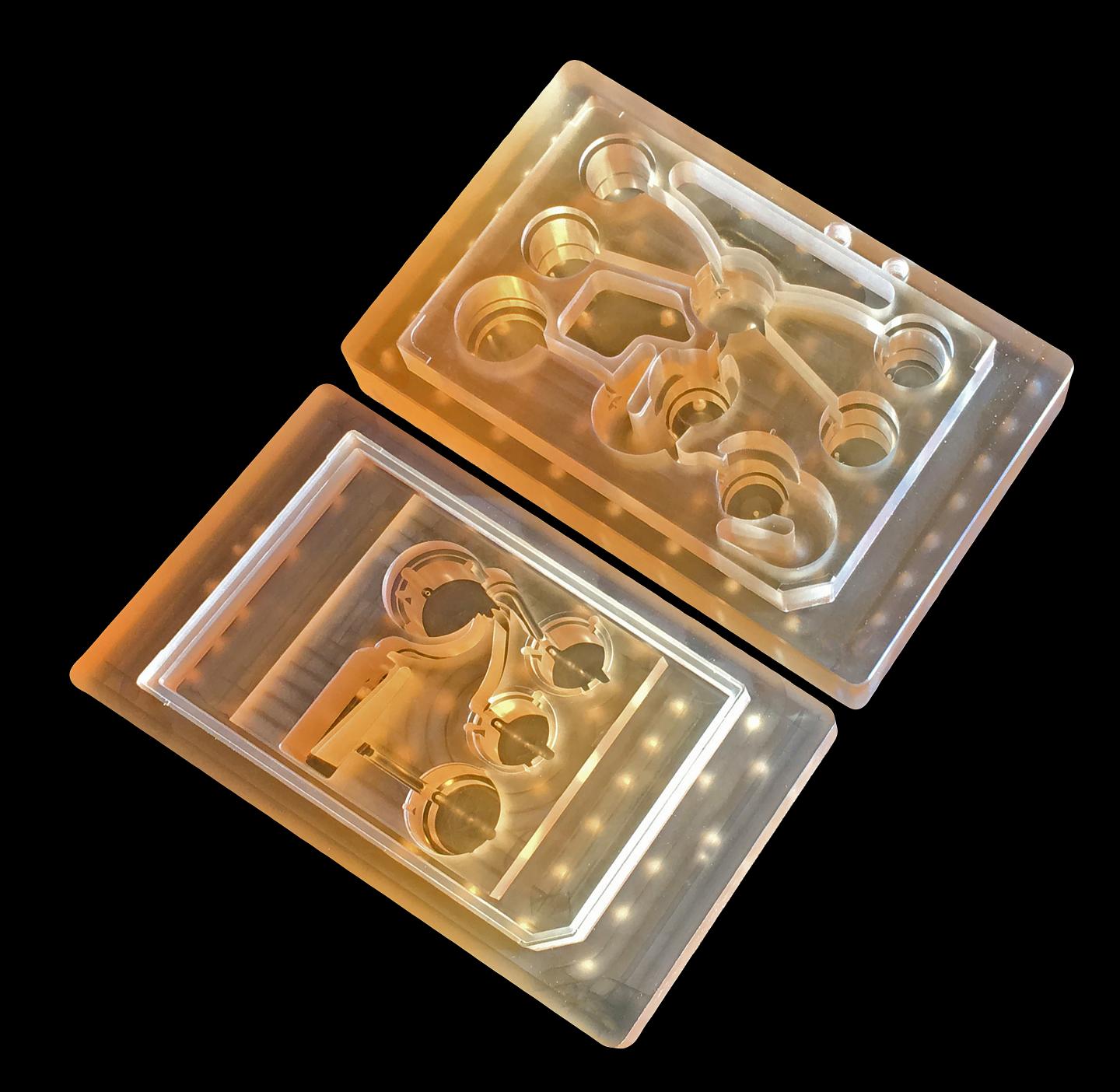
Body-on-a-chip (10 organs)

Also known as human-on-a-chip, the 10-organ body-on-a-chip was being discussed at the 9th World Congress on Alternatives to Animal Testing in the Life Sciences in 2014 in Prague, Czech Republic (see this July 1, 2015 posting for more). At the time, scientists were predicting success at achieving their goal of 10 organs on-a-chip in 2017 (the best at the time was four organs). Only a few months past that deadline, scientists from the Massachusetts Institute of Technology (MIT) seem to have announced a ’10 organ chip’ in a March 14, 2018 news item on ScienceDaily,

MIT engineers have developed new technology that could be used to evaluate new drugs and detect possible side effects before the drugs are tested in humans. Using a microfluidic platform that connects engineered tissues from up to 10 organs, the researchers can accurately replicate human organ interactions for weeks at a time, allowing them to measure the effects of drugs on different parts of the body.

Such a system could reveal, for example, whether a drug that is intended to treat one organ will have adverse effects on another.

A March 14, 2018 MIT news release (also on EurekAlert), which originated the news item, expands on the theme,

“Some of these effects are really hard to predict from animal models because the situations that lead to them are idiosyncratic,” says Linda Griffith, the School of Engineering Professor of Teaching Innovation, a professor of biological engineering and mechanical engineering, and one of the senior authors of the study. “With our chip, you can distribute a drug and then look for the effects on other tissues, and measure the exposure and how it is metabolized.”

These chips could also be used to evaluate antibody drugs and other immunotherapies, which are difficult to test thoroughly in animals because they are designed to interact with the human immune system.

David Trumper, an MIT professor of mechanical engineering, and Murat Cirit, a research scientist in the Department of Biological Engineering, are also senior authors of the paper, which appears in the journal Scientific Reports. The paper’s lead authors are former MIT postdocs Collin Edington and Wen Li Kelly Chen.

Modeling organs

When developing a new drug, researchers identify drug targets based on what they know about the biology of the disease, and then create compounds that affect those targets. Preclinical testing in animals can offer information about a drug’s safety and effectiveness before human testing begins, but those tests may not reveal potential side effects, Griffith says. Furthermore, drugs that work in animals often fail in human trials.

“Animals do not represent people in all the facets that you need to develop drugs and understand disease,” Griffith says. “That is becoming more and more apparent as we look across all kinds of drugs.”

Complications can also arise due to variability among individual patients, including their genetic background, environmental influences, lifestyles, and other drugs they may be taking. “A lot of the time you don’t see problems with a drug, particularly something that might be widely prescribed, until it goes on the market,” Griffith says.

As part of a project spearheaded by the Defense Advanced Research Projects Agency (DARPA), Griffith and her colleagues decided to pursue a technology that they call a “physiome on a chip,” which they believe could offer a way to model potential drug effects more accurately and rapidly. To achieve this, the researchers needed new equipment — a platform that would allow tissues to grow and interact with each other — as well as engineered tissue that would accurately mimic the functions of human organs.

Before this project was launched, no one had succeeded in connecting more than a few different tissue types on a platform. Furthermore, most researchers working on this kind of chip were working with closed microfluidic systems, which allow fluid to flow in and out but do not offer an easy way to manipulate what is happening inside the chip. These systems also require external pumps.

The MIT team decided to create an open system, which essentially removes the lid and makes it easier to manipulate the system and remove samples for analysis. Their system, adapted from technology they previously developed and commercialized through U.K.-based CN BioInnovations, also incorporates several on-board pumps that can control the flow of liquid between the “organs,” replicating the circulation of blood, immune cells, and proteins through the human body. The pumps also allow larger engineered tissues, for example tumors within an organ, to be evaluated.

Complex interactions

The researchers created several versions of their chip, linking up to 10 organ types: liver, lung, gut, endometrium, brain, heart, pancreas, kidney, skin, and skeletal muscle. Each “organ” consists of clusters of 1 million to 2 million cells. These tissues don’t replicate the entire organ, but they do perform many of its important functions. Significantly, most of the tissues come directly from patient samples rather than from cell lines that have been developed for lab use. These so-called “primary cells” are more difficult to work with but offer a more representative model of organ function, Griffith says.

Using this system, the researchers showed that they could deliver a drug to the gastrointestinal tissue, mimicking oral ingestion of a drug, and then observe as the drug was transported to other tissues and metabolized. They could measure where the drugs went, the effects of the drugs on different tissues, and how the drugs were broken down. In a related publication, the researchers modeled how drugs can cause unexpected stress on the liver by making the gastrointestinal tract “leaky,” allowing bacteria to enter the bloodstream and produce inflammation in the liver.

Kevin Healy, a professor of bioengineering and materials science and engineering at the University of California at Berkeley, says that this kind of system holds great potential for accurate prediction of complex adverse drug reactions.

“While microphysiological systems (MPS) featuring single organs can be of great use for both pharmaceutical testing and basic organ-level studies, the huge potential of MPS technology is revealed by connecting multiple organ chips in an integrated system for in vitro pharmacology. This study beautifully illustrates that multi-MPS “physiome-on-a-chip” approaches, which combine the genetic background of human cells with physiologically relevant tissue-to-media volumes, allow accurate prediction of drug pharmacokinetics and drug absorption, distribution, metabolism, and excretion,” says Healy, who was not involved in the research.

Griffith believes that the most immediate applications for this technology involve modeling two to four organs. Her lab is now developing a model system for Parkinson’s disease that includes brain, liver, and gastrointestinal tissue, which she plans to use to investigate the hypothesis that bacteria found in the gut can influence the development of Parkinson’s disease.

Other applications include modeling tumors that metastasize to other parts of the body, she says.

“An advantage of our platform is that we can scale it up or down and accommodate a lot of different configurations,” Griffith says. “I think the field is going to go through a transition where we start to get more information out of a three-organ or four-organ system, and it will start to become cost-competitive because the information you’re getting is so much more valuable.”

The research was funded by the U.S. Army Research Office and DARPA.

Caption: MIT engineers have developed new technology that could be used to evaluate new drugs and detect possible side effects before the drugs are tested in humans. Using a microfluidic platform that connects engineered tissues from up to 10 organs, the researchers can accurately replicate human organ interactions for weeks at a time, allowing them to measure the effects of drugs on different parts of the body. Credit: Felice Frankel

Here’s a link to and a citation for the paper,

Interconnected Microphysiological Systems for Quantitative Biology and Pharmacology Studies by Collin D. Edington, Wen Li Kelly Chen, Emily Geishecker, Timothy Kassis, Luis R. Soenksen, Brij M. Bhushan, Duncan Freake, Jared Kirschner, Christian Maass, Nikolaos Tsamandouras, Jorge Valdez, Christi D. Cook, Tom Parent, Stephen Snyder, Jiajie Yu, Emily Suter, Michael Shockley, Jason Velazquez, Jeremy J. Velazquez, Linda Stockdale, Julia P. Papps, Iris Lee, Nicholas Vann, Mario Gamboa, Matthew E. LaBarge, Zhe Zhong, Xin Wang, Laurie A. Boyer, Douglas A. Lauffenburger, Rebecca L. Carrier, Catherine Communal, Steven R. Tannenbaum, Cynthia L. Stokes, David J. Hughes, Gaurav Rohatgi, David L. Trumper, Murat Cirit, Linda G. Griffith. Scientific Reports, 2018; 8 (1) DOI: 10.1038/s41598-018-22749-0 Published online:

This paper which describes testing for four-, seven-, and ten-organs-on-a-chip, is open access. From the paper’s Discussion,

In summary, we have demonstrated a generalizable approach to linking MPSs [microphysiological systems] within a fluidic platform to create a physiome-on-a-chip approach capable of generating complex molecular distribution profiles for advanced drug discovery applications. This adaptable, reusable system has unique and complementary advantages to existing microfluidic and PDMS-based approaches, especially for applications involving high logD substances (drugs and hormones), those requiring precise and flexible control over inter-MPS flow partitioning and drug distribution, and those requiring long-term (weeks) culture with reliable fluidic and sampling operation. We anticipate this platform can be applied to a wide range of problems in disease modeling and pre-clinical drug development, especially for tractable lower-order (2–4) interactions.

Congratulations to the researchers!

Discovering how the liver prevents nanoparticles from reaching cancer cells

There’s a lot of excitement about nanoparticles as enabling a precise drug delivery system but to date results have been disappointing as a team of researchers at the University of Toronto (Canada) noted recently (see my April 27, 2016 posting). According to those researchers, one of the main problems with the proposed nanoparticle drug delivery system is that we don’t understand how the body delivers materials to cells and disappointingly few nanoparticles (less than 1%) make their way to tumours. That situation may be changing.

An Aug. 19, 2016 news item on Nanowerk announces the latest research from the University of Toronto,

The emerging field of nanomedicine holds great promise in the battle against cancer. Particles the size of protein molecules can be customized to carry tumour-targeting drugs and destroy cancer cells without harming healthy tissue.

But here’s the problem: when nanoparticles are administered into the body, more than 99 per cent of them become trapped in non-targeted organs, such as the liver and spleen. These nanoparticles are not delivered to the site of action to carry out their intended function.

To solve this problem, researchers at the University of Toronto and the University Health Network have figured out how the liver and spleen trap intact nanoparticles as they move through the organ. “If you want to unlock the promise of nanoparticles, you have to understand and solve the problem of the liver,” says Dr. Ian McGilvray, a transplant surgeon at the Toronto General Hospital and scientist at the Toronto General Research Institute (TGRI).

An Aug. 15, 2016 University of Toronto news release by Luke Ng, which originated the news item, expands on the theme,

In a recent paper in the journal Nature Materials, the researchers say that as nanoparticles move through the liver sinusoid, the flow rate slows down 1,000 times, which increases the interaction of the nanoparticles all of types of liver cells. This was a surprising finding because the current thought is that Kupffer cells, responsible for toxin breakdown in the liver, are the ones that gobbles [sic] up the particles.  This study found that liver B-cells and liver sinusoidal endothelial cells are also involved and that the cell phenotype also matters.

“We know that the liver is the principle organ controlling what gets absorbed by our bodies and what gets filtered out—it governs our everyday biological functions,” says Dr. Kim Tsoi (… [and] research partner Sonya MacParland), a U of T orthopaedic surgery resident, and a first author of the paper, who completed her PhD in biomedical engineering with Warren Chan (IBBME). “But nanoparticle drug delivery is a newer approach and we haven’t had a clear picture of how they interact with the liver—until now.”

Tsoi and MacParland first examined both the speed and location of their engineered nanoparticles as they moved through the liver.

“This gives us a target to focus on,” says MacParland, an immunology post-doctoral fellow at U of T and TGRI. “Knowing the specific cells to modify will allow us to eventually deliver more of the nanoparticles to their intended target, attacking only the pathogens or tumours, while bypassing healthy cells.”

“Many prior studies that have tried to reduce nanomaterial clearance in the liver have focused on the particle design itself,” says Chan. “But our work now gives greater insight into the biological mechanisms underpinning our experimental observations — now we hope to use our fundamental findings to help design nanoparticles that work with the body, rather than against it.”

Here’s a link to and a citation for the paper,

Mechanism of hard-nanomaterial clearance by the liver by Kim M. Tsoi, Sonya A. MacParland, Xue-Zhong Ma, Vinzent N. Spetzler, Juan Echeverri, Ben Ouyang, Saleh M. Fadel, Edward A. Sykes, Nicolas Goldaracena, Johann M. Kaths, John B. Conneely, Benjamin A. Alman, Markus Selzner, Mario A. Ostrowski, Oyedele A. Adeyi, Anton Zilman, Ian D. McGilvray, & Warren C. W. Chan. Nature Materials (2016) doi:10.1038/nmat4718 Published online 15 August 2016

This paper is behind a paywall.

University of Toronto (Canada) researchers and lab-grown heart and liver tissue (person-on-a-chip)

Usually called ‘human-on-a-chip’, a team at the University of Toronto have developed a two-organ ‘person on a chip’ according to a March 7, 2016 news item on phys.org (Note: Links have been removed),

Researchers at U of T [University of Toronto] Engineering have developed a new way of growing realistic human tissues outside the body. Their “person-on-a-chip” technology, called AngioChip, is a powerful platform for discovering and testing new drugs, and could eventually be used to repair or replace damaged organs.

Professor Milica Radisic (IBBME, ChemE), graduate student Boyang Zhang and the rest of the team are among those research groups around the world racing to find ways to grow human tissues in the lab, under conditions that mimic a real person’s body. They have developed unique methods for manufacturing small, intricate scaffolds for individual cells to grow on. These artificial environments produce cells and tissues that resemble the real thing more closely than those grown lying flat in a petri dish.

The team’s recent creations have included BiowireTM—an innovative method of growing heart cells around a silk suture—as well as a scaffold for heart cells that snaps together like sheets of Velcro. But AngioChip takes tissue engineering to a whole new level. “It’s a fully three-dimensional structure complete with internal blood vessels,” says Radisic. “It behaves just like vasculature, and around it there is a lattice for other cells to attach and grow.” …

A March 7, 2016 University of Toronto news release (also on EurekAlert), which originated the news item, provides more detail about the AngioChip,

Zhang built the scaffold out of POMaC, a polymer that is both biodegradable and biocompatible. The scaffold is built out of a series of thin layers, stamped with a pattern of channels that are each about 50 to 100 micrometres wide. The layers, which resemble the computer microchips, are then stacked into a 3D structure of synthetic blood vessels. As each layer is added, UV light is used to cross-link the polymer and bond it to the layer below.

When the structure is finished, it is bathed in a liquid containing living cells. The cells quickly attach to the inside and outside of the channels and begin growing just as they would in the human body.

“Previously, people could only do this using devices that squish the cells between sheets of silicone and glass,” says Radisic. “You needed several pumps and vacuum lines to run just one chip. Our system runs in a normal cell culture dish, and there are no pumps; we use pressure heads to perfuse media through the vasculature. The wells are open, so you can easily access the tissue.”

Using the platform, the team has built model versions of both heart and liver tissues that function like the real thing. “Our liver actually produced urea and metabolized drugs,” says Radisic. They can connect the blood vessels of the two artificial organs, thereby modelling not just the organs themselves, but the interactions between them. They’ve even injected white blood cells into the vessels and watched as they squeezed through gaps in the vessel wall to reach the tissue on the other side, just as they do in the human body.

The news release also mentions potential markets and the work that needs to be accomplished before AngioChip is available for purchase,

AngioChip has great potential in the field of pharmaceutical testing. Current drug-testing methods, such as animal testing and controlled clinical trials, are costly and fraught with ethical concerns. Testing on lab-grown human tissues would provide a realistic model at a fraction of the cost, but this area of research is still in its infancy. “In the last few years, it has become possible to order cultures of human cells for testing, but they’re grown on a plate, a two-dimensional environment,” says Radisic. “They don’t capture all the functional hallmarks of a real heart muscle, for example.”

A more realistic platform like AngioChip could enable drug companies to detect dangerous side effects and interactions between organ compartments long before their products reach the market, saving countless lives. It could also be used to understand and validate the effectiveness of current drugs and even to screen libraries of chemical compounds to discover new drugs. Through TARA Biosystems Inc., a spin-off company co-founded by Radisic, the team is already working on commercializing the technology.

In future, Radisic envisions her lab-grown tissues being implanted into the body to repair organs damaged by disease. Because the cells used to seed the platform can come from anyone, the new tissues could be genetically identical to the intended host, reducing the risk of organ rejection. Even in its current form, the team has shown that the AngioChip can be implanted into a living animal, its artificial blood vessels connected to a real circulatory system. The polymer scaffolding itself simply biodegrades after several months.

The team still has much work to do. Each AngioChip is currently made by hand; if the platform is to be used industrially, the team will need to develop high-throughput manufacturing methods to create many copies at once. Still, the potential is obvious. “It really is multifunctional, and solves many problems in the tissue engineering space,” says Radisic. “It’s truly next-generation.”

Here’s a link to and a citation for the paper,

Biodegradable scaffold with built-in vasculature for organ-on-a-chip engineering and direct surgical anastomosis by Boyang Zhang, Miles Montgomery, M. Dean Chamberlain, Shinichiro Ogawa, Anastasia Korolj, Aric Pahnke, Laura A. Wells, Stéphane Massé, Jihye Kim, Lewis Reis, Abdul Momen, Sara S. Nunes, Aaron R. Wheeler, Kumaraswamy Nanthakumar, Gordon Keller, Michael V. Sefton, & Milica Radisic. Nature Materials (2016) doi:10.1038/nmat4570 Published online 07 March 2016

This paper is behind a paywall.

The researchers have made two images illustrating their work available. There’s this still image,

These tiny polymer scaffolds contain channels that are about 100 micrometres wide, about the same diameter as a human hair. When seeded with cells, the channels act as artificial blood vessels. By mimicking tissues in the human heart and other organs, these scaffolds provide a new way to test drugs for potentially dangerous side effects. (Image: Tyler Irving/Boyang Zhang/Kevin Soobrian)

These tiny polymer scaffolds contain channels that are about 100 micrometres wide, about the same diameter as a human hair. When seeded with cells, the channels act as artificial blood vessels. By mimicking tissues in the human heart and other organs, these scaffolds provide a new way to test drugs for potentially dangerous side effects. (Image: Tyler Irving/Boyang Zhang/Kevin Soobrian)

Perhaps more intriguing is this one,

UofT_AngioChipMoving

When seeded with heart cells, the flexible polymer scaffold contracts with a regular rhythm, just like real heart tissue. (Image: Boyang Zhang)

I have mentioned ‘human-on-a-chip’ projects many times here and as the news release writer notes, there is an international race. My July 1, 2015 posting (cross-posted from the June 30, 2015 posting [Testing times: the future of animal alternatives] on the International Innovation blog [a CORDIS-listed project dissemination partner for FP7 and H2020 projects]) notes a couple of those projects,

Organ-on-a-chip projects use stem cells to create human tissues that replicate the functions of human organs. Discussions about human-on-a-chip activities – a phrase used to describe 10 interlinked organ chips – were a highlight of the 9th World Congress on Alternatives to Animal Testing held in Prague, Czech Republic, last year. One project highlighted at the event was a joint US National Institutes of Health (NIH), US Food and Drug Administration (FDA) and US Defense Advanced Research Projects Agency (DARPA) project led by Dan Tagle that claimed it would develop functioning human-on-a-chip by 2017. However, he and his team were surprisingly close-mouthed and provided few details making it difficult to assess how close they are to achieving their goal.

By contrast, Uwe Marx – Leader of the ‘Multi-Organ-Chip’ programme in the Institute of Biotechnology at the Technical University of Berlin and Scientific Founder of TissUse, a human-on-a-chip start-up company – claims to have sold two-organ chips. He also claims to have successfully developed a four-organ chip and that he is on his way to building a human-on-a-chip. Though these chips remain to be seen, if they are, they will integrate microfluidics, cultured cells and materials patterned at the nanoscale to mimic various organs, and will allow chemical testing in an environment that somewhat mirrors a human.

As for where the University of Toronto efforts fit into the race, I don’t know for sure. It’s the first time I’ve come across a reference to liver tissue producing urea but I believe there’s at least one other team in China which has achieved a three-dimensional, more lifelike aspect for liver tissue in my Jan. 29, 2016 posting ‘Constructing a liver’.

Faster, cheaper, pseudo-organs (also known as organoids)

There’ve been any number of ‘organoid’ stories recently, here and elsewhere. This one is special due to a quasi extra-cellular matrix (cells have a type of skeletal structure known as an extra-cellular matrix or ECM). From a Sept. 11, 2015 news item on Azonano,

Scientists have developed a new technique that produces a user friendly, low cost, tissue-engineered pseudo-organ. The chip-based model produces a faithful mimic of the in vivo liver inside a scalable fluid-handling device, demonstrating proof of principle for toxicology tests and opening up potential use in drug testing and personalised medicine.

The work was done by researchers based at the Wake Forest Institute for Regenerative Medicine and the Virginia Tech-Wake Forest University School of Biomedical Engineering and Sciences. They created a device architecture within which were a series of 3D liver cell constructs enclosed in a biopolymer that closely mimics the extra-cellular matrix (ECM). Surrounding the printed cells with this ECM – which the body uses to support cells in the liver – makes this model a more realistic model of the cells in vivo.

A Sept. 10, 2015 Institute of Physics (IOP) press release, which originated the news item, provides more details about the technology,

The technique uses photopatterning to produce defined 3D constructs in a microfluidic system to probe the construct quickly. “It’s basically scaled-down pluming” explains Adam Hall, an author on the paper. “This paper describes fairly hefty devices – a few mm – but we’re working to scale this down considerably.”

Collaboration proved to be the key to success; “The challenges were not too significant once Adam and I merged our areas of expertise.” adds Aleksander Skardal, another author on the paper. “With his background in devices and microfabrication, and my background in biomaterials and biofabrication, the two technologies integrated rather well.”

The 3D construct device offers a new tool in the development of drug treatments. At present, 2D testing in vitro doesn’t replicate the activity of the cells, and until now 3D systems have not provided adequate interactions of cells with the ECM, or offered particularly high-throughput testing.

This is where the combination of technologies has proven vital. “3D constructs are less effective if you can’t probe them quickly” continues Hall. “And without some important task, microfluidics are just a fun party trick.”

The researchers were also happy how quickly the techniques fell into place.

“The first time we attempted to perform the in situ photopatterning – it just worked” says Skardal. “Science isn’t always that easy, so we knew we might be onto something.”

“Yes – this was one of those rare occasions where things seemed to fall into place” adds Hall.

The researchers are now working to reduce the size of the system allowing for multiple constructs that could be tested individually. This would open potential usage in drug testing and personalised medicine.

“Imagine being able to put, for example, tumor cells from a patient on a chip and test different drug cocktails on them” they conclude. “You could determine the effectiveness and side effects of different treatments on an individual basis without endangering the patient.”

Here’s a link to and a citation for the paper,

In situ patterned micro 3D liver constructs for parallel toxicology testing in a fluidic device by Aleksander Skardal, Mahesh Devarasetty, Shay Soker, and Adam R Hall. Biofabrication, Volume 7, Number 3 DOI: 10.1088/1758-5090/7/3/032001 Published 11 September 2015

© 2015 IOP Publishing Ltd

This is an open access paper.

Two-organ tests (body-on-a-chip) show liver damage possible from nanoparticles

This is the first time I’ve seen testing of two organs for possible adverse effects from nanoparticles. In this case, the researchers were especially interested in the liver. From an Aug. 12, 2014 news item on Azonano,

Nanoparticles in food, sunscreen and other everyday products have many benefits. But Cornell [University] biomedical scientists are finding that at certain doses, the particles might cause human organ damage.

A recently published study in Lab on a Chip by the Royal Society of Chemistry and led by senior research associate Mandy Esch shows that nanoparticles injure liver cells when they are in microfluidic devices designed to mimic organs of the human body. The injury was worse when tested in two-organ systems, as opposed to single organs – potentially raising concerns for humans and animals.

Anne Ju’s Aug. 11, 2014 article for Cornell University’s Chronicle describes the motivation for this work and the research itself in more detail,

“We are looking at the effects of what are considered to be harmless nanoparticles in humans,” Esch said. “These particles are not necessarily lethal, but … are there other consequences? We’re looking at the non-lethal consequences.”

She used 50-nanometer carboxylated polystyrene nanoparticles, found in some animal food sources and considered model inert particles. Shuler’s lab specializes in “body-on-a-chip” microfluidics, which are engineered chips with carved compartments that contain cell cultures to represent the chemistry of individual organs.

In Esch’s experiment, she made a human intestinal compartment, a liver compartment and a compartment to represent surrounding tissues in the body. She then observed the effects of fluorescently labeled nanoparticles as they traveled through the system.

Esch found that both single nanoparticles as well as small clusters crossed the gastrointestinal barrier and reached liver cells, and the liver cells released an enzyme called aspartate transaminase, known to be released during cell death or damage.

It’s unclear exactly what damage is occurring or why, but the results indicate that the nanoparticles must be undergoing changes as they cross the gastrointestinal barrier, and that these alterations may change their toxic potential, Esch said. Long-term consequences for organs in proximity could be a concern, she said.

“The motivation behind this study was twofold: one, to show that multi-organ, in vitro systems give us more information when testing for the interaction of a substance with the human body, and two … to look at nanoparticles because they have a huge potential for medicine, yet adverse effects have not been studied in detail yet,” Esch said.

Mary Macleod’s July 3, 2014 article for Chemistry World features a diagram of the two-organ system and more technical details about the research,

Schematic of the two-organ system [downloaded from http://www.rsc.org/chemistryworld/2014/07/nanoparticle-liver-gastrointestinal-tract-microfluidic-chip]

Schematic of the two-organ system [downloaded from http://www.rsc.org/chemistryworld/2014/07/nanoparticle-liver-gastrointestinal-tract-microfluidic-chip]

HepG2/C3A cells were used to represent the liver, with the intestinal cell co-culture consisting of enterocytes (Caco-2) and mucin-producing (HT29-MTX) cells. Carboxylated polystyrene nanoparticles were fluorescently labelled so their movement between the chambers could be tracked. Levels of aspartate transaminase, a cytosolic enzyme released into the culture medium upon cell death, were measured to give an indication of liver damage.

The study saw that single nanoparticles and smaller nanoparticle aggregates were able to cross the GI barrier and reach the liver cells. The increased zeta potentials of these nanoparticles suggest that crossing the barrier may raise their toxic potential. However, larger nanoparticles, which interact with cell membranes and aggregate into clusters, were stopped much more effectively by the GI tract barrier.

The gastrointestinal tract is an important barrier preventing ingested substances crossing into systemic circulation. Initial results indicate that soluble mediators released upon low-level injury to liver cells may enhance the initial injury by damaging the cells which form the GI tract. These adverse effects were not seen in conventional single-organ tests.

Here’s a link to and a citation for the paper,

Body-on-a-chip simulation with gastrointestinal tract and liver tissues suggests that ingested nanoparticles have the potential to cause liver injury by Mandy B. Esch, Gretchen J. Mahler, Tracy Stokol, and Michael L. Shuler. Lab Chip, 2014,14, 3081-3092 DOI: 10.1039/C4LC00371C First published online 27 Jun 2014

This paper is open access until Aug. 12, 2014.

While this research is deeply concerning, it should be noted the researchers are being very careful in their conclusions as per Ju’s article, “It’s unclear exactly what damage is occurring or why, but the results indicate that the nanoparticles must be undergoing changes as they cross the gastrointestinal barrier, and that these alterations may change their toxic potential … Long-term consequences for organs in proximity could be a concern … .”