Tag Archives: Lund University

2023 Nobel prizes (medicine, physics, and chemistry)

For the first time in the 15 years this blog has been around, the Nobel prizes awarded in medicine, physics, and chemistry all are in areas discussed here at one or another. As usual where people are concerned, some of these scientists had a tortuous journey to this prestigious outcome.

Medicine

Two people (Katalin Karikó and Drew Weissman) were awarded the prize in medicine according to the October 2, 2023 Nobel Prize press release, Note: Links have been removed,

The Nobel Assembly at Karolinska Institutet [Sweden]

has today decided to award

the 2023 Nobel Prize in Physiology or Medicine

jointly to

Katalin Karikó and Drew Weissman

for their discoveries concerning nucleoside base modifications that enabled the development of effective mRNA vaccines against COVID-19

The discoveries by the two Nobel Laureates were critical for developing effective mRNA vaccines against COVID-19 during the pandemic that began in early 2020. Through their groundbreaking findings, which have fundamentally changed our understanding of how mRNA interacts with our immune system, the laureates contributed to the unprecedented rate of vaccine development during one of the greatest threats to human health in modern times.

Vaccines before the pandemic

Vaccination stimulates the formation of an immune response to a particular pathogen. This gives the body a head start in the fight against disease in the event of a later exposure. Vaccines based on killed or weakened viruses have long been available, exemplified by the vaccines against polio, measles, and yellow fever. In 1951, Max Theiler was awarded the Nobel Prize in Physiology or Medicine for developing the yellow fever vaccine.

Thanks to the progress in molecular biology in recent decades, vaccines based on individual viral components, rather than whole viruses, have been developed. Parts of the viral genetic code, usually encoding proteins found on the virus surface, are used to make proteins that stimulate the formation of virus-blocking antibodies. Examples are the vaccines against the hepatitis B virus and human papillomavirus. Alternatively, parts of the viral genetic code can be moved to a harmless carrier virus, a “vector.” This method is used in vaccines against the Ebola virus. When vector vaccines are injected, the selected viral protein is produced in our cells, stimulating an immune response against the targeted virus.

Producing whole virus-, protein- and vector-based vaccines requires large-scale cell culture. This resource-intensive process limits the possibilities for rapid vaccine production in response to outbreaks and pandemics. Therefore, researchers have long attempted to develop vaccine technologies independent of cell culture, but this proved challenging.
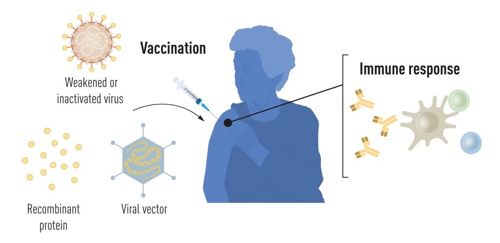
Illustration of methods for vaccine production before the COVID-19 pandemic.
Figure 1. Methods for vaccine production before the COVID-19 pandemic. © The Nobel Committee for Physiology or Medicine. Ill. Mattias Karlén

mRNA vaccines: A promising idea

In our cells, genetic information encoded in DNA is transferred to messenger RNA (mRNA), which is used as a template for protein production. During the 1980s, efficient methods for producing mRNA without cell culture were introduced, called in vitro transcription. This decisive step accelerated the development of molecular biology applications in several fields. Ideas of using mRNA technologies for vaccine and therapeutic purposes also took off, but roadblocks lay ahead. In vitro transcribed mRNA was considered unstable and challenging to deliver, requiring the development of sophisticated carrier lipid systems to encapsulate the mRNA. Moreover, in vitro-produced mRNA gave rise to inflammatory reactions. Enthusiasm for developing the mRNA technology for clinical purposes was, therefore, initially limited.

These obstacles did not discourage the Hungarian biochemist Katalin Karikó, who was devoted to developing methods to use mRNA for therapy. During the early 1990s, when she was an assistant professor at the University of Pennsylvania, she remained true to her vision of realizing mRNA as a therapeutic despite encountering difficulties in convincing research funders of the significance of her project. A new colleague of Karikó at her university was the immunologist Drew Weissman. He was interested in dendritic cells, which have important functions in immune surveillance and the activation of vaccine-induced immune responses. Spurred by new ideas, a fruitful collaboration between the two soon began, focusing on how different RNA types interact with the immune system.

The breakthrough

Karikó and Weissman noticed that dendritic cells recognize in vitro transcribed mRNA as a foreign substance, which leads to their activation and the release of inflammatory signaling molecules. They wondered why the in vitro transcribed mRNA was recognized as foreign while mRNA from mammalian cells did not give rise to the same reaction. Karikó and Weissman realized that some critical properties must distinguish the different types of mRNA.

RNA contains four bases, abbreviated A, U, G, and C, corresponding to A, T, G, and C in DNA, the letters of the genetic code. Karikó and Weissman knew that bases in RNA from mammalian cells are frequently chemically modified, while in vitro transcribed mRNA is not. They wondered if the absence of altered bases in the in vitro transcribed RNA could explain the unwanted inflammatory reaction. To investigate this, they produced different variants of mRNA, each with unique chemical alterations in their bases, which they delivered to dendritic cells. The results were striking: The inflammatory response was almost abolished when base modifications were included in the mRNA. This was a paradigm change in our understanding of how cells recognize and respond to different forms of mRNA. Karikó and Weissman immediately understood that their discovery had profound significance for using mRNA as therapy. These seminal results were published in 2005, fifteen years before the COVID-19 pandemic.
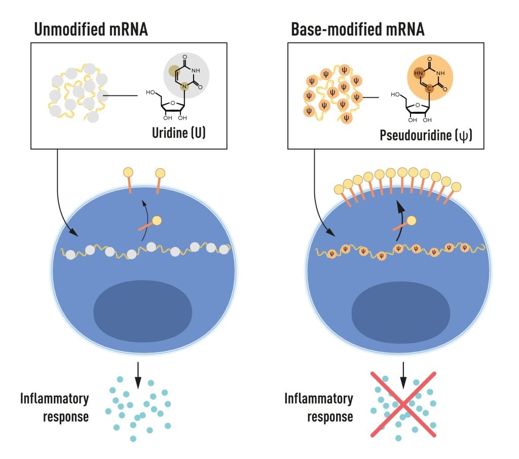
Illustration of the four different bases mRNA contains.
Figure 2. mRNA contains four different bases, abbreviated A, U, G, and C. The Nobel Laureates discovered that base-modified mRNA can be used to block activation of inflammatory reactions (secretion of signaling molecules) and increase protein production when mRNA is delivered to cells.  © The Nobel Committee for Physiology or Medicine. Ill. Mattias Karlén

In further studies published in 2008 and 2010, Karikó and Weissman showed that the delivery of mRNA generated with base modifications markedly increased protein production compared to unmodified mRNA. The effect was due to the reduced activation of an enzyme that regulates protein production. Through their discoveries that base modifications both reduced inflammatory responses and increased protein production, Karikó and Weissman had eliminated critical obstacles on the way to clinical applications of mRNA.

mRNA vaccines realized their potential

Interest in mRNA technology began to pick up, and in 2010, several companies were working on developing the method. Vaccines against Zika virus and MERS-CoV were pursued; the latter is closely related to SARS-CoV-2. After the outbreak of the COVID-19 pandemic, two base-modified mRNA vaccines encoding the SARS-CoV-2 surface protein were developed at record speed. Protective effects of around 95% were reported, and both vaccines were approved as early as December 2020.

The impressive flexibility and speed with which mRNA vaccines can be developed pave the way for using the new platform also for vaccines against other infectious diseases. In the future, the technology may also be used to deliver therapeutic proteins and treat some cancer types.

Several other vaccines against SARS-CoV-2, based on different methodologies, were also rapidly introduced, and together, more than 13 billion COVID-19 vaccine doses have been given globally. The vaccines have saved millions of lives and prevented severe disease in many more, allowing societies to open and return to normal conditions. Through their fundamental discoveries of the importance of base modifications in mRNA, this year’s Nobel laureates critically contributed to this transformative development during one of the biggest health crises of our time.

Read more about this year’s prize

Scientific background: Discoveries concerning nucleoside base modifications that enabled the development of effective mRNA vaccines against COVID-19

Katalin Karikó was born in 1955 in Szolnok, Hungary. She received her PhD from Szeged’s University in 1982 and performed postdoctoral research at the Hungarian Academy of Sciences in Szeged until 1985. She then conducted postdoctoral research at Temple University, Philadelphia, and the University of Health Science, Bethesda. In 1989, she was appointed Assistant Professor at the University of Pennsylvania, where she remained until 2013. After that, she became vice president and later senior vice president at BioNTech RNA Pharmaceuticals. Since 2021, she has been a Professor at Szeged University and an Adjunct Professor at Perelman School of Medicine at the University of Pennsylvania.

Drew Weissman was born in 1959 in Lexington, Massachusetts, USA. He received his MD, PhD degrees from Boston University in 1987. He did his clinical training at Beth Israel Deaconess Medical Center at Harvard Medical School and postdoctoral research at the National Institutes of Health. In 1997, Weissman established his research group at the Perelman School of Medicine at the University of Pennsylvania. He is the Roberts Family Professor in Vaccine Research and Director of the Penn Institute for RNA Innovations.

The University of Pennsylvania October 2, 2023 news release is a very interesting announcement (more about why it’s interesting afterwards), Note: Links have been removed,

The University of Pennsylvania messenger RNA pioneers whose years of scientific partnership unlocked understanding of how to modify mRNA to make it an effective therapeutic—enabling a platform used to rapidly develop lifesaving vaccines amid the global COVID-19 pandemic—have been named winners of the 2023 Nobel Prize in Physiology or Medicine. They become the 28th and 29th Nobel laureates affiliated with Penn, and join nine previous Nobel laureates with ties to the University of Pennsylvania who have won the Nobel Prize in Medicine.

Nearly three years after the rollout of mRNA vaccines across the world, Katalin Karikó, PhD, an adjunct professor of Neurosurgery in Penn’s Perelman School of Medicine, and Drew Weissman, MD, PhD, the Roberts Family Professor of Vaccine Research in the Perelman School of Medicine, are recipients of the prize announced this morning by the Nobel Assembly in Solna, Sweden.

After a chance meeting in the late 1990s while photocopying research papers, Karikó and Weissman began investigating mRNA as a potential therapeutic. In 2005, they published a key discovery: mRNA could be altered and delivered effectively into the body to activate the body’s protective immune system. The mRNA-based vaccines elicited a robust immune response, including high levels of antibodies that attack a specific infectious disease that has not previously been encountered. Unlike other vaccines, a live or attenuated virus is not injected or required at any point.

When the COVID-19 pandemic struck, the true value of the pair’s lab work was revealed in the most timely of ways, as companies worked to quickly develop and deploy vaccines to protect people from the virus. Both Pfizer/BioNTech and Moderna utilized Karikó and Weissman’s technology to build their highly effective vaccines to protect against severe illness and death from the virus. In the United States alone, mRNA vaccines make up more than 655 million total doses of SARS-CoV-2 vaccines that have been administered since they became available in December 2020.

Editor’s Note: The Pfizer/BioNTech and Moderna COVID-19 mRNA vaccines both use licensed University of Pennsylvania technology. As a result of these licensing relationships, Penn, Karikó and Weissman have received and may continue to receive significant financial benefits in the future based on the sale of these products. BioNTech provides funding for Weissman’s research into the development of additional infectious disease vaccines.

Science can be brutal

Now for the interesting bit: it’s in my March 5, 2021 posting (mRNA, COVID-19 vaccines, treating genetic diseases before birth, and the scientist who started it all),

Before messenger RNA was a multibillion-dollar idea, it was a scientific backwater. And for the Hungarian-born scientist behind a key mRNA discovery, it was a career dead-end.

Katalin Karikó spent the 1990s collecting rejections. Her work, attempting to harness the power of mRNA to fight disease, was too far-fetched for government grants, corporate funding, and even support from her own colleagues.

“Every night I was working: grant, grant, grant,” Karikó remembered, referring to her efforts to obtain funding. “And it came back always no, no, no.”

By 1995, after six years on the faculty at the University of Pennsylvania, Karikó got demoted. [emphasis mine] She had been on the path to full professorship, but with no money coming in to support her work on mRNA, her bosses saw no point in pressing on.

She was back to the lower rungs of the scientific academy.

“Usually, at that point, people just say goodbye and leave because it’s so horrible,” Karikó said.

There’s no opportune time for demotion, but 1995 had already been uncommonly difficult. Karikó had recently endured a cancer scare, and her husband was stuck in Hungary sorting out a visa issue. Now the work to which she’d devoted countless hours was slipping through her fingers.

In time, those better experiments came together. After a decade of trial and error, Karikó and her longtime collaborator at Penn — Drew Weissman [emphasis mine], an immunologist with a medical degree and Ph.D. from Boston University — discovered a remedy for mRNA’s Achilles’ heel.

You can get the whole story from my March 5, 2021 posting, scroll down to the “mRNA—it’s in the details, plus, the loneliness of pioneer researchers, a demotion, and squabbles” subhead. If you are very curious about mRNA and the rough and tumble of the world of science, there’s my August 20, 2021 posting “Getting erased from the mRNA/COVID-19 story” where Ian MacLachlan is featured as a researcher who got erased and where Karikó credits his work.

‘Rowing Mom Wins Nobel’ (credit: rowing website Row 2K)

Karikó’s daughter is a two-time gold medal Olympic athlete as the Canadian Broadcasting Corporation’s (CBC) radio programme, As It Happens, notes in an interview with the daughter (Susan Francia). From an October 4, 2023 As It Happens article (with embedded audio programme excerpt) by Sheena Goodyear,

Olympic gold medallist Susan Francia is coming to terms with the fact that she’s no longer the most famous person in her family.

That’s because the retired U.S. rower’s mother, Katalin Karikó, just won a Nobel Prize in Medicine. The biochemist was awarded alongside her colleague, vaccine researcher Drew Weissman, for their groundbreaking work that led to the development of COVID-19 vaccines. 

“Now I’m like, ‘Shoot! All right, I’ve got to work harder,'” Francia said with a laugh during an interview with As It Happens host Nil Köksal. 

But in all seriousness, Francia says she’s immensely proud of her mother’s accomplishments. In fact, it was Karikó’s fierce dedication to science that inspired Francia to win Olympic gold medals in 2008 and 2012.

“Sport is a lot like science in that, you know, you have a passion for something and you just go and you train, attain your goal, whether it be making this discovery that you truly believe in, or for me, it was trying to be the best in the world,” Francia said.

“It’s a grind and, honestly, I love that grind. And my mother did too.”

… one of her [Karikó] favourite headlines so far comes from a little blurb on the rowing website Row 2K: “Rowing Mom Wins Nobel.”

Nowadays, scientists are trying to harness the power of mRNA to fight cancer, malaria, influenza and rabies. But when Karikó first began her work, it was a fringe concept. For decades, she toiled in relative obscurity, struggling to secure funding for her research.

“That’s also that same passion that I took into my rowing,” Francia said.

But even as Karikó struggled to make a name for herself, she says her own mother, Zsuzsanna, always believed she would earn a Nobel Prize one day.

Every year, as the Nobel Prize announcement approached, she would tell Karikó she’d be watching for her name. 

“I was laughing [and saying] that, ‘Mom, I am not getting anything,'” she said. 

But her mother, who died a few years ago, ultimately proved correct. 

Congratulations to both Katalin Karikó and Drew Weissman and thank you both for persisting!

Physics

This prize is for physics at the attoscale.

Aaron W. Harrison (Assistant Professor of Chemistry, Austin College, Texas, US) attempts an explanation of an attosecond in his October 3, 2023 essay (in English “What is an attosecond? A physical chemist explains the tiny time scale behind Nobel Prize-winning research” and in French “Nobel de physique : qu’est-ce qu’une attoseconde?”) for The Conversation, Note: Links have been removed,

“Atto” is the scientific notation prefix that represents 10-18, which is a decimal point followed by 17 zeroes and a 1. So a flash of light lasting an attosecond, or 0.000000000000000001 of a second, is an extremely short pulse of light.

In fact, there are approximately as many attoseconds in one second as there are seconds in the age of the universe.

Previously, scientists could study the motion of heavier and slower-moving atomic nuclei with femtosecond (10-15) light pulses. One thousand attoseconds are in 1 femtosecond. But researchers couldn’t see movement on the electron scale until they could generate attosecond light pulses – electrons move too fast for scientists to parse exactly what they are up to at the femtosecond level.

Harrison does a very good job of explaining something that requires a leap of imagination. He also explains why scientists engage in attosecond research. h/t October 4, 2023 news item on phys.org

Amelle Zaïr (Imperial College London) offers a more technical explanation in her October 4, 2023 essay about the 2023 prize winners for The Conversation. h/t October 4, 2023 news item on phys.org

Main event

Here’s the October 3, 2023 Nobel Prize press release, Note: A link has been removed,

The Royal Swedish Academy of Sciences has decided to award the Nobel Prize in Physics 2023 to

Pierre Agostini
The Ohio State University, Columbus, USA

Ferenc Krausz
Max Planck Institute of Quantum Optics, Garching and Ludwig-Maximilians-Universität München, Germany

Anne L’Huillier
Lund University, Sweden

“for experimental methods that generate attosecond pulses of light for the study of electron dynamics in matter”

Experiments with light capture the shortest of moments

The three Nobel Laureates in Physics 2023 are being recognised for their experiments, which have given humanity new tools for exploring the world of electrons inside atoms and molecules. Pierre Agostini, Ferenc Krausz and Anne L’Huillier have demonstrated a way to create extremely short pulses of light that can be used to measure the rapid processes in which electrons move or change energy.

Fast-moving events flow into each other when perceived by humans, just like a film that consists of still images is perceived as continual movement. If we want to investigate really brief events, we need special technology. In the world of electrons, changes occur in a few tenths of an attosecond – an attosecond is so short that there are as many in one second as there have been seconds since the birth of the universe.

The laureates’ experiments have produced pulses of light so short that they are measured in attoseconds, thus demonstrating that these pulses can be used to provide images of processes inside atoms and molecules.

In 1987, Anne L’Huillier discovered that many different overtones of light arose when she transmitted infrared laser light through a noble gas. Each overtone is a light wave with a given number of cycles for each cycle in the laser light. They are caused by the laser light interacting with atoms in the gas; it gives some electrons extra energy that is then emitted as light. Anne L’Huillier has continued to explore this phenomenon, laying the ground for subsequent breakthroughs.

In 2001, Pierre Agostini succeeded in producing and investigating a series of consecutive light pulses, in which each pulse lasted just 250 attoseconds. At the same time, Ferenc Krausz was working with another type of experiment, one that made it possible to isolate a single light pulse that lasted 650 attoseconds.

The laureates’ contributions have enabled the investigation of processes that are so rapid they were previously impossible to follow.

“We can now open the door to the world of electrons. Attosecond physics gives us the opportunity to understand mechanisms that are governed by electrons. The next step will be utilising them,” says Eva Olsson, Chair of the Nobel Committee for Physics.

There are potential applications in many different areas. In electronics, for example, it is important to understand and control how electrons behave in a material. Attosecond pulses can also be used to identify different molecules, such as in medical diagnostics.

Read more about this year’s prize

Popular science background: Electrons in pulses of light (pdf)
Scientific background: “For experimental methods that generate attosecond pulses of light for the study of electron dynamics in matter” (pdf)

Pierre Agostini. PhD 1968 from Aix-Marseille University, France. Professor at The Ohio State University, Columbus, USA.

Ferenc Krausz, born 1962 in Mór, Hungary. PhD 1991 from Vienna University of Technology, Austria. Director at Max Planck Institute of Quantum Optics, Garching and Professor at Ludwig-Maximilians-Universität München, Germany.

Anne L’Huillier, born 1958 in Paris, France. PhD 1986 from University Pierre and Marie Curie, Paris, France. Professor at Lund University, Sweden.

A Canadian connection?

An October 3, 2023 CBC online news item from the Associated Press reveals a Canadian connection of sorts ,

Three scientists have won the Nobel Prize in physics Tuesday for giving us the first split-second glimpse into the superfast world of spinning electrons, a field that could one day lead to better electronics or disease diagnoses.

The award went to French-Swedish physicist Anne L’Huillier, French scientist Pierre Agostini and Hungarian-born Ferenc Krausz for their work with the tiny part of each atom that races around the centre, and that is fundamental to virtually everything: chemistry, physics, our bodies and our gadgets.

Electrons move around so fast that they have been out of reach of human efforts to isolate them. But by looking at the tiniest fraction of a second possible, scientists now have a “blurry” glimpse of them, and that opens up whole new sciences, experts said.

“The electrons are very fast, and the electrons are really the workforce in everywhere,” Nobel Committee member Mats Larsson said. “Once you can control and understand electrons, you have taken a very big step forward.”

L’Huillier is the fifth woman to receive a Nobel in Physics.

L’Huillier was teaching basic engineering physics to about 100 undergraduates at Lund when she got the call that she had won, but her phone was on silent and she didn’t pick up. She checked it during a break and called the Nobel Committee.

Then she went back to teaching.

Agostini, an emeritus professor at Ohio State University, was in Paris and could not be reached by the Nobel Committee before it announced his win to the world

Here’s the Canadian connection (from the October 3, 2023 CBC online news item),

Krausz, of the Max Planck Institute of Quantum Optics and Ludwig Maximilian University of Munich, told reporters that he was bewildered.

“I have been trying to figure out since 11 a.m. whether I’m in reality or it’s just a long dream,” the 61-year-old said.

Last year, Krausz and L’Huillier won the prestigious Wolf prize in physics for their work, sharing it with University of Ottawa scientist Paul Corkum [emphasis mine]. Nobel prizes are limited to only three winners and Krausz said it was a shame that it could not include Corkum.

Corkum was key to how the split-second laser flashes could be measured [emphasis mine], which was crucial, Krausz said.

Congratulations to Pierre Agostini, Ferenc Krausz and Anne L’Huillier and a bow to Paul Corkum!

For those who are curious. a ‘Paul Corkum’ search should bring up a few postings on this blog but I missed this piece of news, a May 4, 2023 University of Ottawa news release about Corkum and the 2022 Wolf Prize, which he shared with Krausz and L’Huillier,

Chemistry

There was a little drama where this prize was concerned, It was announced too early according to an October 4, 2023 news item on phys.org and, again, in another October 4, 2023 news item on phys.org (from the Oct. 4, 2023 news item by Karl Ritter for the Associated Press),

Oops! Nobel chemistry winners are announced early in a rare slip-up

The most prestigious and secretive prize in science ran headfirst into the digital era Wednesday when Swedish media got an emailed press release revealing the winners of the Nobel Prize in chemistry and the news prematurely went public.

Here’s the fully sanctioned October 4, 2023 Nobel Prize press release, Note: A link has been removed,

The Royal Swedish Academy of Sciences has decided to award the Nobel Prize in Chemistry 2023 to

Moungi G. Bawendi
Massachusetts Institute of Technology (MIT), Cambridge, MA, USA

Louis E. Brus
Columbia University, New York, NY, USA

Alexei I. Ekimov
Nanocrystals Technology Inc., New York, NY, USA

“for the discovery and synthesis of quantum dots”

They planted an important seed for nanotechnology

The Nobel Prize in Chemistry 2023 rewards the discovery and development of quantum dots, nanoparticles so tiny that their size determines their properties. These smallest components of nanotechnology now spread their light from televisions and LED lamps, and can also guide surgeons when they remove tumour tissue, among many other things.

Everyone who studies chemistry learns that an element’s properties are governed by how many electrons it has. However, when matter shrinks to nano-dimensions quantum phenomena arise; these are governed by the size of the matter. The Nobel Laureates in Chemistry 2023 have succeeded in producing particles so small that their properties are determined by quantum phenomena. The particles, which are called quantum dots, are now of great importance in nanotechnology.

“Quantum dots have many fascinating and unusual properties. Importantly, they have different colours depending on their size,” says Johan Åqvist, Chair of the Nobel Committee for Chemistry.

Physicists had long known that in theory size-dependent quantum effects could arise in nanoparticles, but at that time it was almost impossible to sculpt in nanodimensions. Therefore, few people believed that this knowledge would be put to practical use.

However, in the early 1980s, Alexei Ekimov succeeded in creating size-dependent quantum effects in coloured glass. The colour came from nanoparticles of copper chloride and Ekimov demonstrated that the particle size affected the colour of the glass via quantum effects.

A few years later, Louis Brus was the first scientist in the world to prove size-dependent quantum effects in particles floating freely in a fluid.

In 1993, Moungi Bawendi revolutionised the chemical production of quantum dots, resulting in almost perfect particles. This high quality was necessary for them to be utilised in applications.

Quantum dots now illuminate computer monitors and television screens based on QLED technology. They also add nuance to the light of some LED lamps, and biochemists and doctors use them to map biological tissue.

Quantum dots are thus bringing the greatest benefit to humankind. Researchers believe that in the future they could contribute to flexible electronics, tiny sensors, thinner solar cells and encrypted quantum communication – so we have just started exploring the potential of these tiny particles.

Read more about this year’s prize

Popular science background: They added colour to nanotechnology (pdf)
Scientific background: Quantum dots – seeds of nanoscience (pdf)

Moungi G. Bawendi, born 1961 in Paris, France. PhD 1988 from University of Chicago, IL, USA. Professor at Massachusetts Institute of Technology (MIT), Cambridge, MA, USA.

Louis E. Brus, born 1943 in Cleveland, OH, USA. PhD 1969 from Columbia University, New York, NY, USA. Professor at Columbia University, New York, NY, USA.

Alexei I. Ekimov, born 1945 in the former USSR. PhD 1974 from Ioffe Physical-Technical Institute, Saint Petersburg, Russia. Formerly Chief Scientist at Nanocrystals Technology Inc., New York, NY, USA.


The most recent ‘quantum dot’ (a particular type of nanoparticle) story here is a January 5, 2023 posting, “Can I have a beer with those carbon quantum dots?

Proving yet again that scientists can have a bumpy trip to a Nobel prize, an October 4, 2023 news item on phys.org describes how one of the winners flunked his first undergraduate chemistry test, Note: Links have been removed,

Talk about bouncing back. MIT professor Moungi Bawendi is a co-winner of this year’s Nobel chemistry prize for helping develop “quantum dots”—nanoparticles that are now found in next generation TV screens and help illuminate tumors within the body.

But as an undergraduate, he flunked his very first chemistry exam, recalling that the experience nearly “destroyed” him.

The 62-year-old of Tunisian and French heritage excelled at science throughout high school, without ever having to break a sweat.

But when he arrived at Harvard University as an undergraduate in the late 1970s, he was in for a rude awakening.

You can find more about the winners and quantum dots in an October 4, 2023 news item on Nanowerk and in Dr. Andrew Maynard’s (Professor of Advanced Technology Transitions, Arizona State University) October 4, 2023 essay for The Conversation (h/t October 4, 2023 news item on phys.org), Note: Links have been removed,

This year’s prize recognizes Moungi Bawendi, Louis Brus and Alexei Ekimov for the discovery and development of quantum dots. For many years, these precisely constructed nanometer-sized particles – just a few hundred thousandths the width of a human hair in diameter – were the darlings of nanotechnology pitches and presentations. As a researcher and adviser on nanotechnology [emphasis mine], I’ve [Dr. Andrew Maynard] even used them myself when talking with developers, policymakers, advocacy groups and others about the promise and perils of the technology.

The origins of nanotechnology predate Bawendi, Brus and Ekimov’s work on quantum dots – the physicist Richard Feynman speculated on what could be possible through nanoscale engineering as early as 1959, and engineers like Erik Drexler were speculating about the possibilities of atomically precise manufacturing in the the 1980s. However, this year’s trio of Nobel laureates were part of the earliest wave of modern nanotechnology where researchers began putting breakthroughs in material science to practical use.

Quantum dots brilliantly fluoresce: They absorb one color of light and reemit it nearly instantaneously as another color. A vial of quantum dots, when illuminated with broad spectrum light, shines with a single vivid color. What makes them special, though, is that their color is determined by how large or small they are. Make them small and you get an intense blue. Make them larger, though still nanoscale, and the color shifts to red.

The wavelength of light a quantum dot emits depends on its size. Maysinger, Ji, Hutter, Cooper, CC BY

There’s also an October 4, 2023 overview article by Tekla S. Perry and Margo Anderson for the IEEE Spectrum about the magazine’s almost twenty-five years of reporting on quantum dots
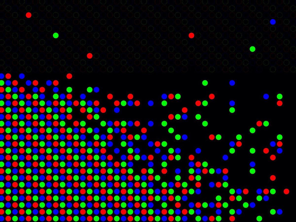
Red blue and green dots mass in rows, with some dots moving away

Image credit: Brandon Palacio/IEEE Spectrum

Your Guide to the Newest Nobel Prize: Quantum Dots

What you need to know—and what we’ve reported—about this year’s Chemistry award

It’s not a long article and it has a heavy focus on the IEEEE’s (Institute of Electrical and Electtronics Engineers) the road quantum dots have taken to become applications and being commercialized.

Congratulations to Moungi Bawendi, Louis Brus, and Alexei Ekimov!
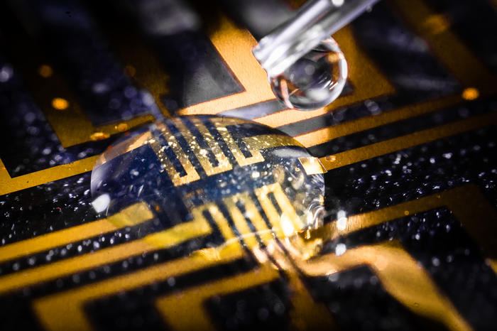
Growing electrodes in your brain?

This isn’t for everybody. From a February 23, 2023 news item on Nanowerk, Note: A link has been removed,

The boundaries between biology and technology are becoming blurred. Researchers at Linköping, Lund, and Gothenburg universities in Sweden have successfully grown electrodes in living tissue using the body’s molecules as triggers. The result, published in the journal Science (“Metabolite-induced in vivo fabrication of substrate-free organic bioelectronics”), paves the way for the formation of fully integrated electronic circuits in living organisms.

Caption: The injectable gel being tested on a microfabricated circuit. Credit: Thor Balkhed

I have two news releases for this research. First, the February 23, 2023 American Association for the Advancement of Science (AAAS) news release on EurekAlert,

Researchers have developed a way to make bioelectronics directly inside living tissues, an approach they tested by making electrodes in the brain, heart, and fin tissue of living zebrafish, as well as in isolated mammalian muscle tissues. According to the authors, the new method paves the way for in vivo fabrication of fully integrated electronic circuits within the nervous system and other living tissue. “Safety and stability analyses over long periods will be essential to determining whether such technology is useful for chronic implantations,” writes Sahika Inal in a related Perspective. “However, the strategy … suggests that any living tissue can turn into electronic matter and brings the field closer to generating seamless biotic-abiotic interfaces with a potentially long lifetime and minimum harm to tissues.” Implantable electronic devices that can interface with soft biological neural tissues offer a valuable approach to studying the complex electrical signaling of the nervous system and enable the therapeutic modulation of neural circuitry to prevent or treat various diseases and disorders. However, conventional bioelectronic implants often require the use of rigid electronic substrates that are incompatible with delicate living tissues and can provoke injury and inflammation that can affect a device’s electrical properties and long-term performance. Overcoming the incompatibility between static, solid-state electronic materials and dynamic, soft biological tissues has proven challenging. Here, Xenofon Strakosas and colleagues present a method to fabricate polymer-based, substrate-free electronic conducting materials directly inside a tissue. Strakosas et al. developed a complex molecular precursor cocktail that, when injected into a tissue, uses endogenous metabolites (glucose and lactate) to induce polymerization of organic precursors to form conducting polymer gels. To demonstrate the approach, the authors “grew” gel electrodes in the brain, heart, and fin tissue of living zebrafish, with no signs of tissue damage, and in isolated mammalian muscle tissues, including beef, pork and chicken. In medicinal leeches, they showed how the conducting gel could interface nervous tissue with electrodes on a tiny flexible probe.

The second is the February 23, 2023 Linköping University press release on EurekAlert, which originated the news item, and it provides further insight,

“For several decades, we have tried to create electronics that mimic biology. Now we let biology create the electronics for us,” says Professor Magnus Berggren at the Laboratory for Organic Electronics, LOE, at Linköping University.

Linking electronics to biological tissue is important to understand complex biological functions, combat diseases in the brain, and develop future interfaces between man and machine. However, conventional bioelectronics, developed in parallel with the semiconductor industry, have a fixed and static design that is difficult, if not impossible, to combine with living biological signal systems.

To bridge this gap between biology and technology, researchers have developed a method for creating soft, substrate-free, electronically conductive materials in living tissue. By injecting a gel containing enzymes as the “assembly molecules”, the researchers were able to grow electrodes in the tissue of zebrafish and medicinal leeches.

“Contact with the body’s substances changes the structure of the gel and makes it electrically conductive, which it isn’t before injection. Depending on the tissue, we can also adjust the composition of the gel to get the electrical process going,” says Xenofon Strakosas, researcher at LOE and Lund University and one of the study’s main authors.

The body’s endogenous molecules are enough to trigger the formation of electrodes. There is no need for genetic modification or external signals, such as light or electrical energy, which has been necessary in previous experiments. The Swedish researchers are the first in the world to succeed in this.

Their study paves the way for a new paradigm in bioelectronics. Where it previously took implanted physical objects to start electronic processes in the body, injection of a viscous gel will be enough in the future.

In their study, the researchers further show that the method can target the electronically conducting material to specific biological substructures and thereby create suitable interfaces for nerve stimulation. In the long term, the fabrication of fully integrated electronic circuits in living organisms may be possible.

In experiments conducted at Lund University, the team successfully achieved electrode formation in the brain, heart, and tail fins of zebrafish and around the nervous tissue of medicinal leeches. The animals were not harmed by the injected gel and were otherwise not affected by the electrode formation. One of the many challenges in these trials was to take the animals’ immune system into account.

“By making smart changes to the chemistry, we were able to develop electrodes that were accepted by the brain tissue and immune system. The zebrafish is an excellent model for the study of organic electrodes in brains,” says Professor Roger Olsson at the Medical Faculty at Lund University, who also has a chemistry laboratory at the University of Gothenburg.

It was Professor Roger Olsson who took the initiative for the study, after he read about the electronic rose developed by researchers at Linköping University in 2015. One research problem, and an important difference between plants and animals, was the difference in cell structure. Whereas plants have rigid cell walls which allow for the formation of electrodes, animal cells are more like a soft mass. Creating a gel with enough structure and the right combination of substances to form electrodes in such surroundings was a challenge that took many years to solve.

“Our results open up for completely new ways of thinking about biology and electronics. We still have a range of problems to solve, but this study is a good starting point for future research,” says Hanne Biesmans, PhD student at LOE and one of the main authors.

Here’s a link to and a citation for the paper,

Metabolite-induced in vivo fabrication of substrate-free organic bioelectronics by Xenofon Strakosas, Hanne Biesmans, Tobias Abrahamsson, Karin Hellman, Malin Silverå Ejneby, Mary J. Donahue, Peter Ekström, Fredrik Ek, Marios Savvakis, Martin Hjort, David Bliman, Mathieu Linares, Caroline Lindholm, Eleni Stavrinidou, Jennifer Y. Gerasimov, Daniel T. Simon, Roger Olsson, and Magnus Berggren. Science 23 Feb 2023 Vol 379, Issue 6634 pp. 795-802 DOI: 10.1126/science.adc9998

This paper is behind a paywall.

Cyborg soil?

Edith Hammer, lecturer (Biology) at Lund University (Sweden) has written a July 22, 2021 essay for The Conversation (h/t July 23, 2021 news item on phys.org) that has everything.: mystery, cyborgs, unexpected denizens, and a phenomenon explored for the first time (Note: Links have been removed),

Dig a teaspoon into your nearest clump of soil, and what you’ll emerge with will contain more microorganisms than there are people on Earth. We know this from lab studies that analyse samples of earth scooped from the microbial wild to determine which forms of microscopic life exist in the world beneath our feet.

The problem is, such studies can’t actually tell us how this subterranean kingdom of fungi, flagellates and amoebae operates in the ground. Because they entail the removal of soil from its environment, these studies destroy the delicate structures of mud, water and air in which the soil microbes reside.

This prompted my lab to develop a way to spy on these underground workers, who are indispensable in their role as organic matter recycling agents, without disturbing their micro-habitats.

Our study revealed the dark, dank cities in which soil microbes reside [emphasis mine]. We found labyrinths of tiny highways, skyscrapers, bridges and rivers which are navigated by microorganisms to find food, or to avoid becoming someone’s next meal. This new window into what’s happening underground could help us better appreciate and preserve Earth’s increasingly damaged soils.

Here’s how the soil scientists probed the secrets buried in soil (Note: A link has been removed),

In our study, we developed a new kind of “cyborg soil”, which is half natural and half artificial. It consists of microengineered chips that we either buried in the wild, or surrounded with soil in the lab for enough time for the microbial cities to emerge within the mud.

The chips literally act like windows to the underground. A transparent patch in the otherwise opaque soil, the chip is cut to mimic the pore structures of actual soil, which are often strange and counter-intuitive at the scale that microbes experience them.

Different physical laws become dominant at the micro scale compared to what we’re acquainted to in our macro world. Water clings to surfaces, and resting bacteria get pushed around by the movement of water molecules. Air bubbles form insurmountable barriers for many microorganisms, due to the surface tension of the water around them.

Here’s some of the what they found,

When we excavated our first chips, we were met with the full variety of single-celled organisms, nematodes, tiny arthropods and species of bacteria that exist in our soils. Fungal hyphae, which burrow like plant roots underground, had quickly grown into the depths of our cyborg soil pores, creating a direct living connection between the real soil and our chips.

This meant we could study a phenomenon known only from lab studies: the “fungal highways” along which bacteria “hitchhike” to disperse through soil. Bacteria usually disperse through water, so by making some of our chips air-filled we could watch how bacteria smuggle themselves into new pores by following the groping arms of fungal hyphae.

Unexpectedly, we also found a high number of protists – enigmatic single-celled organisms which are neither animal, plant or fungus – in the spaces around hyphae. Clearly they too hitch a ride on the fungal highway – a so-far completely unexplored phenomenon.

The essay has a number of embedded videos and images illustrating a fascinating world in a ‘teaspoon of soil’.

Here’s a link to and a citation for the study by the researchers at Lund University,

Microfluidic chips provide visual access to in situ soil ecology by Paola Micaela Mafla-Endara, Carlos Arellano-Caicedo, Kristin Aleklett, Milda Pucetaite, Pelle Ohlsson & Edith C. Hammer. Communications Biology volume 4, Article number: 889 (2021) DOI: https://doi.org/10.1038/s42003-021-02379-5 Published: 20 July 2021

This paper is open access.

Accelerated healing of the tissue in the blood-brain barrier with gelatin

It’s been a few years since my last brain and gelatin story (Dec. 24, 2014 posting: Gelatin nanoparticles for drug delivery after stroke) and this time they’re trying to make brain surgery easier and to reduce any attendant brain damage according to a Nov. 6, 2017 news item on ScienceDaily,

Researchers already know that gelatin-covered electrode implants cause less damage to brain tissue than electrodes with no gelatin coating. Researchers at the Neuronano Research Centre (NRC) at Lund University in Sweden have now shown that microglia, the brain’s cleansing cells, and the enzymes that the cells use in the cleaning process, change in the presence of gelatin.

“Knowledge about the beneficial effects of gelatin could be significant for brain surgery, but also in the development of brain implants,” say the researchers behind the study.

Our brains are surrounded by a blood brain barrier which protects the brain from harmful substances that could enter it via the bloodstream. When the barrier is penetrated, as in the case of biopsy or brain surgery for example, leaks can occur and cause serious inflammation. Researchers at the NRC have previously shown that gelatin accelerates brain tissue healing and reduces damage to nerve cells in the case of electrode implants, but only now are they starting to understand how.

A November 6, 2017 Lund University press release, which originated the news item, provides more details,

The researchers used sedated rats to investigate how the brain is repaired after an injury. Gelatin-coated needles were used in one group, and needles without gelatin in the other.

“The use of gelatin-coated needles reduced or eliminated the leakage of molecules (which normally don’t get through) through the blood brain barrier within twenty-four hours. Without gelatin, the leakage continued for up to three days”, says Lucas Kumosa, one of the researchers behind the study, which was recently published in the research journal Acta Biomaterialia.

Gelatin

The images in the left-hand column show the healing of an injury caused by a stainless steel needle. The images in the right-hand column show what the process looked like when the researchers used a gelatin-coated needle. Gelatin accelerated the healing process and reduced the leakage of blood-borne molecules capable of passing through the blood brain barrier into the brain and causing inflammation.

FEWER INFLAMMATORY CLEANING CELLS

When there is an injury to the brain, microglial cells – the brain’s cleaning cells – gather at the site. They clean up, but can also damage the nerve cell tissue through enzymes they release. In their study, the researchers observed a change in which cleaning cells moved towards the injury site.

“When we used gelatin, we saw only a small number of the inflammatory microglial cells. Instead, we observed cells of a different kind, that are anti-inflammatory, which we believe could be significant in accelerating healing”, explains Lucas Kumosa.

The hypothesis is that the potentially damaging enzymes are occupied with the gelatin instead.

“Gelatin is a protein and its decomposition releases amino-acids that we believe could promote the reconstruction of blood vessels and tissue”, explains Jens Schouenborg, professor of neurophysiology at Lund University.

SURGICAL SIGNIFICANCE

Research is currently underway on how electrodes implanted in the brain could be used in the treatment of various diseases, such as epilepsy or Parkinson’s. A major challenge has been to find ways of reducing damage to the area when using such implants.

“Although the research field of brain electrodes is promising, it has been a challenge to find solutions that don’t damage the brain tissue. Knowledge of how injuries heal faster with gelatin could therefore be significant for the development of surgical treatment as well,” says Jens Schouenborg.

The research is funded by the Knut and Alice Wallenberg Foundation, the Swedish Research Council, Lund University and the Sven-Olof Jansons livsverk Foundation.

Here’s a link to and a citation for the paper,

Gelatin promotes rapid restoration of the blood brain barrier after acute brain injury by Lucas S. Kumosa, Valdemar Zetterberg, Jens Schouenborg. Acta Biomaterialia https://doi.org/10.1016/j.actbio.2017.10.020 Available online 14 October 2017

This paper is open access.

Plastic nanoparticles and brain damage in fish

Researchers in Sweden suggest plastic nanoparticles may cause brain damage in fish according to a Sept. 25, 2017 news item on phys.org,

Calculations have shown that 10 per cent of all plastic produced around the world ultimately ends up in the oceans. As a result, a large majority of global marine debris is in fact plastic waste. Human production of plastics is a well-known environmental concern, but few studies have studied the effects of tiny plastic particles, known as nanoplastic particles.

“Our study is the first to show that nanosized plastic particles can accumulate in fish brains”, says Tommy Cedervall, a chemistry researcher at Lund University.

A Sept. 25, 2017 Lund University press release, which originated the news item, provides more detail about the research,

The Lund University researchers studied how nanoplastics may be transported through different organisms in the aquatic ecosystem, i.e. via algae and animal plankton to larger fish. Tiny plastic particles in the water are eaten by animal plankton, which in turn are eaten by fish.

According to Cedervall, the study includes several interesting results on how plastic of different sizes affects aquatic organisms. Most importantly, it provides evidence that nanoplastic particles can indeed cross the blood-brain barrier in fish and thus accumulate inside fish’s brain tissue.

In addition, the researchers involved in the present study have demonstrated the occurrence of behavioural disorders in fish that are affected by nanoplastics. They eat slower and explore their surroundings less. The researchers believe that these behavioural changes may be linked to brain damage caused by the presence of nanoplastics in the brain.

Another result of the study is that animal plankton die when exposed to nanosized plastic particles, while larger plastic particles do not affect them. Overall, these different effects of nanoplastics may have an impact on the ecosystem as a whole.

“It is important to study how plastics affect ecosystems and that nanoplastic particles likely have a more dangerous impact on aquatic ecosystems than larger pieces of plastics”, says Tommy Cedervall.

However, he does not dare to draw the conclusion that plastic nanoparticles could accumulate in other tissues in fish and thus potentially be transmitted to humans through consumption.

“No, we are not aware of any such studies and are therefore very cautious about commenting on it”, says Tommy Cedervall.

Here’s a link to and a citation for the paper,

Brain damage and behavioural disorders in fish induced by plastic nanoparticles delivered through the food chain by Karin Mattsson, Elyse V. Johnson, Anders Malmendal, Sara Linse, Lars-Anders Hansson & Tommy Cedervall. Scientific Reports 7, Article number: 11452 (2017) doi:10.1038/s41598-017-10813-0 Published online: 13 September 2017

This paper is open access.

Implications of nanoplastic in the aquatic food chain

As plastic breaks down in the oceans into plastic nanoparticles, they enter the food chain when they are ingested by plankton. Researchers in Sweden have published a study about the process. From a May 23, 2016 news item on ScienceDaily,

Plastic accounts for nearly eighty per cent of all waste found in our oceans, gradually breaking down into smaller and smaller particles. New research from Lund University in Sweden investigates how nanosized plastic particles affect aquatic animals in different parts of the food chain.

“Not very many studies have been done on this topic before. Plastic particles of such a small size are difficult to study,” says Karin Mattsson.

A May 23, 2016 Lund University press release, which originated the news item, provides more detail,

“We tested how polystyrene plastic particles of different sizes, charge and surface affect the zooplankton Daphnia. It turned out that the size of the nanoparticles that were most toxic to the Daphnia in our study was 50 nanometers”, says Karin Mattsson.

Because zooplankton like Daphnia are also food for many other aquatic animals, the researchers wanted to study the effect of plastic particles higher up in the food chain. They found that fish that ate Daphnia containing nanoplastics experienced a change in their predatory behaviour and poor appetite. In several studies, researchers also discovered that the nanoparticles had the ability to cross biological barriers, such as the intestinal wall and brain.

“Although in our study we used much larger amounts of nanoplastic than those present in oceans today, we suspect that plastic particles may be accumulated inside the fish. This means that even low doses could ultimately have a negative effect”, says Karin Mattsson.

Plastic breaks down very slowly in nature, and once the microscopically small plastic particles reach lakes and oceans they are difficult to remove. Plastic particles also bind environmental toxins that can become part of the food chain when consumed accidentally.

“Our research indicates the need for more studies and increased caution in the use of nanoplastics”, she says.

Karin Mattsson is a physicist and her research project was produced in collaboration between the Centre for Environmental and Climate Research, the Division Biochemistry and Structural Biology and the Division of Aquatic Biology at Lund University. Karin Mattsson is also affiliated with NanoLund, where several studies are currently conducted to evaluate the safety of nanoparticles.

Here’s a link to and a citation for a paper published online in 2014 and in print in 2015,

Altered Behavior, Physiology, and Metabolism in Fish Exposed to Polystyrene Nanoparticles by Karin Mattsson, Mikael T. Ekvall, Lars-Anders Hansson, Sara Linse, Anders Malmendal, and Tommy Cedervall. Environ. Sci. Technol., 2015, 49 (1), pp 553–561 DOI: 10.1021/es5053655
Publication Date (Web): November 07, 2014

Copyright © 2014 American Chemical Society

More recently, Karin Mattson has published her PhD thesis on the topic (I believe it is written in Swedish).

Better neuroprostheses for brain diseases and mental illneses

I don’t often get news releases from Sweden but I do on occasion and, sometimes, they even come in their original Swedish versions. In this case, Lund University sent me an English language version about their latest work making brain implants (neural prostheses) safer and effective. From a Sept. 29, 2015 Lund University news release (also on EurekAlert),

Neurons thrive and grow in a new type of nanowire material developed by researchers in Nanophysics and Ophthalmology at Lund University in Sweden. In time, the results might improve both neural and retinal implants, and reduce the risk of them losing their effectiveness over time, which is currently a problem

By implanting electrodes in the brain tissue one can stimulate or capture signals from different areas of the brain. These types of brain implants, or neuro-prostheses as they are sometimes called, are used to treat Parkinson’s disease and other neurological diseases.

They are currently being tested in other areas, such as depression, severe cases of autism, obsessive-compulsive disorders and paralysis. Another research track is to determine whether retinal implants are able to replace light-sensitive cells that die in cases of Retinitis Pigmentosa and other eye diseases.

However, there are severe drawbacks associated with today’s implants. One problem is that the body interprets the implants as foreign objects, resulting in an encapsulation of the electrode, which in turn leads to loss of signal.

One of the researchers explains the approach adopted by the research team (from the news release),

“Our nanowire structure prevents the cells that usually encapsulate the electrodes – glial cells – from doing so”, says Christelle Prinz, researcher in Nanophysics at Lund University in Sweden, who developed this technique together with Maria Thereza Perez, a researcher in Ophthalmology.

“I was very pleasantly surprised by these results. In previous in-vitro experiments, the glial cells usually attach strongly to the electrodes”, she says.

To avoid this, the researchers have developed a small substrate where regions of super thin nanowires are combined with flat regions. While neurons grow and extend processes on the nanowires, the glial cells primarily occupy the flat regions in between.

“The different types of cells continue to interact. This is necessary for the neurons to survive because the glial cells provide them with important molecules.”

So far, tests have only been done with cultured cells (in vitro) but hopefully they will soon be able to continue with experiments in vivo.

The substrate is made from the semiconductor material gallium phosphide where each outgrowing nanowire has a diameter of only 80 nanometres (billionths of a metre).

Here’s a link to and a citation for the paper,

Support of Neuronal Growth Over Glial Growth and Guidance of Optic Nerve Axons by Vertical Nanowire Arrays by Gaëlle Piret, Maria-Thereza Perez, and Christelle N. Prinz. ACS Appl. Mater. Interfaces, 2015, 7 (34), pp 18944–18948 DOI: 10.1021/acsami.5b03798 Publication Date (Web): August 11, 2015

Copyright © 2015 American Chemical Society

This paper appears to be open access as I was able to link to the PDF version.

Swedish nano plans in Lund

It was a bit a surprise to learn a few years ago that Chalmers University of Technology (Sweden) was the lead in the European Union’s Graphene Flagship project. I was expecting the lead to be one of the British universities, specifically, the University of Manchester seeing that graphene was first isolated there by Nobel Laureates Andre Geim and Konstantan (Kostya) Novoselov, Since then, I’ve kept an eye on the Swedish nanotechnology enterprise and am pleased to have received a Feb. 13, 2014 announcement about hopes for establishing a new nano centre in Lund, Sweden,

A production facility for start-ups in the field of nanotechnology may be built in the Science Village in Lund, a world-class research and innovation village that is also home to ESS, the European Spallation Source.

“With this new facility, we want to create the conditions to enable new companies to develop from the R&D phase to full production, without needing to leave Sweden,” says Lars Samuelson, Professor of Nanophysics at Lund University.

The project originates from the successful research into nanowires at Lund University, which has resulted in nanotechnology companies like Glo AB and Sol Voltaics AB. Glo was forced to move to Silicon Valley, however, to launch large-scale mass production.

The infrastructure would be intended for companies and researchers in the whole of Sweden who want to develop products with industry standards without needing to invest in expensive equipment themselves.

Samuelson sees more business opportunities for nanowires. In addition to Glo’s light-emitting diodes and Sol Voltaics’ solar cells, Lars Samuelson believes there is potential for new companies focused on applications within electronics, UV light-emitting diodes and biomedicine.

Alongside this project, Lund University is working to extend the Lund Nano Lab which is a pure research laboratory for research on nanowires. This is run by Lund University, whereas the industrial facility is a project outside the University. Together, these two initiatives constitute a way of generating the whole value chain from research to market.

The preliminary study into the facility, funded by Vinnova [Sweden’s innovation agency] and Region Skåne and initiated by the Nanometer Structure Consortium at Lund University, is to result in an estimate of investment requirements and market potential, as well as a proposal for a business model. The aim is to become internationally competitive and financially self-sufficient.

A cluster of companies and services, close to the University’s research, is expected to develop around the common equipment for nanoproduction.

About the Nanometer Structure Consortium at Lund University nmC@LU

The Nanometer Structure Consortium at Lund University was founded in 1989. Today, it is one of Sweden’s Strategic Research Areas, engaging more than 250 researchers at the Faculties of Engineering, Science and Medicine. The research focuses on the materials science of nanostructures and its applications within fundamental science, electronics, optoelectronics, energy conversion and life sciences. Former start-ups from the Nanometer Structure Consortium currently employ around 150 people and have attracted private investments of over one billion Swedish crowns.

I suspect this announcement is intended to both raise awareness and, more importantly, attract potential investors as it goes on to provide a number of contacts,

Initiator: Lars Samuelson, Professor, Nanometer Structure Consortium, Lund University, tel. +46 46 222 76 79, lars.samuelson@ftf.lth.se

Chair of the project’s steering group: Heiner Linke, Professor, Coordinator of the Nanometer Structure Consortium, Lund University, tel. +46 46 222 42 45, heiner.linke@ftf.lth.se

Project manager: Yvonne Mårtensson, Nanova, tel. +46 708 337782, yvonne.martensson@nanova.se

Daniel Kronmann, Innovation Systems Unit, Region Skåne, 040-675 34 36, 0706-15 28 10, Daniel.Kronmann@skane.se

International Media Officer
lotte.billing@er.lu.se
+46 72 7074546

I wish them good luck with their plans.

US Air Force wants to merge classical and quantum physics

The US Air Force wants to merge classical and quantum physics for practical purposes according to a May 5, 2014 news item on Azonano,

The Air Force Office of Scientific Research has selected the Harvard School of Engineering and Applied Sciences (SEAS) to lead a multidisciplinary effort that will merge research in classical and quantum physics and accelerate the development of advanced optical technologies.

Federico Capasso, Robert L. Wallace Professor of Applied Physics and Vinton Hayes Senior Research Fellow in Electrical Engineering, will lead this Multidisciplinary University Research Initiative [MURI] with a world-class team of collaborators from Harvard, Columbia University, Purdue University, Stanford University, the University of Pennsylvania, Lund University, and the University of Southampton.

The grant is expected to advance physics and materials science in directions that could lead to very sophisticated lenses, communication technologies, quantum information devices, and imaging technologies.

“This is one of the world’s strongest possible teams,” said Capasso. “I am proud to lead this group of people, who are internationally renowned experts in their fields, and I believe we can really break new ground.”

A May 1, 2014 Harvard University School of Engineering and Applied Sciences news release, which originated the news item, provides a description of project focus: nanophotonics and metamaterials along with some details of Capasso’s work in these areas (Note: Links have been removed),

The premise of nanophotonics is that light can interact with matter in unusual ways when the material incorporates tiny metallic or dielectric features that are separated by a distance shorter than the wavelength of the light. Metamaterials are engineered materials that exploit these phenomena, producing strange effects, enabling light to bend unnaturally, twist into a vortex, or disappear entirely. Yet the fabrication of thick, or bulk, metamaterials—that manipulate light as it passes through the material—has proven very challenging.

Recent research by Capasso and others in the field has demonstrated that with the right device structure, the critical manipulations can actually be confined to the very surface of the material—what they have dubbed a “metasurface.” These metasurfaces can impart an instantaneous shift in the phase, amplitude, and polarization of light, effectively controlling optical properties on demand. Importantly, they can be created in the lab using fairly common fabrication techniques.

At Harvard, the research has produced devices like an extremely thin, flat lens, and a material that absorbs 99.75% of infrared light. But, so far, such devices have been built to order—brilliantly suited to a single task, but not tunable.

This project, however,is focused on the future (Note: Links have been removed),

“Can we make a rapidly configurable metasurface so that we can change it in real time and quickly? That’s really a visionary frontier,” said Capasso. “We want to go all the way from the fundamental physics to the material building blocks and then the actual devices, to arrive at some sort of system demonstration.”

The proposed research also goes further. A key thrust of the project involves combining nanophotonics with research in quantum photonics. By exploiting the quantum effects of luminescent atomic impurities in diamond, for example, physicists and engineers have shown that light can be captured, stored, manipulated, and emitted as a controlled stream of single photons. These types of devices are essential building blocks for the realization of secure quantum communication systems and quantum computers. By coupling these quantum systems with metasurfaces—creating so-called quantum metasurfaces—the team believes it is possible to achieve an unprecedented level of control over the emission of photons.

“Just 20 years ago, the notion that photons could be manipulated at the subwavelength scale was thought to be some exotic thing, far fetched and of very limited use,” said Capasso. “But basic research opens up new avenues. In hindsight we know that new discoveries tend to lead to other technology developments in unexpected ways.”

The research team includes experts in theoretical physics, metamaterials, nanophotonic circuitry, quantum devices, plasmonics, nanofabrication, and computational modeling. Co-principal investigator Marko Lončar is the Tiantsai Lin Professor of Electrical Engineering at Harvard SEAS. Co-PI Nanfang Yu, Ph.D. ’09, developed expertise in metasurfaces as a student in Capasso’s Harvard laboratory; he is now an assistant professor of applied physics at Columbia. Additional co-PIs include Alexandra Boltasseva and Vladimir Shalaev at Purdue, Mark Brongersma at Stanford, and Nader Engheta at the University of Pennsylvania. Lars Samuelson (Lund University) and Nikolay Zheludev (University of Southampton) will also participate.

The bulk of the funding will support talented graduate students at the lead institutions.

The project, titled “Active Metasurfaces for Advanced Wavefront Engineering and Waveguiding,” is among 24 planned MURI awards selected from 361 white papers and 88 detailed proposals evaluated by a panel of experts; each award is subject to successful negotiation. The anticipated amount of the Harvard-led grant is up to $6.5 million for three to five years.

For anyone who’s not familiar (that includes me, anyway) with MURI awards, there’s this from Wikipedia (Note: links have been removed),

Multidisciplinary University Research Initiative (MURI) is a basic research program sponsored by the US Department of Defense (DoD). Currently each MURI award is about $1.5 million a year for five years.

I gather that in addition to the Air Force, the Army and the Navy also award MURI funds.

INFERNOS: realizing Maxwell’s Demon

Before getting to the INFERNOS project and its relationship to Maxwell’s demon, I want to share a pretty good example of this ‘demon’ thought experiment which, as recently as Feb. 4, 2013, I featured in a piece about quantum dots,

James Clerk Maxwell, physicist,  has entered the history books for any number reasons but my personal favourite is Maxwell’s demon, a thought experiment he proposed in the 1800s to violate the 2nd law of thermodynamics. Lisa Zyga in her Feb. 1, 2013 article for phys.org provides an explanation,

When you open your door on a cold winter day, the warm air from your home and the cold air from outside begin to mix and evolve toward thermal equilibrium, a state of complete entropy where the temperatures outside and inside are the same. This situation is a rough example of the second law of thermodynamics, which says that entropy in a closed system never decreases. If you could control the air flow in a way that uses a sufficiently small amount of energy, so that the entropy of the system actually decreases overall, you would have a hypothetical mechanism called Maxwell’s demon.

An Oct. 9, 2013 news item on Nanowerk ties together INFERNOS and the ‘demon’,

Maxwell’s Demon is an imaginary creature that the mathematician James Clerk Maxwell created in 1897. The creature could turn heat into work without causing any other change, which violates the second law of thermodynamics. The primary goal of the European project INFERNOS (Information, fluctuations, and energy control in small systems) is to realize experimentally Maxwell’s Demon; in other words, to develop the electronic and biomolecular nanodevices that support this principle.

The Universitat de Barcelona (University of Barcelona) Oct. 7, 2013 news release, which originated the news item, provides more details about the project,

Although Maxwell’s Demon is one of the cornerstones of theoretical statistical mechanisms, little has been done about its definite experimental realization. Marco Ribezzi, researcher from the Department of Fundamental Physics, explains that “the principal novelty of INFERNOS is to bring a robust and rigorous experimental base for this field of knowledge. We aim at creating a device that can use information to supply/extract energy to/from a system”. In this sense, the UB group, in which researcher Fèlix Ritort from the former department also participates, focuses their activity on understanding how information and temperature changes are used in individual molecules manipulation.

From the theory side, researchers will work in order to develop a theory of the fluctuation processes in small systems, which would then facilitate efficient algorithms for the Maxwell’s Demon operation.

INFERNOS is a three-year European project of the programme Future and Emerging Technologies (FET). Besides the University of Barcelona, INFERNOS partners are: Aalto University (Finland), project coordinator, Lund University (Sweden), the University of Oslo (Norway), Delf University of Technology (Netherlands), the National Center for Scientific Research (France) and the Research Foundation of State University of New York.

I like the INFERNOS logo, demon and all,

Logo of the European project INFERNOS (Information, fluctuations, and energy control in small systems).

Logo of the European project INFERNOS (Information, fluctuations, and energy control in small systems).

The INFERNOS project website can be found here.

And for anyone who finds that music is the best way to learn, here are Flanders & Swann* performing ‘First and Second Law’ from a 1964 show,

Enjoy!

* ‘Swan’ corrected to ‘Swann’ on April 1, 2014.