Tag Archives: Mathias Kolle

Tough colour and the flower beetle

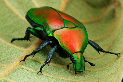
The flower beetle Torynorrhina flammea. [downloaded from https://www.nanowerk.com/nanotechnology-news2/newsid=58269.php]

That is one gorgeous beetle and a June 17, 2021 news item on Nanowerk reveals that it features in a structural colour story (i.e, how structures rather than pigments create colour),

The unique mechanical and optical properties found in the exoskeleton of a humble Asian beetle has the potential to offer a fascinating new insight into how to develop new, effective bio-inspired technologies.

Pioneering new research by a team of international scientists, including Professor Pete Vukusic from the University of Exeter, has revealed a distinctive, and previously unknown property within the carapace of the flower beetle – a member of the scarab beetle family.

The study showed that the beetle has small micropillars within the carapace – or the upper section of the exoskeleton – that give the insect both strength and flexibility to withstand damage very effectively.

Crucially, these micropillars are incorporated into highly regular layering in the exoskeleton that concurrently give the beetle an intensely bright metallic colour appearance.

A June 18, 2021 University of Exeter press release (also on EurekAlert but published June 17, 2021), delves further into the researchers’ new insights,

For this new study, the scientists used sophisticated modelling techniques to determine which of the two functions – very high mechanical strength or conspicuously bright colour – were more important to the survival of the beetle.

They found that although these micropillars do create a highly enhanced toughness of the beetle shell, they were most beneficial for optimising the scattering of coloured light that generates its conspicuous appearance.

The research is published this week in the leading journal, Proceedings of the National Academy of Sciences, PNAS.

Professor Vukusic, one of three leads of the research along with Professor Li at Virginia Tech and Professor Kolle at MIT [Massachusetts Institute of Technology], said: “The astonishing insights generated by this research have only been possible through close collaborative work between Virginia Tech, MIT, Harvard and Exeter, in labs that trailblaze the fields of materials, mechanics and optics. Our follow-up venture to make use of these bio-inspired principles will be an even more exciting journey.”.

The seeds of the pioneering research were sown more than 16 years ago as part of a short project created by Professor Vukusic in the Exeter undergraduate Physics labs. Those early tests and measurements, made by enthusiastic undergraduate students, revealed the possibility of intriguing multifunctionality.

The original students examined the form and structure of beetles’ carapce to try to understand the simple origin of their colour. They noticed for the first time, however, the presence of strength-inducing micropillars.

Professor Vukusic ultimately carried these initial findings to collaborators Professor Ling Li at Virginia Tech and Professor Mathias Kolle at Harvard and then MIT who specialise in the materials sciences and applied optics. Using much more sophisticated measurement and modelling techniques, the combined research team were also to confirm the unique role played by the micropillars in enhancing the beetles’ strength and toughness without compromising its intense metallic colour.

The results from the study could also help inspire a new generation of bio-inspired materials, as well as the more traditional evolutionary research.

By understanding which of the functions provides the greater benefit to these beetles, scientists can develop new techniques to replicate and reproduce the exoskeleton structure, while ensuring that it has brilliant colour appearance with highly effective strength and toughness.

Professor Vukusic added: “Such natural systems as these never fail to impress with the way in which they perform, be it optical, mechanical or in another area of function. The way in which their optical or mechanical properties appear highly tolerant of all manner of imperfections too, continues to offer lessons to us about scientific and technological avenues we absolutely should explore. There is exciting science ahead of us on this journey.”

Here’s a link to and a citation for the paper,

Microstructural design for mechanical–optical multifunctionality in the exoskeleton of the flower beetle Torynorrhina flammea by Zian Jia, Matheus C. Fernandes, Zhifei Deng, Ting Yang, Qiuting Zhang, Alfie Lethbridge, Jie Yin, Jae-Hwang Lee, Lin Han, James C. Weaver, Katia Bertoldi, Joanna Aizenberg, Mathias Kolle, Pete Vukusic, and Ling Li. PNAS June 22, 2021 118 (25) e2101017118; DOI: https://doi.org/10.1073/pnas.2101017118

This paper is behind a paywall.
Colo(u)r-changing bandage for better compression

This is a structural colo(u)r story, from a May 29, 2018 news item on Nanowerk,

Compression therapy is a standard form of treatment for patients who suffer from venous ulcers and other conditions in which veins struggle to return blood from the lower extremities. Compression stockings and bandages, wrapped tightly around the affected limb, can help to stimulate blood flow. But there is currently no clear way to gauge whether a bandage is applying an optimal pressure for a given condition.

Now engineers at MIT {Massachusetts Institute of Technology] have developed pressure-sensing photonic fibers that they have woven into a typical compression bandage. As the bandage is stretched, the fibers change color. Using a color chart, a caregiver can stretch a bandage until it matches the color for a desired pressure, before, say, wrapping it around a patient’s leg.

The photonic fibers can then serve as a continuous pressure sensor — if their color changes, caregivers or patients can use the color chart to determine whether and to what degree the bandage needs loosening or tightening.

A May 29, 2018 MIT news release (also on EurekAlert), which originated the news item, provides more detail,

“Getting the pressure right is critical in treating many medical conditions including venous ulcers, which affect several hundred thousand patients in the U.S. each year,” says Mathias Kolle, assistant professor of mechanical engineering at MIT. “These fibers can provide information about the pressure that the bandage exerts. We can design them so that for a specific desired pressure, the fibers reflect an easily distinguished color.”

Kolle and his colleagues have published their results in the journal Advanced Healthcare Materials. Co-authors from MIT include first author Joseph Sandt, Marie Moudio, and Christian Argenti, along with J. Kenji Clark of the Univeristy of Tokyo, James Hardin of the United States Air Force Research Laboratory, Matthew Carty of Brigham and Women’s Hospital-Harvard Medical School, and Jennifer Lewis of Harvard University.

Natural inspiration

The color of the photonic fibers arises not from any intrinsic pigmentation, but from their carefully designed structural configuration. Each fiber is about 10 times the diameter of a human hair. The researchers fabricated the fiber from ultrathin layers of transparent rubber materials, which they rolled up to create a jelly-roll-type structure. Each layer within the roll is only a few hundred nanometers thick.

In this rolled-up configuration, light reflects off each interface between individual layers. With enough layers of consistent thickness, these reflections interact to strengthen some colors in the visible spectrum, for instance red, while diminishing the brightness of other colors. This makes the fiber appear a certain color, depending on the thickness of the layers within the fiber.

“Structural color is really neat, because you can get brighter, stronger colors than with inks or dyes just by using particular arrangements of transparent materials,” Sandt says. “These colors persist as long as the structure is maintained.”

The fibers’ design relies upon an optical phenomenon known as “interference,” in which light, reflected from a periodic stack of thin, transparent layers, can produce vibrant colors that depend on the stack’s geometric parameters and material composition. Optical interference is what produces colorful swirls in oily puddles and soap bubbles. It’s also what gives peacocks and butterflies their dazzling, shifting shades, as their feathers and wings are made from similarly periodic structures.

“My interest has always been in taking interesting structural elements that lie at the origin of nature’s most dazzling light manipulation strategies, to try recreating and employing them in useful applications,” Kolle says.

A multilayered approach

The team’s approach combines known optical design concepts with soft materials, to create dynamic photonic materials.

While a postdoc at Harvard in the group of Professor Joanna Aizenberg, Kolle was inspired by the work of Pete Vukusic, professor of biophotonics at the University of Exeter in the U.K., on Margaritaria nobilis, a tropical plant that produces extremely shiny blue berries. The fruits’ skin is made up of cells with a periodic cellulose structure, through which light can reflect to give the fruit its signature metallic blue color.

Together, Kolle and Vukusic sought ways to translate the fruit’s photonic architecture into a useful synthetic material. Ultimately, they fashioned multilayered fibers from stretchable materials, and assumed that stretching the fibers would change the individual layers’ thicknesses, enabling them to tune the fibers’ color. The results of these first efforts were published in Advanced Materials in 2013.

When Kolle joined the MIT faculty in the same year, he and his group, including Sandt, improved on the photonic fiber’s design and fabrication. In their current form, the fibers are made from layers of commonly used and widely available transparent rubbers, wrapped around highly stretchable fiber cores. Sandt fabricated each layer using spin-coating, a technique in which a rubber, dissolved into solution, is poured onto a spinning wheel. Excess material is flung off the wheel, leaving a thin, uniform coating, the thickness of which can be determined by the wheel’s speed.

For fiber fabrication, Sandt formed these two layers on top of a water-soluble film on a silicon wafer. He then submerged the wafer, with all three layers, in water to dissolve the water-soluble layer, leaving the two rubbery layers floating on the water’s surface. Finally, he carefully rolled the two transparent layers around a black rubber fiber, to produce the final colorful photonic fiber.

Reflecting pressure

The team can tune the thickness of the fibers’ layers to produce any desired color tuning, using standard optical modeling approaches customized for their fiber design.

“If you want a fiber to go from yellow to green, or blue, we can say, ‘This is how we have to lay out the fiber to give us this kind of [color] trajectory,'” Kolle says. “This is powerful because you might want to have something that reflects red to show a dangerously high strain, or green for ‘ok.’ We have that capacity.”

The team fabricated color-changing fibers with a tailored, strain-dependent color variation using the theoretical model, and then stitched them along the length of a conventional compression bandage, which they previously characterized to determine the pressure that the bandage generates when it’s stretched by a certain amount.

The team used the relationship between bandage stretch and pressure, and the correlation between fiber color and strain, to draw up a color chart, matching a fiber’s color (produced by a certain amount of stretching) to the pressure that is generated by the bandage.

To test the bandage’s effectiveness, Sandt and Moudio enlisted over a dozen student volunteers, who worked in pairs to apply three different compression bandages to each other’s legs: a plain bandage, a bandage threaded with photonic fibers, and a commercially-available bandage printed with rectangular patterns. This bandage is designed so that when it is applying an optimal pressure, users should see that the rectangles become squares.

Overall, the bandage woven with photonic fibers gave the clearest pressure feedback. Students were able to interpret the color of the fibers, and based on the color chart, apply a corresponding optimal pressure more accurately than either of the other bandages.

The researchers are now looking for ways to scale up the fiber fabrication process. Currently, they are able to make fibers that are several inches long. Ideally, they would like to produce meters or even kilometers of such fibers at a time.

“Currently, the fibers are costly, mostly because of the labor that goes into making them,” Kolle says. “The materials themselves are not worth much. If we could reel out kilometers of these fibers with relatively little work, then they would be dirt cheap.”

Then, such fibers could be threaded into bandages, along with textiles such as athletic apparel and shoes as color indicators for, say, muscle strain during workouts. Kolle envisions that they may also be used as remotely readable strain gauges for infrastructure and machinery.

“Of course, they could also be a scientific tool that could be used in a broader context, which we want to explore,” Kolle says.

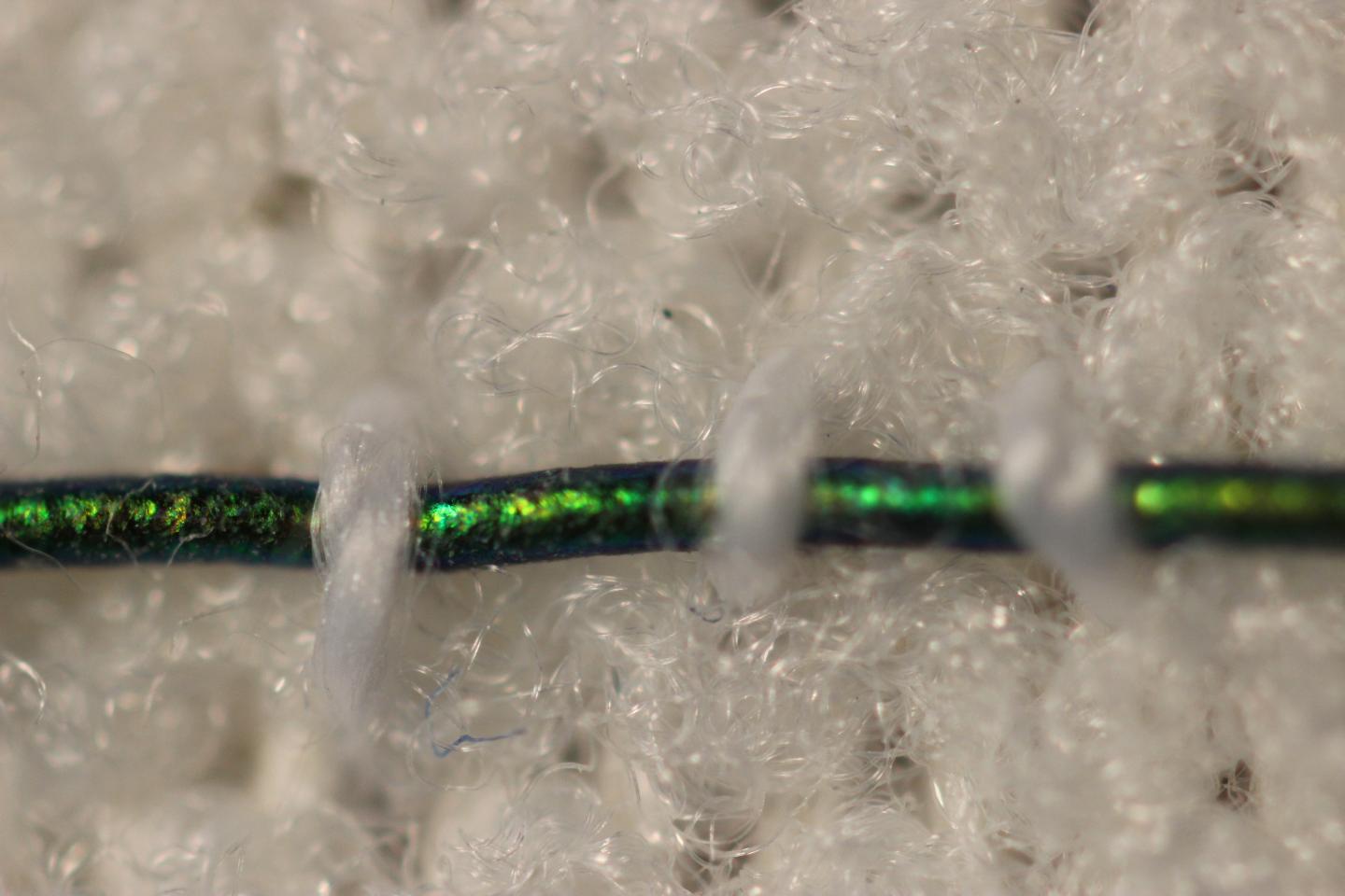
Here’s what the bandage looks like,

Caption: Engineers at MIT have developed pressure-sensing photonic fibers that they have woven into a typical compression bandage. Credit Courtesy of the researchers

Here’s a link to and a citation for the paper,

Stretchable Optomechanical Fiber Sensors for Pressure Determination in Compressive Medical Textiles by Joseph D. Sandt, Marie Moudio, J. Kenji Clark, James Hardin, Christian Argenti, Matthew Carty, Jennifer A. Lewis, Mathias Kolle. Advanced Healthcare Materials https://doi.org/10.1002/adhm.201800293 First published: 29 May 2018

This paper is behind a paywall.

Blue-striped limpets and their nanophotonic features

This is a structural colour story limpets and the Massachusetts Institute of Technology (MIT) and Harvard University. For the impatient here’s a video summary of the work courtesy of the researchers,

A Feb. 26, 2015 news item on ScienceDaily reiterates the details for those who like to read their science,

The blue-rayed limpet is a tiny mollusk that lives in kelp beds along the coasts of Norway, Iceland, the United Kingdom, Portugal, and the Canary Islands. These diminutive organisms — as small as a fingernail — might escape notice entirely, if not for a very conspicuous feature: bright blue dotted lines that run in parallel along the length of their translucent shells. Depending on the angle at which light hits, a limpet’s shell can flash brilliantly even in murky water.

Now scientists at MIT and Harvard University have identified two optical structures within the limpet’s shell that give its blue-striped appearance. The structures are configured to reflect blue light while absorbing all other wavelengths of incoming light. The researchers speculate that such patterning may have evolved to protect the limpet, as the blue lines resemble the color displays on the shells of more poisonous soft-bodied snails.

A Feb. 26, 2015 MIT news release (also on EurekAlert), which originated the news item, explains why this discovery is special,

The findings, reported this week in the journal Nature Communications, represent the first evidence of an organism using mineralized structural components to produce optical displays. While birds, butterflies, and beetles can display brilliant blues, among other colors, they do so with organic structures, such as feathers, scales, and plates. The limpet, by contrast, produces its blue stripes through an interplay of inorganic, mineral structures, arranged in such a way as to reflect only blue light.

The researchers say such natural optical structures may serve as a design guide for engineering color-selective, controllable, transparent displays that require no internal light source and could be incorporated into windows and glasses.

“Let’s imagine a window surface in a car where you obviously want to see the outside world as you’re driving, but where you also can overlay the real world with an augmented reality that could involve projecting a map and other useful information on the world that exists on the other side of the windshield,” says co-author Mathias Kolle, an assistant professor of mechanical engineering at MIT. “We believe that the limpet’s approach to displaying color patterns in a translucent shell could serve as a starting point for developing such displays.”

The news release then reveals how this research came about,

Kolle, whose research is focused on engineering bioinspired, optical materials — including color-changing, deformable fibers — started looking into the optical features of the limpet when his brother Stefan, a marine biologist now working at Harvard, brought Kolle a few of the organisms in a small container. Stefan Kolle was struck by the mollusk’s brilliant patterning, and recruited his brother, along with several others, to delve deeper into the limpet shell’s optical properties.

To do this, the team of researchers — which also included Ling Li and Christine Ortiz at MIT and James Weaver and Joanna Aizenberg at Harvard — performed a detailed structural and optical analysis of the limpet shells. They observed that the blue stripes first appear in juveniles, resembling dashed lines. The stripes grow more continuous as a limpet matures, and their shade varies from individual to individual, ranging from deep blue to turquoise.

The researchers scanned the surface of a limpet’s shell using scanning electron microscopy, and found no structural differences in areas with and without the stripes — an observation that led them to think that perhaps the stripes arose from features embedded deeper in the shell.

To get a picture of what lay beneath, the researchers used a combination of high-resolution 2-D and 3-D structural analysis to reveal the 3-D nanoarchitecture of the photonic structures embedded in the limpets’ translucent shells.

What they found was revealing: In the regions with blue stripes, the shells’ top and bottom layers were relatively uniform, with dense stacks of calcium carbonate platelets and thin organic layers, similar to the shell structure of other mollusks. However, about 30 microns beneath the shell surface the researchers noted a stark difference. In these regions, the researchers found that the regular plates of calcium carbonate morphed into two distinct structural features: a multilayered structure with regular spacing between calcium carbonate layers resembling a zigzag pattern, and beneath this, a layer of randomly dispersed, spherical particles.

The researchers measured the dimensions of the zigzagging plates, and found the spacing between them was much wider than the more uniform plates running through the shell’s unstriped sections. They then examined the potential optical roles of both the multilayer zigzagging structure and the spherical particles.

Kolle and his colleagues used optical microscopy, spectroscopy, and diffraction microscopy to quantify the blue stripe’s light-reflection properties. They then measured the zigzagging structures and their angle with respect to the shell surface, and determined that this structure is optimized to reflect blue and green light.

The researchers also determined that the disordered arrangement of spherical particles beneath the zigzag structures serves to absorb transmitted light that otherwise could de-saturate the reflected blue color.

From these results, Kolle and his team deduced that the zigzag pattern acts as a filter, reflecting only blue light. As the rest of the incoming light passes through the shell, the underlying particles absorb this light — an effect that makes a shell’s stripes appear even more brilliantly blue.

And, for those who can never get enough detail, the news release provides a bit more than the video,

The team then sought to tackle a follow-up question: What purpose do the blue stripes serve? The limpets live either concealed at the base of kelp plants, or further up in the fronds, where they are visually exposed. Those at the base grow a thicker shell with almost no stripes, while their blue-striped counterparts live higher on the plant.

Limpets generally don’t have well-developed eyes, so the researchers reasoned that the blue stripes must not serve as a communication tool, attracting one organism to another. Rather, they think that the limpet’s stripes may be a defensive mechanism: The mollusk sits largely exposed on a frond, so a plausible defense against predators may be to appear either invisible or unappetizing. The researchers determined that the latter is more likely the case, as the limpet’s blue stripes resemble the patterning of poisonous marine snails that also happen to inhabit similar kelp beds.

Kolle says the group’s work has revealed an interesting insight into the limpet’s optical properties, which may be exploited to engineer advanced transparent optical displays. The limpet, he points out, has evolved a microstructure in its shell to satisfy an optical purpose without overly compromising the shell’s mechanical integrity. Materials scientists and engineers could take inspiration from this natural balancing act.

“It’s all about multifunctional materials in nature: Every organism — no matter if it has a shell, or skin, or feathers — interacts in various ways with the environment, and the materials with which it interfaces to the outside world frequently have to fulfill multiple functions simultaneously,” Kolle says. “[Engineers] are more and more focusing on not only optimizing just one single property in a material or device, like a brighter screen or higher pixel density, but rather on satisfying several … design and performance criteria simultaneously. We can gain inspiration and insight from nature.”

Peter Vukusic, an associate professor of physics at the University of Exeter in the United Kingdom, says the researchers “have done an exquisite job” in uncovering the optical mechanism behind the limpet’s conspicuous appearance.

“By using multiple and complementary analysis techniques they have elucidated, in glorious detail, the many structural and physiological factors that have given rise to the optical signature of this highly evolved system,” says Vukusic, who was not involved in the study. “The animal’s complex morphology is highly interesting for photonics scientists and technologists interested in manipulating light and creating specialized appearances.”

Here’s a link to and a citation for the paper,

A highly conspicuous mineralized composite photonic architecture in the translucent shell of the blue-rayed limpet by Ling Li, Stefan Kolle, James C. Weaver, Christine Ortiz, Joanna Aizenberg & Mathias Kolle. Nature Communications 6, Article number: 6322 doi:10.1038/ncomms7322 Published 26 February 2015

This article is open access.

White beetles and complex photonic nanostructures

At least one species of white beetles which have excited scientists with their complex nanostructures are native to Southeast Asia according to an Aug. 15, 2014 news item on Nanowerk,

The physical properties of the ultra-white scales on certain species of beetle could be used to make whiter paper, plastics and paints, while using far less material than is used in current manufacturing methods.

The Cyphochilus beetle, which is native to South-East Asia, is whiter than paper, thanks to ultra-thin scales which cover its body. A new investigation of the optical properties of these scales has shown that they are able to scatter light more efficiently than any other biological tissue known, which is how they are able to achieve such a bright whiteness.

An Aug. 15, 2014 University of Cambridge press release (also on EurekAlert), which originated the news item, describes the properties needed to create the optical conditions necessary for the colour white to be seen,

Animals produce colours for several purposes, from camouflage to communication, to mating and thermoregulation. Bright colours are usually produced using pigments, which absorb certain wavelengths of light and reflect others, which our eyes then perceive as colour.

To appear as white, however, a tissue needs to reflect all wavelengths of light with the same efficiency. The ultra-white Cyphochilus and L. Stigma beetles produce this colouration by exploiting the geometry of a dense complex network of chitin – a molecule similar in structure to cellulose, which is found throughout nature, including in the shells of molluscs, the exoskeletons of insects and the cell walls of fungi. The chitin filaments are just a few billionths of a metre thick, and on their own are not particularly good at reflecting light.

The research, a collaboration between the University of Cambridge and the European Laboratory for non-Linear Spectroscopy in Italy has shown that the beetles have optimised their internal structure in order to produce maximum white with minimum material, like a painter who needs to whiten a wall with a very small quantity of paint. This efficiency is particularly important for insects that fly, as it makes them lighter.

Here’s what the Cyphochilus beetle looks like,

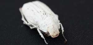
Cyphochilus beetle Credit: Lorenzo Cortese and Silvia Vignolini

Cyphochilus beetle Credit: Lorenzo Cortese and Silvia Vignolini Courtesy University of Cambridge

The press release goes on to describe the beetle’s optical properties in greater detail,

Over millions of years of evolution the beetles have developed a compressed network of chitin filaments. This network is directionally-dependent, or anisotropic, which allows high intensities of reflected light for all colours at the same time, resulting in a very intense white with very little material.

“Current technology is not able to produce a coating as white as these beetles can in such a thin layer,” said Dr Silvia Vignolini of the University’s Cavendish Laboratory, who led the research. “In order to survive, these beetles need to optimise their optical response but this comes with the strong constraint of using as little material as possible in order to save energy and to keep the scales light enough in order to fly. Curiously, these beetles succeed in this task using chitin, which has a relatively low refractive index.”

The secret lies in the beetles’ nanostructures,

Exactly how this could be possible remained unclear up to now. The researchers studied how light propagates in the white scales, quantitatively measuring their scattering strength for the first time and demonstrating that they scatter light more efficiently than any other low-refractive-index material yet known.

“These scales have a structure that is truly complex since it gives rise to something that is more than the sum of its parts,” said co-author Dr Matteo Burresi of the Italian National Institute of Optics in Florence. “Our simulations show that a randomly packed collection of its constituent elements by itself is not sufficient to achieve the degree of brightness that we observe.”

Here’s a link to and a citation for the paper,

Bright-White Beetle Scales Optimise Multiple Scattering of Light by Matteo Burresi, Lorenzo Cortese, Lorenzo Pattelli, Mathias Kolle, Peter Vukusic, Diederik S. Wiersma, Ullrich Steiner, & Silvia Vignolini.  Scientific Reports 4, Article number: 6075 doi:10.1038/srep06075 Published 15 August 2014

This paper is open access.

Thermal control of windows with artificial vasculature

Ben Hatton, a professor of Engineering at the University of Toronto, and his colleagues at Harvard University are proposing a ‘bio-inspired’ alternative to commonly proposed techniques for gaining  thermal control over windows. From an Aug. 2, 2013 news item on ScienceDaily (Note: A link has been removed),

In a recent article in Solar Energy Materials & Solar Cells, Hatton and colleagues at Harvard University describe a novel process to cut down on heat loss during the winter and keep buildings cool during the summer. Their “bio-inspired approach to thermal control for cooling (or heating) building window surfaces” calls for attaching optically clear, flexible elastomer sheets, bonded to regular glass window panes.

The elastomer sheets, made from polydimethylsiloxane (PDMS) have channels running through them through which room temperature water flows. The technique has resulted in 7 to 9 degrees of cooling in laboratory experiments and is effective both at small and large scales, Hatton and his colleagues said.

“Our results show that an artificial vascular network within a transparent layer, composed of channels on the micrometer to millimeter scale, and extending over the surface of a window, offers an additional and novel cooling mechanism for building windows and a new thermal control tool for building design,” he said.

Here’s a link to and a citation for the research paper,

An artificial vasculature for adaptive thermal control of windows by Benjamin D. Hatton, Ian Wheeldon, Matthew J. Hancock, Mathias Kolle, Joanna Aizenberg, and Donald E. Ingber. Solar Energy Materials and Solar Cells, 2013; 117: 429 DOI: 10.1016/j.solmat.2013.06.027; Volume 117, October 2013, Pages 429–436.

I have written about thermal control of windows before as per this Sept. 4, 2012 posting which features an excerpt of an article discussing thermochromic, electrochromic, and gasochromic windows.