Tag Archives: mechanical properties

Tough colour and the flower beetle

The flower beetle Torynorrhina flammea. [downloaded from https://www.nanowerk.com/nanotechnology-news2/newsid=58269.php]

That is one gorgeous beetle and a June 17, 2021 news item on Nanowerk reveals that it features in a structural colour story (i.e, how structures rather than pigments create colour),

The unique mechanical and optical properties found in the exoskeleton of a humble Asian beetle has the potential to offer a fascinating new insight into how to develop new, effective bio-inspired technologies.

Pioneering new research by a team of international scientists, including Professor Pete Vukusic from the University of Exeter, has revealed a distinctive, and previously unknown property within the carapace of the flower beetle – a member of the scarab beetle family.

The study showed that the beetle has small micropillars within the carapace – or the upper section of the exoskeleton – that give the insect both strength and flexibility to withstand damage very effectively.

Crucially, these micropillars are incorporated into highly regular layering in the exoskeleton that concurrently give the beetle an intensely bright metallic colour appearance.

A June 18, 2021 University of Exeter press release (also on EurekAlert but published June 17, 2021), delves further into the researchers’ new insights,

For this new study, the scientists used sophisticated modelling techniques to determine which of the two functions – very high mechanical strength or conspicuously bright colour – were more important to the survival of the beetle.

They found that although these micropillars do create a highly enhanced toughness of the beetle shell, they were most beneficial for optimising the scattering of coloured light that generates its conspicuous appearance.

The research is published this week in the leading journal, Proceedings of the National Academy of Sciences, PNAS.

Professor Vukusic, one of three leads of the research along with Professor Li at Virginia Tech and Professor Kolle at MIT [Massachusetts Institute of Technology], said: “The astonishing insights generated by this research have only been possible through close collaborative work between Virginia Tech, MIT, Harvard and Exeter, in labs that trailblaze the fields of materials, mechanics and optics. Our follow-up venture to make use of these bio-inspired principles will be an even more exciting journey.”.

The seeds of the pioneering research were sown more than 16 years ago as part of a short project created by Professor Vukusic in the Exeter undergraduate Physics labs. Those early tests and measurements, made by enthusiastic undergraduate students, revealed the possibility of intriguing multifunctionality.

The original students examined the form and structure of beetles’ carapce to try to understand the simple origin of their colour. They noticed for the first time, however, the presence of strength-inducing micropillars.

Professor Vukusic ultimately carried these initial findings to collaborators Professor Ling Li at Virginia Tech and Professor Mathias Kolle at Harvard and then MIT who specialise in the materials sciences and applied optics. Using much more sophisticated measurement and modelling techniques, the combined research team were also to confirm the unique role played by the micropillars in enhancing the beetles’ strength and toughness without compromising its intense metallic colour.

The results from the study could also help inspire a new generation of bio-inspired materials, as well as the more traditional evolutionary research.

By understanding which of the functions provides the greater benefit to these beetles, scientists can develop new techniques to replicate and reproduce the exoskeleton structure, while ensuring that it has brilliant colour appearance with highly effective strength and toughness.

Professor Vukusic added: “Such natural systems as these never fail to impress with the way in which they perform, be it optical, mechanical or in another area of function. The way in which their optical or mechanical properties appear highly tolerant of all manner of imperfections too, continues to offer lessons to us about scientific and technological avenues we absolutely should explore. There is exciting science ahead of us on this journey.”

Here’s a link to and a citation for the paper,

Microstructural design for mechanical–optical multifunctionality in the exoskeleton of the flower beetle Torynorrhina flammea by Zian Jia, Matheus C. Fernandes, Zhifei Deng, Ting Yang, Qiuting Zhang, Alfie Lethbridge, Jie Yin, Jae-Hwang Lee, Lin Han, James C. Weaver, Katia Bertoldi, Joanna Aizenberg, Mathias Kolle, Pete Vukusic, and Ling Li. PNAS June 22, 2021 118 (25) e2101017118; DOI: https://doi.org/10.1073/pnas.2101017118

This paper is behind a paywall.
Getting a more complete picture of aerosol particles at the nanoscale

What is in the air we breathe? In addition to the gases we learned about in school there are particles, not just the dust particles you can see, but micro- and nanoparticles too and scientists would like to know more about them.

An August 23, 2017 news item on Nanowerk features work which may help scientists in their quest,

They may be tiny and invisible, says Xiaoji Xu, but the aerosol particles suspended in gases play a role in cloud formation and environmental pollution and can be detrimental to human health.

Aerosol particles, which are found in haze, dust and vehicle exhaust, measure in the microns. One micron is one-millionth of a meter; a thin human hair is about 30 microns thick.

The particles, says Xu, are among the many materials whose chemical and mechanical properties cannot be fully measured until scientists develop a better method of studying materials at the microscale as well as the much smaller nanoscale (1 nm is one-billionth of a meter).

Xu, an assistant professor of chemistry, has developed such a method and utilized it to perform noninvasive chemical imaging of a variety of materials, as well as mechanical mapping with a spatial resolution of 10 nanometers.

The technique, called peak force infrared (PFIR) microscopy, combines spectroscopy and scanning probe microscopy. In addition to shedding light on aerosol particles, Xu says, PFIR will help scientists study micro- and nanoscale phenomena in a variety of inhomogeneous materials.

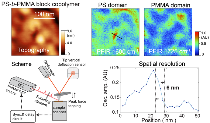
The lower portion of this image by Xiaoji Xu’s group shows the operational scheme of peak force infrared (PFIR) microscopy. The upper portion shows the topography of nanoscale PS-b-PMMA polymer islands on a gold substrate. (Image courtesy of Xiaoji Xu)

An August 22, 2017 Lehih University news release by Kurt Pfitzer (also on EurekAlert), which originated the news item, explains the research in more detail (Note: A link has been removed),

“Materials in nature are rarely homogeneous,” says Xu. “Functional polymer materials often consist of nanoscale domains that have specific tasks. Cellular membranes are embedded with proteins that are nanometers in size. Nanoscale defects of materials exist that affect their mechanical and chemical properties.

“PFIR microscopy represents a fundamental breakthrough that will enable multiple innovations in areas ranging from the study of aerosol particles to the investigation of heterogeneous and biological materials,” says Xu.

Xu and his group recently reported their results in an article titled “Nanoscale simultaneous chemical and mechanical imaging via peak force infrared microscopy.” The article was published in Science Advances, a journal of the American Association for the Advancement of Science, which also publishes Science magazine.

The article’s lead author is Le Wang, a Ph.D. student at Lehigh. Coauthors include Xu and Lehigh Ph.D. students Haomin Wang and Devon S. Jakob, as well as Martin Wagner of Bruker Nano in Santa Barbara, Calif., and Yong Yan of the New Jersey Institute of Technology.

“PFIR microscopy enables reliable chemical imaging, the collection of broadband spectra, and simultaneous mechanical mapping in one simple setup with a spatial resolution of ~10 nm,” the group wrote.

“We have investigated three types of representative materials, namely, soft polymers, perovskite crystals and boron nitride nanotubes, all of which provide a strong PFIR resonance for unambiguous nanochemical identification. Many other materials should be suited as well for the multimodal characterization that PFIR microscopy has to offer.

“In summary, PFIR microscopy will provide a powerful analytical tool for explorations at the nanoscale across wide disciplines.”

Xu and Le Wang also published a recent article about the use of PFIR to study aerosols. Titled “Nanoscale spectroscopic and mechanical characterization of individual aerosol particles using peak force infrared microscopy,” the article appeared in an “Emerging Investigators” issue of Chemical Communications, a journal of the Royal Society of Chemistry. Xu was featured as one of the emerging investigators in the issue. The article was coauthored with researchers from the University of Macau and the City University of Hong Kong, both in China.

PFIR simultaneously obtains chemical and mechanical information, says Xu. It enables researchers to analyze a material at various places, and to determine its chemical compositions and mechanical properties at each of these places, at the nanoscale.

“A material is not often homogeneous,” says Xu. “Its mechanical properties can vary from one region to another. Biological systems such as cell walls are inhomogeneous, and so are materials with defects. The features of a cell wall measure about 100 nanometers in size, placing them well within range of PFIR and its capabilities.”

PFIR has several advantages over scanning near-field optical microscopy (SNOM), the current method of measuring material properties, says Xu. First, PFIR obtains a fuller infrared spectrum and a sharper image—6-nm spatial resolution—of a wider variety of materials than does SNOM. SNOM works well with inorganic materials, but does not obtain as strong an infrared signal as the Lehigh technique does from softer materials such as polymers or biological materials.

“Our technique is more robust,” says Xu. “It works better with soft materials, chemical as well as biological.”

The second advantage of PFIR is that it can perform what Xu calls point spectroscopy.

“If there is something of interest chemically on a surface,” Xu says, “I put an AFM [atomic force microscopy] probe to that location to measure the peak-force infrared response.

“It is very difficult to obtain these spectra with current scattering-type scanning near-field optical microscopy. It can be done, but it requires very expensive light sources. Our method uses a narrow-band infrared laser and costs about $100,000. The existing method uses a broadband light source and costs about $300,000.”

A third advantage, says Xu, is that PFIR obtains a mechanical as well as a chemical response from a material.

“No other spectroscopy method can do this,” says Xu. “Is a material rigid or soft? Is it inhomogeneous—is it soft in one area and rigid in another? How does the composition vary from the soft to the rigid areas? A material can be relatively rigid and have one type of chemical composition in one area, and be relatively soft with another type of composition in another area.

“Our method simultaneously obtains chemical and mechanical information. It will be useful for analyzing a material at various places and determining its compositions and mechanical properties at each of these places, at the nanoscale.”

A fourth advantage of PFIR is its size, says Xu.

“We use a table-top laser to get infrared spectra. Ours is a very compact light source, as opposed to the much larger sizes of competing light sources. Our laser is responsible for gathering information concerning chemical composition. We get mechanical information from the AFM [atomic force microscope]. We integrate the two types of measurements into one device to simultaneously obtain two channels of information.”

Although PFIR does not work with liquid samples, says Xu, it can measure the properties of dried biological samples, including cell walls and protein aggregates, achieving a 10-nm spatial resolution without staining or genetic modification.

This looks like very exciting work.

Here are links and citations for both studies mentioned in the news release (the most recently published being cited first),

Nanoscale simultaneous chemical and mechanical imaging via peak force infrared microscopy by Le Wang, Haomin Wang, Martin Wagner, Yong Yan, Devon S. Jakob, and Xiaoji G. Xu. Science Advances 23 Jun 2017: Vol. 3, no. 6, e1700255 DOI: 10.1126/sciadv.1700255

Nanoscale spectroscopic and mechanical characterization of individual aerosol particles using peak force infrared microscopy by Le Wang, Dandan Huang, Chak K. Chan, Yong Jie Li, and Xiaoji G. Xu. Chem. Commun., 2017,53, 7397-7400 DOI: 10.1039/C7CC02301D First published on 16 Jun 2017

The June 23, 2017 paper is open access while the June 16, 2017 paper is behind a paywall.

A DNA origami-based nanoscopic force clamp

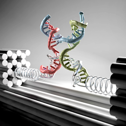
Nanoclamp made of DNA strands. Illustration: Christoph Hohmann

Nanoclamp made of DNA strands. Illustration: Christoph Hohmann

An Oct. 21, 2016 news item on ScienceDaily announces a new nanotool,

Physicists at Ludwig-Maximilians-Universitat (LMU) in Munich have developed a novel nanotool that provides a facile means of characterizing the mechanical properties of biomolecules.

An Oct. 21, 2016 Ludwig-Maximilians-Universitat (LMU) press release (also on EurekAlert), which originated the news item, explains the work in more detail (Note: A link has been removed),

Faced with the thousands of proteins and genes found in virtually every cell in the body, biologists want to know how they all work exactly: How do they interact to carry out their specific functions and how do they respond and adapt to perturbations? One of the crucial factors in all of these processes is the question of how biomolecules react to the minuscule forces that operate at the molecular level. LMU physicists led by Professor Tim Liedl, in collaboration with researchers at the Technical University in Braunschweig and at Regensburg University, have come up with a method that allows them to exert a constant force on a single macromolecule with dimensions of a few nanometers, and to observe the molecule’s response. The researchers can this way test whether or not a protein or a gene is capable of functioning normally when its structure is deformed by forces of the magnitude expected in the interior of cells. This new method of force spectroscopy uses self-assembled nanoscopic power gauges, requires no macroscopic tools and can analyze large numbers of molecules in parallel, which speeds up the process of data acquisition enormously.

With their new approach, the researchers have overcome two fundamental limitations of the most commonly used force spectroscopy instruments. In the case of force microscopy and methodologies based on optical or magnetic tweezers, the molecules under investigation are always directly connected to a macroscopic transducer. They require precise control of the position of an object – a sphere or a sharp metal tip on the order of a micrometer in size – that exerts a force on molecules that are anchored to that object. This strategy is technically extremely demanding and the data obtained is often noisy. Furthermore, these procedures can only be used to probe molecules one at a time. The new method dispenses with all these restrictions. “The structures we use operate completely autonomously“, explains Philipp Nickels, a member of Tim Liedl’s research group. “And we can use them to study countless numbers of molecules simultaneously.”

A feather-light touch

The members of the Munich group, which is affiliated with the Cluster of Excellence NIM (Nanosystems Initiative Munich), are acknowledged masters of “DNA origami”. This methodology exploits the base-pairing properties of DNA for the construction of nanostructures from strands that fold up and pair locally in a manner determined by their nucleotide sequences. In the present case, the DNA sequences are programmed to interact with each other in such a way that the final structure is a molecular clamp that can be programmed to exert a defined force on a test molecule. To this end, a single-stranded DNA that contains a specific sequence capable of recruiting the molecule of interest spans from one arm of the clamp to the other. The applied force can then be tuned by changing the length of the single strand base by base. “That is equivalent to stretching a spring ever so-o-o slightly,” says Nickels. Indeed, by this means it is possible to apply extremely tiny forces between 1 and 15 pN (1 pN = one billionth of a Newton) – comparable in magnitude to those that act on proteins and genes in cells. “In principle, we can capture any type of biomolecule with these clamps and investigate its physical properties,” says Tim Liedl.

The effect of the applied force is read out by taking advantage of the phenomenon of Förster Resonant Energy Transfer (FRET). “FRET involves the transfer of energy between two fluorescent dyes and is strongly dependent on the distance between them.” explains Professor Philip Tinnefeld from TU Braunschweig. When the force applied to the test molecule is sufficient to deform it, the distance between the fluorescent markers changes and the magnitude of energy transfer serves as an exquisitely precise measure of the distortion of the test molecule on the nanometer scale.

Together with Dina Grohmann from Universität Regensburg, the team has used the new technique to investigate the properties of the so-called TATA Binding Protein, an important gene regulator which binds to a specific upstream nucleotide sequence in genes and helps to trigger their expression. They found that the TATA protein can no longer perform its normal function if its target sequence is subjected to a force of more than 6 pN. – The new technology has just made its debut. But since the clamps are minuscule and operate autonomously, it may well be possible in the future to use them to study molecular processes in living cells in real time.

Sometimes reading these news releases, my mind is boggled. What an extraordinary time to live.

Here’s a link to and a citation for the paper,

Molecular force spectroscopy with a DNA origami–based nanoscopic force clamp by Philipp C. Nickels, Bettina Wünsch, Phil Holzmeister, Wooli Bae, Luisa M. Kneer, Dina Grohmann, Philip Tinnefeld, Tim Lied. Science  21 Oct 2016: Vol. 354, Issue 6310, pp. 305-307 DOI: 10.1126/science.aah5974

This paper is behind a paywall.