Tag Archives: Michael F. Crommie

A graphene ‘camera’ and your beating heart: say cheese

Comparing it to a ‘camera’, even with the quotes, is a bit of a stretch for my taste but I can’t come up with a better comparison. Here’s a video so you can judge for yourself,

Caption: This video repeats three times the graphene camera images of a single beat of an embryonic chicken heart. The images, separated by 5 milliseconds, were measured by a laser bouncing off a graphene sheet lying beneath the heart. The images are about 2 millimeters on a side. Credit: UC Berkeley images by Halleh Balch, Alister McGuire and Jason Horng

A June 16, 2021 news item on ScienceDaily announces the research,

Bay Area [San Francisco, California] scientists have captured the real-time electrical activity of a beating heart, using a sheet of graphene to record an optical image — almost like a video camera — of the faint electric fields generated by the rhythmic firing of the heart’s muscle cells.

A University of California at Berkeley (UC Berkeley) June 16, 2021 news release (also on EurekAlert) by Robert Sanders, which originated the news item, provides more detail,

The graphene camera represents a new type of sensor useful for studying cells and tissues that generate electrical voltages, including groups of neurons or cardiac muscle cells. To date, electrodes or chemical dyes have been used to measure electrical firing in these cells. But electrodes and dyes measure the voltage at one point only; a graphene sheet measures the voltage continuously over all the tissue it touches.

The development, published online last week in the journal Nano Letters, comes from a collaboration between two teams of quantum physicists at the University of California, Berkeley, and physical chemists at Stanford University.

“Because we are imaging all cells simultaneously onto a camera, we don’t have to scan, and we don’t have just a point measurement. We can image the entire network of cells at the same time,” said Halleh Balch, one of three first authors of the paper and a recent Ph.D. recipient in UC Berkeley’s Department of Physics.

While the graphene sensor works without having to label cells with dyes or tracers, it can easily be combined with standard microscopy to image fluorescently labeled nerve or muscle tissue while simultaneously recording the electrical signals the cells use to communicate.

“The ease with which you can image an entire region of a sample could be especially useful in the study of neural networks that have all sorts of cell types involved,” said another first author of the study, Allister McGuire, who recently received a Ph.D. from Stanford and. “If you have a fluorescently labeled cell system, you might only be targeting a certain type of neuron. Our system would allow you to capture electrical activity in all neurons and their support cells with very high integrity, which could really impact the way that people do these network level studies.”

Graphene is a one-atom thick sheet of carbon atoms arranged in a two-dimensional hexagonal pattern reminiscent of honeycomb. The 2D structure has captured the interest of physicists for several decades because of its unique electrical properties and robustness and its interesting optical and optoelectronic properties.

“This is maybe the first example where you can use an optical readout of 2D materials to measure biological electrical fields,” said senior author Feng Wang, UC Berkeley professor of physics. “People have used 2D materials to do some sensing with pure electrical readout before, but this is unique in that it works with microscopy so that you can do parallel detection.”

The team calls the tool a critically coupled waveguide-amplified graphene electric field sensor, or CAGE sensor.

“This study is just a preliminary one; we want to showcase to biologists that there is such a tool you can use, and you can do great imaging. It has fast time resolution and great electric field sensitivity,” said the third first author, Jason Horng, a UC Berkeley Ph.D. recipient who is now a postdoctoral fellow at the National Institute of Standards and Technology. “Right now, it is just a prototype, but in the future, I think we can improve the device.”

Graphene is sensitive to electric fields

Ten years ago, Wang discovered that an electric field affects how graphene reflects or absorbs light. Balch and Horng exploited this discovery in designing the graphene camera. They obtained a sheet of graphene about 1 centimeter on a side produced by chemical vapor deposition in the lab of UC Berkeley physics professor Michael Crommie and placed on it a live heart from a chicken embryo, freshly extracted from a fertilized egg. These experiments were performed in the Stanford lab of Bianxiao Cui, who develops nanoscale tools to study electrical signaling in neurons and cardiac cells.

The team showed that when the graphene was tuned properly, the electrical signals that flowed along the surface of the heart during a beat were sufficient to change the reflectance of the graphene sheet.

“When cells contract, they fire action potentials that generate a small electric field outside of the cell,” Balch said. “The absorption of graphene right under that cell is modified, so we will see a change in the amount of light that comes back from that position on the large area of graphene.”

In initial studies, however, Horng found that the change in reflectance was too small to detect easily. An electric field reduces the reflectance of graphene by at most 2%; the effect was much less from changes in the electric field when the heart muscle cells fired an action potential.

Together, Balch, Horng and Wang found a way to amplify this signal by adding a thin waveguide below graphene, forcing the reflected laser light to bounce internally about 100 times before escaping. This made the change in reflectance detectable by a normal optical video camera.

“One way of thinking about it is that the more times that light bounces off of graphene as it propagates through this little cavity, the more effects that light feels from graphene’s response, and that allows us to obtain very, very high sensitivity to electric fields and voltages down to microvolts,” Balch said.

The increased amplification necessarily lowers the resolution of the image, but at 10 microns, it is more than enough to study cardiac cells that are several tens of microns across, she said.

Another application, McGuire said, is to test the effect of drug candidates on heart muscle before these drugs go into clinical trials to see whether, for example, they induce an unwanted arrhythmia. To demonstrate this, he and his colleagues observed the beating chicken heart with CAGE and an optical microscope while infusing it with a drug, blebbistatin, that inhibits the muscle protein myosin. They observed the heart stop beating, but CAGE showed that the electrical signals were unaffected.

Because graphene sheets are mechanically tough, they could also be placed directly on the surface of the brain to get a continuous measure of electrical activity — for example, to monitor neuron firing in the brains of those with epilepsy or to study fundamental brain activity. Today’s electrode arrays measure activity at a few hundred points, not continuously over the brain surface.

“One of the things that is amazing to me about this project is that electric fields mediate chemical interactions, mediate biophysical interactions — they mediate all sorts of processes in the natural world — but we never measure them. We measure current, and we measure voltage,” Balch said. “The ability to actually image electric fields gives you a look at a modality that you previously had little insight into.”

Here’s a link to and a citation for the paper,

Graphene Electric Field Sensor Enables Single Shot Label-Free Imaging of Bioelectric Potentials by Halleh B. Balch, Allister F. McGuire, Jason Horng, Hsin-Zon Tsai, Kevin K. Qi, Yi-Shiou Duh, Patrick R. Forrester, Michael F. Crommie, Bianxiao Cui, and Feng Wang. Nano Lett. 2021, XXXX, XXX, XXX-XXX OI: https://doi.org/10.1021/acs.nanolett.1c00543 Publication Date: June 8, 2021 © 2021 American Chemical Society

This paper is behind a paywall.
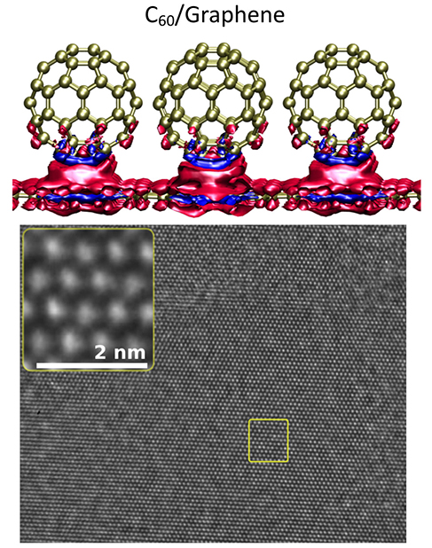
Psst: secret marriage … Buckyballs and Graphene get together!

A March 1, 2018 news item on Nanowerk announces  a new coupling,

Scientists combined buckyballs, [also known as buckminsterfullerenes, fullerenes, or C60] which resemble tiny soccer balls made from 60 carbon atoms, with graphene, a single layer of carbon, on an underlying surface. Positive and negative charges can transfer between the balls and graphene depending on the nature of the surface as well as the structural order and local orientation of the carbon ball. Scientists can use this architecture to develop tunable junctions for lightweight electronic devices.

The researchers have made this illustration of their work available,

Researchers are developing new, lightweight electronics that rapidly conduct electricity by covering a sheet of carbon (graphene) with buckyballs. Electricity is the flow of electrons. On these lightweight structures, electrons as well as positive holes (missing electrons) transfer between the balls and graphene. The team showed that the crystallinity and orientation of the balls, as well as the underlying layer, affected this charge transfer. The top image shows a calculation of the charge density for a specific orientation of the balls on graphene. The blue represents positive charges, while the red is negative. The bottom image shows that the balls are in a close-packed structure. The bright dots correspond to the projected images of columns of buckyball molecules. Courtesy: US Department of Energy Office of Science

A February 28, 2018 US Department of Energy (DoE) Office of Science news release, which originated the news item, provides more detail,

The Impact

Fast-moving electrons and their counterpart, holes, were preserved in graphene with crystalline buckyball overlayers. Significantly, the carbon ball provides charge transfer to the graphene. Scientists expect the transfer to be highly tunable with external voltages. This marriage has ramifications for smart electronics that run longer and do not break as easily, bringing us closer to sensor-embedded smart clothing and robotic skin.

Summary

Charge transfer at the interface between dissimilar materials is at the heart of almost all electronic technologies such as transistors and photovoltaic devices. In this study, scientists studied charge transfer at the interface region of buckyball molecules deposited on graphene, with and without a supporting substrate, such as hexagonal boron nitride. They employed ab initio density functional theory with van der Waals interactions to model the structure theoretically. Van der Waals interactions are weak connections between neutral molecules. The team used high-resolution transmission electron microscopy and electronic transport measurements to characterize experimentally the properties of the interface. The researchers observed that charge transfer between buckyballs and the graphene was sensitive to the nature of the underlying substrate, in addition, to the crystallinity and local orientation of the buckyballs. These studies open an avenue to devices where buckyball layers on top of graphene can serve as electron acceptors and other buckyball layers as electron donors. Even at room temperature, buckyball molecules were orientationally locked into position. This is in sharp contrast to buckyball molecules in un-doped bulk crystalline configurations, where locking occurs only at low temperature. High electron and hole mobilities are preserved in graphene with crystalline buckyball overlayers. This finding has ramifications for the development of organic high-mobility field-effect devices and other high mobility applications.

Here’s a link to and a citation for the paper,

Molecular Arrangement and Charge Transfer in C60 /Graphene Heterostructures by Claudia Ojeda-Aristizabal, Elton J. G. Santos, Seita Onishi, Aiming Yan, Haider I. Rasool, Salman Kahn, Yinchuan Lv, Drew W. Latzke, Jairo Velasco Jr., Michael F. Crommie, Matthew Sorensen, Kenneth Gotlieb, Chiu-Yun Lin, Kenji Watanabe, Takashi Taniguchi, Alessandra Lanzara, and Alex Zettl. ACS Nano, 2017, 11 (5), pp 4686–4693 DOI: 10.1021/acsnano.7b00551 Publication Date (Web): April 24, 2017

Copyright © 2017 American Chemical Society

This paper is behind a paywall.

PlasCarb: producing graphene and renewable hydrogen from food waster

I have two tidbits about PlasCarb the first being an announcement of its existence and the second an announcement of its recently published research. A Jan. 13, 2015 news item on Nanowerk describes the PlasCarb project (Note: A link has been removed),

The Centre for Process Innovation (CPI) is leading a European collaborative project that aims to transform food waste into a sustainable source of significant economic added value, namely graphene and renewable hydrogen.

The project titled PlasCarb will transform biogas generated by the anaerobic digestion of food waste using an innovative low energy microwave plasma process to split biogas (methane and carbon dioxide) into high value graphitic carbon and renewable hydrogen.

A Jan. 13, 2015 CPI press release, which originated the news item, describes the project and its organization in greater detail,

CPI  as the coordinator of the project is responsible for the technical aspects in the separation of biogas into methane and carbon dioxide, and separating of the graphitic carbon produced from the renewable hydrogen. The infrastructure at CPI allows for the microwave plasma process to be trialled and optimised at pilot production scale, with a future technology roadmap devised for commercial scale manufacturing.

Graphene is one of the most interesting inventions of modern times. Stronger than steel, yet light, the material conducts electricity and heat. It has been used for a wide variety of applications, from strengthening tennis rackets, spray on radiators, to building semiconductors, electric circuits and solar cells.

The sustainable creation of graphene and renewable hydrogen from food waste in provides a sustainable method towards dealing with food waste problem that the European Union faces. It is estimated that 90 million tonnes of food is wasted each year, a figure which could rise to approximately 126 million tonnes by 2020. In the UK alone, food waste equates to a financial loss to business of at least £5 billion per year.

Dr Keith Robson, Director of Formulation and Flexible Manufacturing at CPI said, “PlasCarb will provide an innovative solution to the problems associated with food waste, which is one of the biggest challenges that the European Union faces in the strive towards a low carbon economy.  The project will not only seek to reduce food waste but also use new technological methods to turn it into renewable energy resources which themselves are of economic value, and all within a sustainable manner.”

PlasCarb will utilise quality research and specialist industrial process engineering to optimise the quality and economic value of the Graphene and hydrogen, further enhancing the sustainability of the process life cycle.

Graphitic carbon has been identified as one of Europe’s economically critical raw materials and of strategic performance in the development of future emerging technologies. The global market for graphite, either mined or synthetic is worth over €10 billion per annum. Hydrogen is already used in significant quantities by industry and recognised with great potential as a future transport fuel for a low carbon economy. The ability to produce renewable hydrogen also has added benefits as currently 95% of hydrogen is produced from fossil fuels. Moreover, it is currently projected that increasing demand of raw materials from fossil sources will lead to price volatility, accelerated environmental degradation and rising political tensions over resource access.

Therefore, the latter stages of the project will be dedicated to the market uptake of the PlasCarb process and the output products, through the development of an economically sustainable business strategy, a financial risk assessment of the project results and a flexible financial model that is able to act as a primary screen of economic viability. Based on this, an economic analysis of the process will be determined. Through the development of a decentralised business model for widespread trans-European implementation, the valorisation of food waste will have the potential to be undertaken for the benefit of local economies and employment. More specifically, three interrelated post project exploitation markets have been defined: food waste management, high value graphite and RH2 sales.

PlasCarb is a 3-year collaborative project, co-funded under the European Union’s Seventh Framework Programme (FP7) and will further reinforce Europe’s leading position in environmental technologies and innovation in high value Carbon. The consortium is composed of eight partners led by CPI from five European countries, whose complimentary research and industrial expertise will enable the required results to be successfully delivered. The project partners are; The Centre for Process Innovation (UK), GasPlas AS (NO), CNRS (FR), Fraunhofer IBP (DE), Uvasol Ltd (UK), GAP Waste Management (UK), Geonardo Ltd. (HU), Abalonyx AS (NO).

You can find PlasCarb here.

The second announcement can be found in a PlasCarb Jan. 14, 2015 press release announcing the publication of research on heterostructures of graphene ribbons,

Few materials have received as much attention from the scientific world or have raised so many hopes with a view to their potential deployment in new applications as graphene has. This is largely due to its superlative properties: it is the thinnest material in existence, almost transparent, the strongest, the stiffest and at the same time the most strechable, the best thermal conductor, the one with the highest intrinsic charge carrier mobility, plus many more fascinating features. Specifically, its electronic properties can vary enormously through its confinement inside nanostructured systems, for example. That is why ribbons or rows of graphene with nanometric widths are emerging as tremendously interesting electronic components. On the other hand, due to the great variability of electronic properties upon minimal changes in the structure of these nanoribbons, exact control on an atomic level is an indispensable requirement to make the most of all their potential.

The lithographic techniques used in conventional nanotechnology do not yet have such resolution and precision. In the year 2010, however, a way was found to synthesise nanoribbons with atomic precision by means of the so-called molecular self-assembly. Molecules designed for this purpose are deposited onto a surface in such a way that they react with each other and give rise to perfectly specified graphene nanoribbons by means of a highly reproducible process and without any other external mediation than heating to the required temperature. In 2013 a team of scientists from the University of Berkeley and the Centre for Materials Physics (CFM), a mixed CSIC (Spanish National Research Council) and UPV/EHU (University of the Basque Country) centre, extended this very concept to new molecules that were forming wider graphene nanoribbons and therefore with new electronic properties. This same group has now managed to go a step further by creating, through this self-assembly, heterostructures that blend segments of graphene nanoribbons of two different widths.

The forming of heterostructures with different materials has been a concept widely used in electronic engineering and has enabled huge advances to be made in conventional electronics. “We have now managed for the first time to form heterostructures of graphene nanoribbons modulating their width on a molecular level with atomic precision. What is more, their subsequent characterisation by means of scanning tunnelling microscopy and spectroscopy, complemented with first principles theoretical calculations, has shown that it gives rise to a system with very interesting electronic properties which include, for example, the creation of what are known as quantum wells,” pointed out the scientist Dimas de Oteyza, who has participated in this project. This work, the results of which are being published this very week in the journal Nature Nanotechnology, therefore constitutes a significant success towards the desired deployment of graphene in commercial electronic applications.

Here’s a link to and a citation for the paper,

Molecular bandgap engineering of bottom-up synthesized graphene nanoribbon heterojunctions by Yen-Chia Chen, Ting Cao, Chen Chen, Zahra Pedramrazi, Danny Haberer, Dimas G. de Oteyza, Felix R. Fischer, Steven G. Louie, & Michael F. Crommie. Nature Nanotechnology (2015) doi:10.1038/nnano.2014.307 Published online 12 January 2015

This article is behind a paywall but there is a free preview available via ReadCube access.