Tag Archives: microRNAs

Nanoparticles can activate viruses lying dormant in lung cells

The nanoparticles in question are from combustion engines, which means that we are exposed to them. One other note, the testing has not been done on humans but rather on cells. From a Jan. 16, 2017 news item on ScienceDaily,

Nanoparticles from combustion engines can activate viruses that are dormant in in lung tissue cells. This is the result of a study by researchers of Helmholtz Zentrum München, a partner in the German Center for Lung Research (DZL), which has now been published in the journal Particle and Fibre Toxicology.

To evade the immune system, some viruses hide in cells of their host and persist there. In medical terminology, this state is referred to as a latent infection. If the immune system becomes weakened or if certain conditions change, the viruses become active again, begin to proliferate and destroy the host cell. A team of scientists led by Dr. Tobias Stöger of the Institute of Lung Biology and Prof. Dr. Heiko Adler, deputy head of the research unit Lung Repair and Regeneration at Helmholtz Zentrum München, now report that nanoparticles can also trigger this process.

A Jan. 16, 2017 Helmholtz Zentrum München press release (also on EurekAlert), which originated the news item, provides more detail,

“From previous model studies we already knew that the inhalation of nanoparticles has an inflammatory effect and alters the immune system,” said study leader Stöger. Together with his colleagues Heiko Adler and Prof. Dr. Philippe Schmitt-Kopplin, he showed that “an exposure to nanoparticles can reactivate latent herpes viruses in the lung.”

Specifically, the scientists tested the influence of nanoparticles typically generated by fossil fuel combustion in an experimental model for a particular herpes virus infection. They detected a significant increase in viral proteins, which are only produced with active virus proliferation. “Metabolic and gene expression analyses also revealed patterns resembling acute infection,” said Philippe Schmitt-Kopplin, head of the research unit Analytical BioGeoChemistry (BGC). Moreover, further experiments with human cells demonstrated that Epstein-Barr viruses are also ‘awakened’ when they come into contact with the nanoparticles.

Potential approach for chronic lung diseases

In further studies, the research team would like to test whether the results can also be transferred to humans. “Many people carry herpes viruses, and patients with idiopathic pulmonary fibrosis are particularly affected,” said Heiko Adler. “If the results are confirmed in humans, it would be important to investigate the molecular process of the reactivation of latent herpes viruses induced by particle inhalation. Then we could try to influence this pathway therapeutically.”

Special cell culture models shall therefore elucidate the exact mechanism of virus reactivation by nanoparticles. “In addition,” Stöger said, ”in long-term studies we would like to investigate to what extent  repeated nanoparticle exposure with corresponding virus reactivation leads to chronic inflammatory and remodeling processes in the lung.”

Further Information

Background:
In 2015 another group at the Helmholtz Zentrum München demonstrated how the Epstein-Barr virus  hides in human cells. In March 2016 researchers also showed that microRNAs silence immune alarm signals of cells infected with the Epstein-Barr virus.

Original Publication:
Sattler, C. et al. (2016): Nanoparticle exposure reactivates latent herpesvirus and restores a signature of acute infection. Particle and Fibre Toxicology, DOI 10.1186/s12989-016-0181-1

Here’s a link to and a citation for the paper on investigating latent herpes virus,

Nanoparticle exposure reactivates latent herpesvirus and restores a signature of acute infection by Christine Sattler, Franco Moritz, Shanze Chen, Beatrix Steer, David Kutschke, Martin Irmler, Johannes Beckers, Oliver Eickelberg, Philippe Schmitt-Kopplin, Heiko Adler. Particle and Fibre Toxicology201714:2 DOI: 10.1186/s12989-016-0181-1 Published: 10 January 2017

©  The Author(s). 2017

This paper is open access and, so too, is the 2016 paper.
Attomolar cancer detection: measuring microRNAs in blood

The latest research does not lead to a magical disease detector where nanoscale sensors swim through the body continuously monitoring our health and alerting us should something untoward occur (see this Oct. 28, 2014 article on RT.com for more about Google X’s development plans for it and this Nov. 11, 2015 news item on Nanowerk for a measured response from a researcher in the field).

Now onto some real research, a Nov. 17, 2015 news item on ScienceDaily announces an ultrasensitive (attoscale) sensor employing gold nanoparticles for detecting cancer,

A simple, ultrasensitive microRNA sensor developed and tested by researchers from the schools of science and medicine at Indiana University-Purdue University Indianapolis and the Indiana University Melvin and Bren Simon Cancer Center holds promise for the design of new diagnostic strategies and, potentially, for the prognosis and treatment of pancreatic and other cancers.

A Nov. 17, 2015 Indiana University-Purdue University Indianapolis news release on EurekAlert, which originated the news item, provides more detail about research that seems to have focused largely on pancreatic cancer detection (Note: Links have been removed),

In a study published in the Nov. [2015] issue of ACS Nano, a peer-reviewed journal of the American Chemical Society focusing on nanoscience and nanotechnology research, the IUPUI researchers describe their design of the novel, low-cost, nanotechnology-enabled reusable sensor. They also report on the promising results of tests of the sensor’s ability to identify pancreatic cancer or indicate the existence of a benign condition by quantifying changes in levels of microRNA signatures linked to pancreatic cancer. MicroRNAs are small molecules of RNA that regulate how larger RNA molecules lead to protein expression. As such, microRNAs are very important in biology and disease states.

“We used the fundamental concepts of nanotechnology to design the sensor to detect and quantify biomolecules at very low concentrations,” said Rajesh Sardar, Ph.D., who developed the sensor.

“We have designed an ultrasensitive technique so that we can see minute changes in microRNA concentrations in a patient’s blood and confirm the presence of pancreatic cancer.” Sardar is an assistant professor of chemistry and chemical biology in the School of Science at IUPUI and leads an interdisciplinary research program focusing on the intersection of analytical chemistry and the nanoscience of metallic nanoparticles.

“If we can establish that there is cancer in the pancreas because the sensor detects high levels of microRNA-10b or one of the other microRNAs associated with that specific cancer, we may be able to treat it sooner,” said Murray Korc, M.D., the Myles Brand Professor of Cancer Research at the IU School of Medicine and a researcher at the IU Simon Cancer Center. Korc, worked with Sardar to improve the sensor’s capabilities and led the testing of the sensor and its clinical uses as well as advancing the understanding of pancreatic cancer biology.

“That’s especially significant for pancreatic cancer, because for many patients it is symptom-free for years or even a decade or more, by which time it has spread to other organs, when surgical removal is no longer possible and therapeutic options are limited,” said Korc. “For example, diagnosis of pancreatic cancer at an early stage of the disease followed by surgical removal is associated with a 40 percent five-year survival. Diagnosis of metastatic pancreatic cancer, by contrast, is associated with life expectancy that is often only a year or less.

“The beauty of the sensor designed by Dr. Sardar is its ability to accurately detect mild increases in microRNA levels, which could allow for early cancer diagnosis,” Korc added.

Over the past decade, studies have shown that microRNAs play important roles in cancer and other diseases, such as diabetes and cardiovascular disorders. The new IUPUI nanotechnology-based sensor can detect changes in any of these microRNAs.

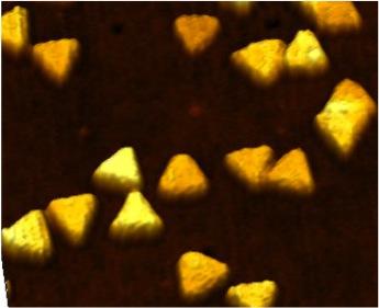
The sensor is a small glass chip that contains triangular-shaped gold nanoparticles called ‘nanoprisms.’ After dipping it in a sample of blood or another body fluid, the scientist measures the change in the nanoprism’s optical property to determine the levels of specific microRNAs.

For anyone concerned about the cost associated with creating sensors based on gold, about patents, or about current techniques for monitoring microRNAs, there’s more from the news release (Note: A link has been removed),

“Using gold nanoprisms may sound expensive, but it isn’t because these particles are so very tiny,” Sardar said. “It’s a rather cheap technique because it uses nanotechnology and needs very little gold. $250 worth of gold makes 4,000 sensors. Four thousand sensors allow you to do at least 4,000 tests. The low cost makes this technique ideal for use anywhere, including in low-resource environments in this country and around the world.”

Indiana University Research and Technology Corporation has filed a patent application on Sardar’s and Korc’s groundbreaking nanotechnology-enabled sensor. The researchers’ ultimate goal is to design ultrasensitive and extremely selective low-cost point-of-care diagnostics enabling individual therapeutic approaches to diseases.

Currently, polymerase chain reaction technology is used to determine microRNA signatures, which requires extraction of the microRNA from blood or other biological fluid and reverse transcription or amplification of the microRNA. PCR provides relative values. By contrast, the process developed at IUPUI is simpler, quantitative, more sensitive and highly specific even when two different microRNAs vary in a single position. The study demonstrated that the IUPUI nanotechnology-enabled sensor is as good as if not better than the most advanced PCR in detection and quantification of microRNA.

Here’s a link to and a citation for the paper,

Label-Free Nanoplasmonic-Based Short Noncoding RNA Sensing at Attomolar Concentrations Allows for Quantitative and Highly Specific Assay of MicroRNA-10b in Biological Fluids and Circulating Exosomes by Gayatri K. Joshi, Samantha Deitz-McElyea, Thakshila Liyanage, Katie Lawrence, Sonali Mali, Rajesh Sardar*, and Murray Korc. ACS Nano, Article ASAP DOI: 10.1021/acsnano.5b04527 Publication Date (Web): October 7, 2015

Copyright © 2015 American Chemical Society

This is an open access paper.

The researchers have provided this illustration of gold nanoprisms,

Caption: Indiana University-Purdue University Indianapolis researchers have developed a novel, low-cost, nanotechnology-enabled reusable sensor for which a patent application has been filed. Credit: Department of Chemistry and Chemical Biology, School of Science, Indiana University-Purdue University Indianapolis

Caption: Indiana University-Purdue University Indianapolis researchers have developed a novel, low-cost, nanotechnology-enabled reusable sensor for which a patent application has been filed. Credit: Department of Chemistry and Chemical Biology, School of Science, Indiana University-Purdue University Indianapolis