Tag Archives: nanogold

Gold nanoparticle tattoo changes medical diagnostics?

The tattoos are in fact implantable sensors. Here’s more from an April 6, 2021 news item on ScienceDaily,

The idea of implantable sensors that continuously transmit information on vital values and concentrations of substances or drugs in the body has fascinated physicians and scientists for a long time. Such sensors enable the constant monitoring of disease progression and therapeutic success. However, until now implantable sensors have not been suitable to remain in the body permanently but had to be replaced after a few days or weeks. On the one hand, there is the problem of implant rejection because the body recognizes the sensor as a foreign object. On the other hand, the sensor’s color which indicates concentration changes has been unstable so far and faded over time. Scientists at Johannes Gutenberg University Mainz (JGU) have developed a novel type of implantable sensor which can be operated in the body for several months. The sensor is based on color-stable gold nanoparticles that are modified with receptors for specific molecules. Embedded into an artificial polymeric tissue, the nanogold is implanted under the skin where it reports changes in drug concentrations by changing its color.

An April 6, 2021 Johannes Gutenberg Universitaet Mainz press release (also on EurekAlert), which originated the news item, provides more detail about the proposed tattoo/implantable sensors,

Implant reports information as an “invisible tattoo”

Professor Carsten Sönnichsen’s research group at JGU has been using gold nanoparticles as sensors to detect tiny amounts of proteins in microscopic flow cells for many years. Gold nanoparticles act as small antennas for light: They strongly absorb and scatter it and, therefore, appear colorful. They react to alterations in their surrounding by changing color. Sönnichsen’s team has exploited this concept for implanted medical sensing.

To prevent the tiny particles from swimming away or being degraded by immune cells, they are embedded in a porous hydrogel with a tissue-like consistency. Once implanted under the skin, small blood vessels and cells grow into the pores. The sensor is integrated in the tissue and is not rejected as a foreign body. “Our sensor is like an invisible tattoo, not much bigger than a penny and thinner than one millimeter,” said Professor Carsten Sönnichsen, head of the Nanobiotechnology Group at JGU. Since the gold nanoparticles are infrared, they are not visible to the eye. However, a special kind of measurement device can detect their color noninvasively through the skin.

In their study published in Nano Letters, the JGU researchers implanted their gold nanoparticle sensors under the skin of hairless rats. Color changes in these sensors were monitored following the administration of various doses of an antibiotic. The drug molecules are transported to the sensor via the bloodstream. By binding to specific receptors on the surface of the gold nanoparticles, they induce color change that is dependent on drug concentration. Thanks to the color-stable gold nanoparticles and the tissue-integrating hydrogel, the sensor was found to remain mechanically and optically stable over several months.

Huge potential of gold nanoparticles as long-lasting implantable medical sensors

“We are used to colored objects bleaching over time. Gold nanoparticles, however, do not bleach but keep their color permanently. As they can be easily coated with various different receptors, they are an ideal platform for implantable sensors,” explained Dr. Katharina Kaefer, first author of the study.

The novel concept is generalizable and has the potential to extend the lifetime of implantable sensors. In future, gold nanoparticle-based implantable sensors could be used to observe concentrations of different biomarkers or drugs in the body simultaneously. Such sensors could find application in drug development, medical research, or personalized medicine, such as the management of chronic diseases.

Interdisciplinary team work brought success

Sönnichsen had the idea of using gold nanoparticles as implanted sensors already in 2004 when he started his research in biophysical chemistry as a junior professor in Mainz. However, the project was not realized until ten years later in cooperation with Dr. Thies Schroeder and Dr. Katharina Kaefer, both scientists at JGU. Schroeder was experienced in biological research and laboratory animal science and had already completed several years of research work in the USA. Kaefer was looking for an exciting topic for her doctorate and was particularly interested in the complex and interdisciplinary nature of the project. Initial results led to a stipend awarded to Kaefer by the Max Planck Graduate Center (MPGC) as well as financial support from Stiftung Rheinland-Pfalz für Innovation. “Such a project requires many people with different scientific backgrounds. Step by step we were able to convince more and more people of our idea,” said Sönnichsen happily. Ultimately, it was interdisciplinary teamwork that resulted in the successful development of the first functional implanted sensor with gold nanoparticles.

The researchers have provided an image which illustrates several elements described in the press release,
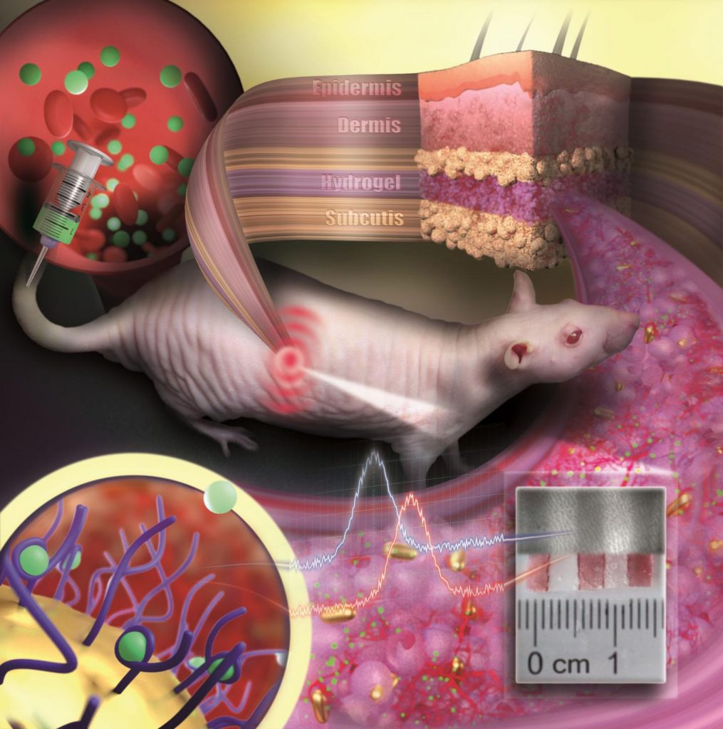
Caption: Gold nanoparticles embedded in a porous hydrogel can be implanted under the skin and used as medical sensors. The sensor is like an invisible tattoo revealing concentration changes of substances in the blood by color change. Credit: ill./©: Nanobiotechnology Group, JGU Department of Chemistry

Here’s a link to and a citation for the paper,

Implantable Sensors Based on Gold Nanoparticles for Continuous Long-Term Concentration Monitoring in the Body by Katharina Kaefer, Katja Krüger, Felix Schlapp, Hüseyin Uzun, Sirin Celiksoy, Bastian Flietel, Axel Heimann, Thies Schroeder, Oliver Kempski, and Carsten Sönnichsen. Nano Lett. 2021, XXXX, XXX, XXX-XXX DOI: https://doi.org/10.1021/acs.nanolett.1c00887 Publication Date:March 30, 2021 © 2021 The Authors. Published by American Chemical Society

This paper is behind a paywall.
Single molecule nanogold-based probe for photoacoustic Imaging and SERS biosensing

As I understand it, the big deal is that A*STAR (Singapore’s Agency for Science, Rechnology and Research) scientists have found a way to make a single molecule probe do the work of a two-molecule probe when imaging tumours. From a July 29, 2015 news item on Nanowerk (Note: A link has been removed),

An organic dye that can light up cancer cells for two powerful imaging techniques providing complementary diagnostic information has been developed and successfully tested in mice by A*STAR researchers (“Single Molecule with Dual Function on Nanogold: Biofunctionalized Construct for In Vivo Photoacoustic Imaging and SERS Biosensing”).

A July 29, 2015 A*STAR news release, which originated the news item, describes the currently used multimodal imaging technique and provides details about the new single molecule technique,

Imaging tumors is vitally important for cancer research, but each imaging technique has its own limitations for studying cancer in living organisms. To overcome the limitations of individual techniques, researchers typically employ a combination of various imaging methods — a practice known as multimodal imaging. In this way, they can obtain complementary information and hence a more complete picture of cancer.

Two very effective methods for imaging tumors are photoacoustic imaging and surface-enhanced Raman scattering (SERS). Photoacoustic imaging can image deep tissue with a good resolution, whereas SERS detects miniscule amounts of a target molecule. To simultaneously use both photoacoustic imaging and SERS, a probe must produce signals for both imaging modalities.

In multimodal imaging, researchers typically combine probes for each imaging modality into a single two-molecule probe. However, the teams of Malini Olivo at the A*STAR Singapore Bioimaging Consortium and Bin Liu at the A*STAR Institute of Materials Research and Engineering, along with overseas collaborator Ben Zhong Tang from the Hong Kong University of Science and Technology, adopted a different approach — they developed single-molecule probes that can be used for both photoacoustic imaging and SERS. The probes are based on organic cyanine dyes that absorb near-infrared light, which has the advantage of being able to deeply penetrate tissue, enabling tumors deep within the body to be imaged.

Once the team had verified that the probes worked for both imaging modalities, they optimized the performances of the probes by adding gold nanoparticles to them to amplify the SERS signal and by encapsulating them in the polymer polyethylene glycol to stabilize their structures.

The researchers then deployed these optimized probes in live mice. By functionalizing the probes with an antibody that recognizes a tumor cell-surface protein, they were able to use them to target tumors. The scientists found that, in photoacoustic imaging, the tumor-targeted probes produced signals that were roughly three times stronger than those of unmodified probes. Using SERS, the team was also able to monitor the concentrations of the probes in the tumor, spleen and liver in real time with a high degree of sensitivity.

U. S. Dinish, a senior scientist in Olivo’s group, recalls the team’s “surprise at the sensitivity and potential of the nanoconstruct.” He anticipates that the probe could be used to guide surgical removal of tumors.

Here’s a link to and a citation for the paper,

Single Molecule with Dual Function on Nanogold: Biofunctionalized Construct for In Vivo Photoacoustic Imaging and SERS Biosensing by U. S. Dinish, Zhegang Song, Chris Jun Hui Ho, Ghayathri Balasundaram, Amalina Binte Ebrahim Attia, Xianmao Lu, Ben Zhong Tang, Bin Liu, and Malini Olivo. Advanced Functional Materials, Vol 25 Issue 15
pages 2316–2325, April 15, 2015 DOI: 10.1002/adfm.201404341 Article first published online: 11 MAR 2015

© 2015 WILEY-VCH Verlag GmbH & Co. KGaA, Weinheim

This paper is behind a paywall.

Research into nanosilver’s antibiotic properties and nanogold’s detection skills

There is a puzzling and exciting announcement from the Canadian Light Source in a May 27, 2015 news item on Nanowerk,

Precious metals like silver and gold have biomedical properties that have been used for centuries, but how do these materials effectively combat the likes of cancer and bacteria without contaminating the patient and the environment?

These are the questions that researchers from Dalhousie University and the Canadian Light Source are trying to find out.

Perhaps I’m misreading the announcement but the statement that nanosilver and nanogold don’t contaminate the patient or the environment is a bit exuberant. There are published studies examining questions about whether or not nanosilver may affect the environment and health and the answer is that no one is certain yet. You can read more about two studies highlighted in my February 28, 2013 posting titled:  Silver nanoparticles, water, the environment, and toxicity. As for nanosilver and nanogold not contaminating patients, that too is a problematic statement. For example, I have this paper which cites several studies on nanogold and possible toxicity. The paper itself is a plea to standardize testing and protocols so researchers can do a better job of establishing toxicity issues with nanogold.

GoldNP_ToxicityMar2015

Reservations aside, it’s good to learn of some Canadian research in this area. From a May 26, 2015 Canadian Light Source news release, which originated the news item, provides more details about the research and its current focus on nanosilver,

“Gold and silver are both exciting materials,” said Peng Zhang, Associate Professor of Chemistry at Dalhousie. “We can use gold to either detect or kill cancer cells. Silver is also excited and a very promising material as an antibacterial agents.”

Zhang said that if you compare silver to current antibiotics, silver does not show drug-resistant behaviour. “But with silver, so far, we are not finding that,” he added.

Finding out why silver is such a great antibacterial agent is the focus of Zhang’s research, recently published in the journal Langmuir.

“We want to understand the relationship between the atomic structure and bioactivity of nanosilver as to why it is so efficient at inhibiting bacterial activity. It’s a big puzzle.”

Zhang said it is very hard to understand what is happening at the atomic level. Using small nanosilver particles is the most effective way, because when you make silver small, you can expect higher activity because of the increased surface area.

This poses another problem however, as the nanosilver needs to be stabilized with a coating or the silver particles will bond together forming large pieces of silver that do not efficiently interact with the bacteria.

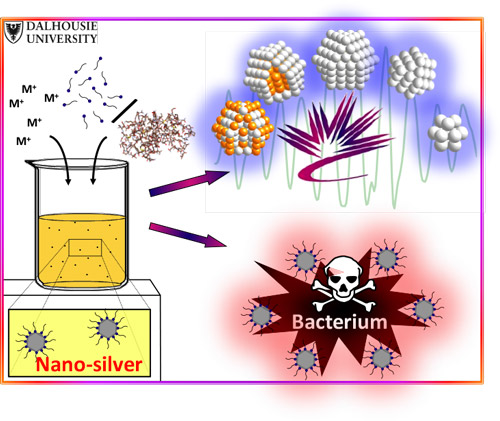
Zhang’s group used two different coatings to compare the effectiveness of the silver as an antibacterial agent. The first was a small amino acid coating and the other was a larger polymer coating. And after testing the interactions between the nanosilver and the bacteria, and looking at the atomic structure of nanosilver using the CLS and the Advanced Photon Source, the researchers were surprised to find that the thicker, larger polymer coating actually created a better delivery method for sliver to inhibit the bacteria.

“We proposed that the small amino acid coating would bind so tightly to the silver surface that it would be difficult for  the silver atoms to interact with the bacteria, whereas the polymers are actually very good at staying in place and still releasing sufficient amount of silver with the bacteria.”

Zhang said the next steps will be to find out if the nanosilver is actually attacking good cells in living systems before they can make any further progress on determining whether nanosilver is an effective and efficient antibactieral agent that could be used to cure human and animal diseases.

Here’s an illustration provided by the researchers,

The atomic structure of nanosilver, revealed by synchrotron X-ray spectroscopy, is proving to be a determinant of silver’s antibacterial activity. Padmos, J. Daniel, et al. "Impact of Protecting Ligands on Surface Structure and Antibacterial Activity of Silver Nanoparticles." Langmuir 31.12 (2015): 3745-3752.

The atomic structure of nanosilver, revealed by synchrotron X-ray spectroscopy, is proving to be a determinant of silver’s antibacterial activity.
Padmos, J. Daniel, et al. “Impact of Protecting Ligands on Surface Structure and Antibacterial Activity of Silver Nanoparticles.” Langmuir 31.12 (2015): 3745-3752.

Here’s a link to and a citation for the paper,

Impact of Protecting Ligands on Surface Structure and Antibacterial Activity of Silver Nanoparticles by J. Daniel Padmos, Robert T. M. Boudreau, Donald F. Weaver, and Peng Zhang. Langmuir, 2015, 31 (12), pp 3745–3752
DOI: 10.1021/acs.langmuir.5b00049 Publication Date (Web): March 15, 2015

Copyright © 2015 American Chemical Society

This paper is behind a paywall.

Nickel-eating plant in the Philippines

For anyone interested in phytoremediation and/or phytomining, this news from the Philippines is quite exciting (from a May 9, 2014 news release on EurekAlert, Note: A link has been removed, (also on ScienceDaily),

Scientists from the University of the Philippines, Los Baños (UPLB) have discovered a new plant species with an unusual lifestyle — it eats nickel for a living — accumulating up to 18,000 ppm of the metal in its leaves without itself being poisoned, says Professor Edwino Fernando, lead author of the report. Such an amount is a hundred to a thousand times higher than in most other plants. The study was published in the open access journal PhytoKeys.

The new species is called Rinorea niccolifera, reflecting its ability to absorb nickel in very high amounts. Nickel hyperaccumulation is such a rare phenomenon with only about 0.5–1% of plant species native to nickel-rich soils having been recorded to exhibit the ability. Throughout the world, only about 450 species are known with this unusual trait, which is still a small proportion of the estimated 300,000 species of vascular plants.

A May 9, 2014 Penfold Publishers news release, which originated the items elsewhere, provides more details and an image of the nickel-eating plant,

The new species, according to Dr Marilyn Quimado, one of the lead scientists of the research team, was discovered on the western part of Luzon Island in the Philippines, an area known for soils rich in heavy metals.

“Hyperacccumulator plants have great potentials for the development of green technologies, for example, ‘phytoremediation’ and ‘phytomining'”, explains Dr Augustine Doronila of the School of Chemistry, University of Melbourne, who is also co-author of the report.

Phytoremediation refers to the use of hyperacccumulator plants to remove heavy metals in contaminated soils. Phytomining, on the other hand, is the use of hyperacccumulator plants to grow and harvest in order to recover commercially valuable metals in plant shoots from metal-rich sites. [emphasis mine]

In a previous piece about phytomining and in contrast to this news release, I suggested that phytoremediation could also function as phytomining (an idea I found elsewhere), my March 5, 2013 posting. There are also a November 22, 2012 posting and a Sept. 26, 2012 posting on the topic of phyto-mining (anyone keen to read everything here on this topic, may want to search the term both with and without hyphens).

Here is the nickel-eating plant,

Caption: This photo shows the newly described metal-eating plant, Rinorea niccolifera. Credit: Dr. Edwino S. Fernando Usage Restrictions: CC-BY 4.0

Caption: This photo shows the newly described metal-eating plant, Rinorea niccolifera.
Credit: Dr. Edwino S. Fernando
Usage Restrictions: CC-BY 4.0

Here’s a link to and a citation for the paper,

Rinorea niccolifera (Violaceae), a new, nickel-hyperaccumulating species from Luzon Island, Philippines by Edwino Fernando, Marilyn Quimado, and Augustine Doronila. PhytoKeys 37: 1–13. doi: 10.3897/phytokeys.37.7136

This paper is open access.

In a burst of curiosity I checked out the University of Philippines website and found some research bearing similarity to today’s (May 9, 2014) piece although in this case the metal being discussed is gold and the researchers are investigating the possibility of using bacteria to produce gold nanoparticles. From an April 16, 2014 article written by Miguel Victor T. Durian for the university’s Horizon magazine,

A pioneering nanotechnology study conducted by scientists at the UPLB National Institute of Molecular Biology and Biotechnology (BIOTECH) is exploring the potentials of plantgrowth- promoting bacteria (PGPB) in the biosynthesis of nanogold.

Dr. Lilia M. Fernando, Dr. Florinia E. Merca, and Dr. Erlinda S. Paterno are looking at how nanogold could be produced in large quantities using PGPB as this could bring down medical diagnostic and treatment costs especially against a dreaded disease – cancer.

“Our study primarily aimed to find a less expensive source of gold through the biosynthesis of the element by microorganisms.” Dr. Fernando explained. “Gold costs around 200 to 300 US dollars (or about Php9,000 to Php14,000), …,” Ms. Fernando added.

Furthermore, PGPB is abundantly available in the soils of the Philippines. In fact, the researchers carried out their collection of PGPB in Tarlac and Bohol. Moreover, cultivation of PGPB does not require any special incubation procedures in order to maintain its nano-size because it can survive at room temperature. This makes the cultivation of PGPB easier and less expensive compared to other microorganisms.

I look forward to hearing more about these projects as they develop.